Major Article
Corresponding author: Dra. Laura Rocha Guerino.
e-mail: [email protected]
Received 18 August 2016
Accepted 6 December 2016
Prevalence and distribution of
Angiostrongylus cantonensis
(Nematoda, Angiostrongylidae) in
Achatina fulica
(Mollusca, Gastropoda) in Baixada Santista,
São Paulo, Brazil
Laura Rocha Guerino
[1],[2], Iracy Lea Pecora
[2], Marcel Sabino Miranda
[3],
Cryslaine Aguiar-Silva
[4], Omar dos Santos Carvalho
[4], Roberta Lima Caldeira
[4]and Reinaldo José da Silva
[1][1]. Laboratório de Parasitologia de Animais Silvestres, Departamento de Parasitologia, Universidade Estadual Paulista Júlio de Mesquita Filho, Botucatu, SP, Brasil. [2]. Laboratório de Moluscos, Universidade Estadual Paulista Júlio de Mesquita Filho, São Vicente, SP, Brasil. [3]. Laboratório de
Malacologia, Departamento de Biologia Animal, Universidade Estadual de Campinas, Campinas, SP, Brasil. [4]. Laboratório de Helmintologia e Malacologia Médica, Centro de Pesquisas René Rachou, Fundação Oswaldo Cruz, Belo Horizonte, MG, Brasil.
Abstract
Introduction
:
Angiostrongylus cantonensis
causes eosinophilic meningoencephalitis in humans. Worldwide expansion of this
nematode is linked to the dispersion of their hosts. This study aimed to determine the prevalence of
A. cantonensis
infection
in
Achatina fulica
in the nine municipalities that make up Baixada Santista, São Paulo, Brazil.
Methods
:
Angiostrongylus
cantonensis
larvae were analyzed using optical microscopy. We performed polymerase chain reaction and restriction fragment
length polymorphism using restriction endonuclease
ClaI
, directed to the internal transcribed spacer region 2 of
A. cantonensis
larval DNA.
Results
: Of the 540 snails analyzed, 117 (21.7%) were infected by
A. cantonensis
. For morphological and
morphometric analyses, 60 larvae were used. Second-stage larvae were, on average, 358.2µm long and 26.4µm wide, while
third-stage larvae were, on average, 450µm long and 21.12µm wide. The tails of the larvae ended in a ine tip.
Conclusions
: All
municipalities comprising Baixada Santista had
A. fulica
that were naturally infected with
A. cantonensis
. All of the observed
characteristics were typical of the species.
Keywords:
Rat lungworm. Giant African snail. Eosinophilic meningitis. Nematode. Emerging parasitosis.
INTRODUCTION
Two of the 19 species from the
Angiostrongylus
genus
can infect humans:
Angiostrongylus costaricensis
(Morera &
Céspedes, 1971), which causes abdominal angiostrongyliasis
1and
Angiostrongylus cantonensis
(Chen, 1935), which is the
etiologic agent of eosinophilic meningoencephalitis, also called
rat lungworm
2.
A. cantonensis
has been observed in several
regions of the world
3-7, and they were distributed from Eastern
Asia to other continents by two main hosts: rats (deinitive hosts)
and
Achatina fulica
Bowdich, 1822 (one of the intermediate
hosts), especially during the Second World War
8. Several species
of land and freshwater snails have also been found to be naturally
infected with
A. cantonensis
9-14.
In Brazil, the occurrence of
A. cantonensis
has been reported
in all states except for Acre
9-17. Man, being an accidental host,
acquires parasitosis when eating foods contaminated with
stage-three larvae (L3), raw or undercooked mollusks, and paratenic
hosts such as shrimp, frogs, ish, and latworms
4,18-20, as well as
crabs and lizards
21,22. In humans, these parasites migrate to the
central nervous system (CNS), where they die in the meninges,
causing inlammatory reactions
23 24.
Achatina fulica
plays a crucial role in the global dispersion
of
A. cantonensis
1,8,22,25,26, since it is present in most areas where
this nematode is endemic. These mollusks are associated with an
anthropic environment, and once established, their population
can signiicantly increase
27. Remains of human activity favor
the adaptation of this mollusk, as such remains provide food
and shelter
28. In Brazil, this mollusk has high potential to be
involved in the transmission of
A. cantonensis
owing to its wide
distribution, including to different ecosystems
29-31.
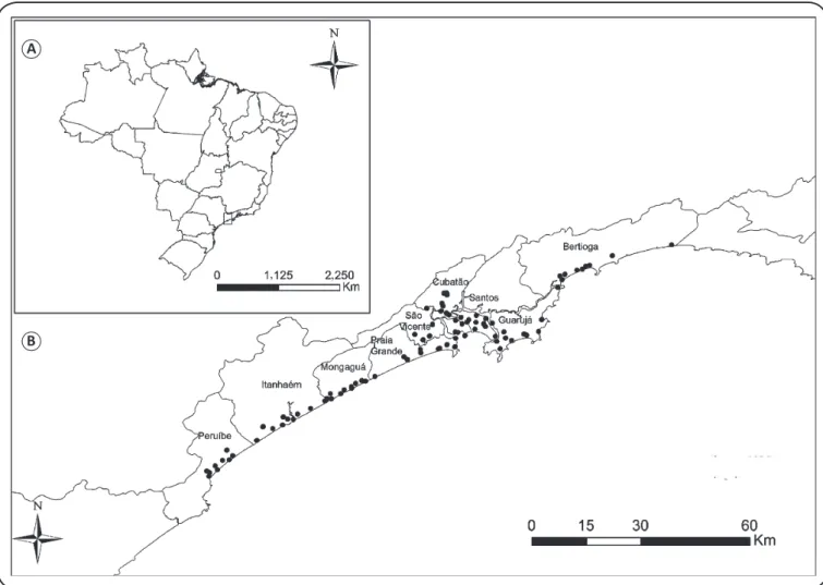
FIGURE 1 – A: Location of the study area and B: collection points in the municipalities comprising Baixada Santista, São Paulo, Brazil.
A
B
METHODS
Samples were collected from January to July, 2012.
Specimens were captured in vacant lots in urban areas or where
there were forest fragments or waste remains from 90 sites in
the nine municipalities comprising Baixada Santista, São Paulo
State: Bertioga, Cubatão, Guarujá, Itanhaém, Mongaguá, Santos,
São Vicente, Peruíbe, and Praia Grande (
Figure 1
). Six adult
snails were collected from ten sites in each municipality, for
a total of 540 individuals. All 90 sites were characterized as
to sanitary and georeferenced conditions. After identiication
of the snail, performed in accordance to Simone
32, the
digestion procedure of mollusks was individually performed
in accordance with methods of Wallace and Rosen
33, followed
by the Baermann method
34.
A. cantonensis
larvae were then
counted and subjected to molecular analysis. The DNA was
extracted from the pool of larvae from each snail using the
Wizard Genomic DNA Puriication Kit (Promega), according
to the manufacturer's instructions. The deoxyribonucleic acid
(DNA) was subjected to polymerase chain reaction associated
with restriction fragment length polymorphism (PCR-RFLP),
and the primers used were directed to the internal transcribed
spacer region 2 (ITS2) of ribossomal DNA (rDNA). NC1
(forward; 5'ACGTCTGGTTCAGGGTTGTT-3') and NC2 primers
(reverse: 5'-TTAGTTTCTTTTCCTCCGCT-3') were designed by
Gasser
35and anchored in the conserved regions in the inal portion
of subunit 5.8S and the initial portion of subunit 28S. Further,
cleavage of this amplicon was performed with endonuclease
ClaI
(Biolabs) and the profiles were compared to those of
A. cantonensis
and
A. costaricensis
established by Caldeira
36. For
morphological and morphometric analysis, 60 larvae were used,
which were ixed in 70% ethanol, clariied with Amann lactophenol,
and analyzed (Leica Application Suite LAS V 3.8 Software and
DMB 5000 Leica
®microscope, Leica Microsystems, Wetzlar,
Germany). The taxonomic identiication of nematodes was based
on morphological and morphometric parameters established
by Ash
37and Lv
10. The SADIE index
38was used to analyze the
spatial patterns of the percentage of infected specimens from
geographical coordinates and the percentage of infected
A. fulica
.
RESULTS
Achatina fulica
was detected in anthropogenic environments,
especially in those with great availability of food and shelter
(82% of evaluated sites). Of the 90 sites analyzed, 73 (81.1%)
had mollusks with nematode larvae, and, of these, 52 (71.2%)
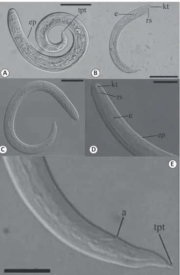
FIGURE 2 - Angiostrongylus cantonensis isolated from Achatina fulica. (A, B) Second-stage larvae (L2): scale, 50µm; (C, D) Third-stage larvae (L3): scale, 25µm; (E) Anterior end of L3 larvae showing anus and tail with pointed tip: scale, 25 µm. Legend: kt: knob-like tips; rs: a rod-like structure;
e: esophagus; ep: excretory pore. Posterior end showing: tpt: a tail with a pointed tip; a: anus.
A
B
C
D
E
were infected with
A. cantonensis
. Of the 540 mollusks, 204
(37.7%) had nematode larvae, of which, 117 (57.3%) were
infected with
A. cantonensis
(21.6% of the total) (
Table 1
).
The prevalence of
A. cantonensis
infection in
A. fulica
for each
municipality and the absolute number of parasite loads per
mollusk are shown in
Table 2
.
The results were negative for the presence of
A. costaricensis
.
Spatial analysis showed that the percentage of
A. fulica
infected with
A. cantonensis
in Baixada Santista had a random
distribution, characterized by the absence of areas with much
higher or much smaller infection percentages within the region
(I = 1:38; p = 0.0957).
Morphological and morphometric analyses revealed that
the larvae showed filiform bodies, striated cuticles in the
transverse direction with rounded anterior ends showing two
well-developed structures in the form of buttons and another
in the form of a rod, followed by a long esophagus (
Figure 2
).
The results of the morphological analyses of second-stage larvae
(L2) and L3 of
A. cantonensis
are shown in
Table 3
.
DISCUSSION
Several snails play roles as intermediate hosts for
A. cantonensis
. Among them, the giant African snail
A. fulica
is
one of the most important due to its abundance and occupation
in different ecosystems. In this study, among 540
A. fulica
specimens analyzed, 204 (37.8%) were found to contain
nematodes, a value similar to that obtained by Rocco
39, who
reported a rate of 34.2%. In both studies, specimens were
obtained in anthropic environments where snails probably
lived with small rodents, which is critical for the maintenance
of parasites in the environment.
Recovered
A. cantonensis
larvae presented two morphotypes
that were visually classiied by morphometry and morphology
as larval stages 2 and 3 (L2 and L3). Although the detail of
the tail ending in a ine tip is a typical feature of the species,
it cannot be used alone as a precise taxonomic identiication
factor
37; however, L3 presented measures compatible with those
obtained by Ash
37and Thiengo
11(
Table 3
).
Lv
10found that, before the second molting, the main
characteristics of L2 were similar to those of L3, as shown in
Figure 2
, which were two structures, similar to buttons and rods
in shape. The founding of these two larval stages in the same
snails is probably due to constant reinfections of the mollusk
in the natural environment and to the method by which the
analyzed material was obtained, in which the entire contents
of the soft parts were processed.
Molecular analysis revealed the presence of
A. fulica
that
were naturally infected with
A. cantonensis
in urban areas of
the nine municipalities of the Baixada Santista region, with
an infection rate of 21.7%. The variation of this rate is broad
and has been observed in several municipalities, such as São
Gonçalo (35.4%) and Barra do Piraí (10.3%), both in the State
of Rio de Janeiro and Joinville/SC (27.4%)
12, China (13.4%
and 28.4%)
40,41, Pernambuco (42%)
11, and Japan (52.79%)
42.
The climatic characteristics of Baixada Santista are appropriate
for the development of
A. fulica
and
A. cantonensis
. In fact,
Ishii
43has observed that the L3 of
A. cantonensis
develop
better at temperatures ranging from 20°C to 30°C. In addition
to environmental factors such as temperature, variations in the
infection rate can be inluenced by biological cycle dynamics of
the parasite in its hosts, by the population density of mollusks
and rodents, and by biological characteristics
22,42,44.
Municipality Number of Achatina fulica naturally infected with nematode larvae (%)
Number of Achatina fulica infected with
Angiostrongylus cantonensis among those with nematode larvae (%)
Bertioga 21/60 (35.0) 10/21 (47.6)
Cubatão 16/60 (26.7) 08/16 (50.0)
Guarujá 14/60 (23.3) 08/14 (57.1)
Itanhaém 28/60 (46.7) 15/28 (53.6)
Mongaguá 25/60 (41.7) 17/25 (68.0)
Peruíbe 34/60 (56.7) 15/34 (44.1)
Praia Grande 30/60 (50.0) 27/30 (90.0)
Santos 16/60 (26.7) 08/16 (50.0)
São Vicente 20/60 (33.0) 09/20 (45.0)
Total 204 (37.7) 117/204 (57.3)
TABLE 1
Prevalence of nematode larvae and Angiostrongylus cantonensis in Achatina fulica mollusks in the nine municipalities comprising Baixada Santista, São Paulo, Brazil (n = 540; 60/municipality).
Municipality Total of snails Number of positive snails (%) Individual parasitic load
Bertioga 60 10 (16.7) 5; 7; 18; 36; 52; 98; 113; 148; 274; 9,723
Cubatão 60 8 (13.3) 4; 5; 17; 22; 30; 53; 82; 147
Guarujá 60 8 (13.3) 6; 11; 36; 187; 526; 703; 1,907; 2,407
Itanhaém 60 15 (25.0) 9; 19; 35; 36; 41; 42; 52; 61; 93; 109;
179; 215; 307; 601; 3,800
Mongaguá 60 17 (28.3) 6; 6; 7; 21; 21; 23; 30; 49; 62; 106; 110;
131; 349; 362; 448; 1,070; 3,213
Peruíbe 60 15 (25.0) 1; 3; 4; 4; 5; 8; 23; 27; 27; 66; 477; 937;
1,251; 1,302; 1,508
Praia Grande 60 27 (45.0)
1; 2; 3; 4; 11; 16; 19; 20; 28; 41; 45; 52; 54; 61; 74; 79; 85; 91; 126; 185; 203; 233; 388; 432; 568; 700; 1,717
Santos 60 8 (13.3) 1; 8; 12; 24; 281; 632; 1,328; 1,675
São Vicente 60 9 (15.0) 6; 14; 54; 61; 69; 160; 193; 285; 2,509
TABLE 2
Characteristics
L2 L3
mean ±
standard deviation variation
mean ±
standard deviation variation
Body length 358.2 ± 27.8 299.5 - 399.2 450.8 ± 23.5 410.5 - 493.6
Width 26.4 ± 2.6 21.9 -34.5 21.1 ± 5.5 13.1 - 38.5
Esophagus length 145.2 ± 22.2 107.4 -236.0 168.7 ± 8.8 149.3 - 185.4
Excretory pore 61.9 ± 7.6 53.9 - 89.9 86.0 ± 4.3 77.9 - 93.2
Tail length 29.1 ± 3.4 21.2 - 39.7 35.3 ± 3.8 28.8 - 44.6
Termination of tail Tapered Tapered
TABLE 3
Measurements (µm) of second- and third-stage larvae and tail characteristics of Angiostrongylus cantonensis retrieved from naturally infected Achatina fulica.
L2: second-stage larvae; L3: third-stage larvae.
studies on the action of
A. fulica
and other species of mollusks
as intermediate hosts of
A. cantonensis
and their role in public
health. For example, previous studies have shown the presence
of other species naturally infected with
A. cantonensis
in
addition to
A. fulica
in Brazil, such as
Bradybaena similares
,
Subulina octona
,
Sarasinula marginata,
and
Sarasinula
linguaeformis
9,11-14.
Most animal populations have aggregate spatial distribution
patterns, generally owing to the distribution and supply of
resources in the environment
45. In this study, a regular spatial
distribution pattern was observed, which is quite rare. The
probability is that this distribution was due to the presence
of high populations of
A. fulica
in urban areas related to their
high adaptability, which makes it not a limiting resource of the
A. cantonensis
distribution. Furthermore, as the parasite can
be found in different species of intermediate hosts, its spatial
distribution becomes more regular. These data are especially
useful and can be used by public health authorities to establish
policies related to surveillance and planning of preventive
actions. Isolated cases of eosinophilic meningoencephalitis
have recently been reported in Brazil
9,11,14,15,46,47. Thus, it is
plausible that
A. cantonensis
continues to spread to new regions,
increasing the risk of eosinophilic meningoencephalitis in
humans.
Conlict of interest
The authors declare that there is no conlict of interest.
Acknowledgements
To Biologist Aparecido Guerino for helping in the collections of molluscs and to Prof Dr Marcos Antonio de Oliveira for giving the LABIMES (Laboratory of Molecular Biology and Structural) from the Biosciences Institute for molecular analysis.
Financial support
This work was supported by the Fundação de Amparo à Pesquisa do Estado de São Paulo (FAPESP), Process number: 2011/05893-8.
REFERENCES
1. Morera P. Life history and redescription of Angiostrongylus costaricensis Morera and Céspedes. Am J Trop Med Hyg. 1973;22(5):613-21.
2. Alicata JE. Angiostrongylus cantonensis (Nematoda: Metastrongylidae) as a causative agent of eosinophilic meningits of man in Hawaii and Tahiti. Can J Zool. 2011;40(1):5-8.
3. Pien FD, Pien BC. Angiostrongylus cantonensis eosinophilic meningitis. Int J Infect Dis. 1999;3(3):161-3.
4. Prociv P, Spratt DM, Carlisle MS. Neuro-angiostrongyliasis: unresolved issues. Int J Parasitol. 2000;30(12-13):1295-1303.
5. Lindo JF, Waugh C, Hall J, Cunningham-Myrie C, Ashley D, Eberhard ML, et al. Enzootic Angiostrongylus cantonensis in rats and snails after an outbreak of human eosinophilic meningitis, Jamaica. Emerg Infect Dis. 2002;8(3):324-6.
6. Raccurt CP, Blaise J, Durette-Desset MC. Presence of
Angiostrongylus cantonensis in Haiti. Trop Med Int Healt. 2003;8(5):423-6.
7. Lai CH, Yen CM, Chin C, Chung HC, Kuo HC, Lin HH. Eosinophilic meningitis caused by Angiostrongylus cantonensis after ingestion of raw frogs. Am J Trop Med Hyg. 2007;76(2):399-402.
8. Kliks MM, Palumbo NE. Eosinophilic meningitis beyond the
Paciic Basin: the global dispersal of a peridomestic zoonosis
caused by Angiostrongylus cantonensis, the nematode lungworm of rats. Soc Sci Med. 1992;34(2):199-212.
10. Lv S, Zhang Y, Liu HX, Zhang CW, Steinmann P, Zhou XN, et al. Angiostrongylus cantonensis: morphological and behavioral investigation within the freshwater snail Pomacea canaliculata. Parasitol Res. 2009;104(6):1351-9.
11. Thiengo SC, Maldonado A, Mota EM, Torres EJL, Caldeira R, Carvalho OS, et al. The giant African snail Achatinafulica as natural intermediate host of Angiostrongyluscantonensis in Pernambuco, northeast Brazil. Acta Trop. 2010;115(3):194-9.
12. Maldonado Jr A, Simões RO, Oliveira APM, Motta EM, Fernandez MA, Pereira ZM, et al. First report of Angiostrongylus cantonensis
(Nematoda: Metastrongylidae) in Achatina fulica (Mollusca: Gastropoda) from Southeast and South Brazil. Mem Inst Oswaldo Cruz. 2010;105(7):938-41.
13. Carvalho OS, Scholte RGC, Mendonça CLF, Passos LKJ, Caldeira RL. Angiostrongylus cantonensis (Nematoda: Metastrongylidae) in molluscs from harbour areas in Brazil. Mem Inst Oswaldo Cruz. 2012;107(6):740-6.
14. Espírito-Santo MCC, Pinto PLS, Mota DJG, Gryschek RCB. The
irst of Angiostrongylus cantonensis eosinophilic meningitis diagnosed in the city of São Paulo, Brazil. Rev Inst Med Trop Sao Paulo. 2013;55(2):129-32.
15. Ciaravolo RMC, Pinto PLS, Mota DJG. Meningite eosinofílica e a infecção por Angiostrongylus cantonensis: um agravo emergente no Brasil. Vector Inf Tec Cient SUCEN. 2010;8:7-8.
16. Cognato BB, Morassutti AL, Silva ACA, Graeff-Teixeira C. First report of Angiostrongylus cantonensis in Porto Alegre, State of Rio Grande do Sul, Southern Brazil. Rev Soc Bras Med Trop. 2013;46(5):664-5.
17. Moreira VLC, Giese EG, Melo FTV, Simões RO, Thiengo SC, Maldonado Jr A, et al. Endemic angiostrongyliasis in the Brazilian Amazon: natural parasitism of Angiostrongylus cantonensis in
Rattus rattus and R. norvegicus, and sympatric giant African land snails, Achatina fulica. Acta Trop. 2013;125(1):90-7.
18. Rachford FW. Potential intermediate and paratenic hosts for
Angiostrongyluscantonensis. J Parasitol. 1975;61(6):1117-9. 19. Ash LR. Observations on the role of mollusks and planarians in the
transmission of Angiostrongylus cantonensis infection to man New Caledonia. Rev Biol Trop. 1976;24(1):163-74.
20. Lv S, Zhang Y, Steinmann P, Zhou XN. Emerging angiostrongyliasis in Mainland China. Emerg Infect Dis. 2008;14(1):161-4.
21. Panackel C, Vishad G, Cherian K, Vijayakumar K, Sharma RN. Eosinophilic meningitis due to Angiostrongyluscantonensis. Indian J Med Microbiol. 2006;24(3):220-1.
22. Wang QP, Lai DH, Zhu XQ, Chen XG, Lun ZR. Human angiostrongyliasis. Lancet Infect Dis. 2008;8(10):621-30.
23. Hughes PA, Magnet AD, Fishbain JT. Eosinophilic meningitis: a case series report and review of the literature. Mil Med. 2003;168(10):817-21.
24. Chotmongkol V, Sawadpanitch K, Sawanyawisuth K, Louhawilai S, Limpawattana P. Treatment of eosinophilic meningitis with a combination of prednisolone and mebendazole. Am J Trop Med Hyg. 2006;74(6):1122-4.
25. Alicata JE. Biology and distribution of the rat lungworm,
Angiostrongylus cantonensis, and its relationship to eosinophilic meningoencephalitis and other neurological disorders of man and animals. Adv Parasitol. 1965;3:223-48.
26. Tokiwa T, Hashimoto T, Yabe T, Komatsu N, Akao N, Ohta N. First report of Angiostrongylus cantonensis (Nematoda:
Angiostrongylidae) infections in invasive Rodents from ive Islands
of the Ogasawara Archipelago, Japan. Plos One. 2013;8(8):e-70729.
27. Simião MC, Fischer ML. Estimativa e inferências do método de controle do molusco exótico Achatina fulica Bowdich 1822 (Stilommatophora; Achatinidae) em Pontal do Paraná, Litoral do Estado do Paraná. Cad Biodivers. 2004;4(2):74-83.
28. Fischer ML, Costa LCM. O caramujo gigante africano Achatina fulica noBrasil. 1st edition. Curitiba/PR: Editora Champagnat; 2010.
269p.
29. Santana Teles HM, Vaz JF, Fontes LR, Domingos MF. Registro de
Achatina fulica Bowdich, 1822 (Mollusca, Gastropoda) no Brasil: caramujo hospedeiro intermediário da angiostrongilíase. Rev Saúde Pública. 1997;31(3):310-2.
30. Vasconcelos MC, Pile E. Ocorrência de Achatina fulica no Vale do Paraíba, Estado do Rio de Janeiro, Brasil. Rev Saúde Pública. 2001;35(6):582-4.
31. Oliveira JCS, Corrêa KJG, Vasconcelos HCG. Ocorrência de
Achatinafulica (Mollusca: Pulmonata: Achatinidae) em três bairros da cidade de Santana, Amapá. Biota Amazônica. 2013;3(1):9-12.
32. Simone LRL. Land and freshwater molluscs of Brazil. São Paulo: EGB/FAPESP; 2006. 390p.
33. Wallace GD, Rosen L. Techniques for recovering and identifying larvae of Angiostrongyluscantonensis. Malacologia. 1969;7:427-38. 34. Moraes RG. Contribuição para o estudo do Strongyloides stercoralis e da estrongiloidíase no Brasil. Rev Serv Esp Saúde Públ. 1948;1:507-624.
35. Gasser RB, Chilton NB, Hoste H, Beveridge I. Rapid sequencing of rDNA from single worms and eggs of parasitic helminths. Nucleic Acids Res. 1993;21(10):2525-6.
36. Caldeira RL, Carvalho OS, Mendonça CLFG, Graeff-Teixeira C, Silva MCF, Ben R, et al. Molecular differentiation of Angiostrongylus costaricensis, A. cantonensis and A. vasorum by polymerase chain reaction and restriction fragment length polymorphism. Mem Inst Oswaldo Cruz. 2003;98(8):1039-43.
37. Ash LR. Diagnostic morphology of the third-stage larvae of Angiostrongylus cantonensis, Angiostrongylus vasorum, Aelurostrongylus abstrusus, and Anailaroides rostratus (Nematoda: Metastrongyloidea). J Parasitol. 1970;56(2):249-53.
38. Perry JN. Spatial analysis by distance indices. J Anim Ecol. 1995;64:303-14.
39. Rocco SC. Infecção natural de nematódeos parasitas em Achatina fulica Ferussac (Gigante africano) (Gastropoda: Pulmonata: Stylommatophora) relacionada com as condições sanitárias em vida livre e em criações comerciais no Estado de São Paulo. Tese de Doutorado. São Paulo: Coordenadoria de Controle de Doenças da Secretaria de Estado da Saúde de São Paulo; 2007. 140p.
40. Lv S, Zhang Y, Liu HX, Hu L, Yang K, Steinmann P, et al. Invasive
snails and an emerging infectious disease: results from the irst
national survey on Angiostrongylus cantonensis in China. Plos Negl Trop Dis. 2009;3(2):e368. doi: 10.1371/journal.pntd.0000368.
41. Hu X, Du J, Tong C, Wang S, Liu J, Li Y, et al. Epidemic status of
Angiostrongylus cantonensis in Hainan Island, China. Asian Pac J Trop Med. 2011 4(4):275-7.
42. Noda S, Uchikawa R, Matayoshi S, Watanabe Y, Sato A. Observations on the transmission of Angiostrongylus cantonensis
from snail to rodent. J Helminthol. 1987;61(3):241-6.
43. Ishii AI. Effects of temperature on the larval development of
Angiostrongylus cantonensis in intermediate host, Biomphalaria glabrata. Z Parasitenkd. 1984;70(3):375-9.
and ac9, have two diferente biological and morphological proiles.
Mem Inst Oswaldo Cruz. 2014;109(8):1057-63.
45. Krebs CJ. Ecological methodology. 3nd edition. New York (NY):
Addison-Wesley Educational Publishers; 2014.
46. Lima ARMC, Mesquita SD, Santos SS, Aquino ERP, Rosa LRS, Duarte FS, et al. Alicata disease; neuroinfestation by
Angiostrongylus cantonensis in Recife, Pernambuco, Brazil. Arq Neuropsiquiatr. 2009;67(4):1093-96.
47. Morassutti AL, Thiengo SC, Fernandez M, Sawanyawisuth
K, Graeff-Teixeira C. Eosinophilic meningitis caused by