A Cephalometric Analysis for Evaluation of
Changes in Soft Tissues in the Regions of the
Upper and Lower Lips and Chin due to
Orthognathic Maxillary Advancement Surgery
Hélcio Tadeu Ribeiro
1Ana Célia Faria
1Alexandre Laguna Terreri
1Francisco Veríssimo de Mello-Filho
11Department of Ophthalmology, Otorhinolaryngology and Head and
Neck Surgery, Faculty of Medicine of Ribeirão Preto, Universidade de São Paulo, Ribeirão Preto, SP, Brazil
Int Arch Otorhinolaryngol 2014;18:57–62.
Address for correspondence Ana Célia Faria, PhD, Department of Ophthalmology, Otorhinolaryngology and Head and Neck Surgery Faculty of Medicine of Ribeirão Preto, Universidade de São Paulo Av. Bandeirantes, 3900–Ribeirão Preto, SP, Brazil 14049-900
(e-mail: [email protected]).
Introduction
Cephalometry as used today took several years to be refined and went through various phases for the establishment of its methodology. In the Renaissance, more elaborate techniques
arose for the measurement of the body and the face. Albrecht Durer (1417–1518) and Leonardo da Vinci (1452–1519) drew human faces on which they traced straight lines between different points joining homologous anatomical structures that divided the head with vertical and horizontal lines. Keywords
►
orthognathic surgery
►
cephalometry
►
maxilla
Abstract
Introduction
There is currently no consensus regarding the best method for
predict-ing the changes in soft tissues due to the modi
fi
cation of hard tissues in orthognathic
surgery.
Objective
To measure the changes in soft tissues of the upper lip, lower lip, and chin
regions due to the modi
fi
cations of hard tissues caused by orthognathic maxillary
advancement surgery using a cephalometric methodology.
Methods
The study was conducted on 35 patients with dentoskeletal and facial
deformities submitted to orthognathic maxillary advancement surgery. Two
teleradio-graphs were taken: one during the preoperative period and the other 1 year after the
surgery, on which the cephalometric tracing was drawn.
Results
A strong correlation (r
¼
0.747) was demonstrated in the horizontal analysis
between the hard A (Ah) point (located in the deepest point of the anterior curvature of
the maxilla) and the soft A (As) point in the advancement of the maxilla, with a mean
variation of 0.859% occurring in As with each 1% variation of the Ah point. A mean
variation of 0.698% occurred in the superior soft prostion point (prolongation of the
superior hard prostion point to its corresponding point on soft tissue) for each 1%
variation in the superior hard prostion point (bone point located at the junction of the
alveolar process with the crown of the upper incisors).
Conclusion
The cephalometric methodology applied here revealed that the soft
tissues of the upper lip accompanied 70 to 80% of the movement of hard tissues in
maxillary advancement and that the soft tissues of the lower lip did not change or
showed no signi
fi
cant changes.
received July 22, 2013 accepted
September 10, 2013
DOI http://dx.doi.org/ 10.1055/s-0033-1361082. ISSN 1809-9777.
Copyright © 2014 by Thieme Publicações Ltda, Rio de Janeiro, Brazil
Variations in the direction of these lines were considered to be deviations of facial structures. These were probably the first evaluations of facial changes or asymmetries. Curiously, both artists used the true vertical line (TVL; plumb line) and the natural head position (NHP) as reference bases.1
The manual method has been long used to execute the cephalometric tracing and to obtain the angular and linear measurements necessary for its interpretation. As the study of cephalometry progressed, new measurements were pre-sented by different investigators and the amount of informa-tion added to the cephalograms became quite extensive.
Legan and Burstone proposed a cephalometric analysis of soft tissues directed at orthosurgical patients to complement previously published analyses of hard tissues.2,3 The soft tissues that cover the teeth and bones are highly variable in thickness, and the measurements of hard tissues may diverge from the facial contour of soft tissues presented by the patient. The use of arbitrary reference planes in skeletal analysis, such as the Frankfurt horizontal plane and the sella-nasion, to evaluate the maxillary and mandibular posi-tioning may also lead to some imprecision because these reference planes may present variations. The main objective of orthosurgical treatment is to improve the appearance of the face rather than to cause the cephalometric measure-ments of the patients to be within normal patterns. Although in many cases the improvement of facial appearance causes the cephalometric measurements to be close to normal, in some cases this may not be true.
A study published in 1999 described a new way to evaluate the lateral cephalometric radiographies using the TVL.4This method was denoted “soft tissue cephalometric analysis,” whereby the horizontal and vertical positions of the reference points of soft tissues are evaluated in relation to the TVL.
There is currently no consensus regarding the best method for predicting the changes in soft tissues due to the modifi -cation of hard tissues in orthognathic surgery.
The objective of the present study was to measure the changes in the soft tissues of the upper lip, lower lip, and chin regions resulting from orthognathic maxillary advancement surgery using a cephalometric methodology.
Methods
Population
The study was conducted on 35 Caucasian patients of both sexes age 18 to 50 years, with dentoskeletal facial deformities. The research protocol was approved by the Ethics Committee of Institution (protocol no. 6849/2007) and all patients gave informed consent to the work.
The inclusion criteria were: adult patients with class III skeletal facial pattern; absence of cleft lip/palate or any other developmental anomaly; patients submitted only to maxil-lary advancement by the Le Fort I technique with no vertical modification or other complementary procedures.
Exclusion criteria were: patients whose surgery generated vertical maxillary movement of more than 3 mm; patients who required some type of graft or interpositional bone substitute during surgery; impossibility of superposition of
structures of the skull base on the pre- and postoperative cephalometric radiographies; patients submitted to any type of mentoplasty so that there would be no additional factor that might alter the movement of the lower lip; patients who did not accept to participate in the study or did not follow the preestablished protocol.
Lateral Cephalometric Radiographies
Lateral cephalometric radiographies were obtained from each patient by the same operator. All patients were in-structed to assume NHP using as reference a mirror posi-tioned in front of them. The following protocol was adopted to obtain NHP: erect position, with the feet10 cm apart; gaze
fixed on the reflected image of one’s eyes using the mirror in front; the auricular olives, when inserted in the tragus, should maintain a slight contact with the skin to prevent possible elevations of the neck or the head. The nasion positioner was adapted in a gentle manner so that the median sagittal plane would remain perpendicular to the horizontal plane. TVL was obtained using a metal wire attached to a plumb line posi-tioned close to the anterior margin of thefilm holder frame, so that it would appear in front of the soft tissue profile of the patient.
Two teleradiographs were taken for each patient, one of them preoperatively and the other after the orthognathic surgery.
Cephalometry
The cephalometric tracings were obtained by a manual method using a 0.5-mm pencil, a millimeter ruler, and acetate paper laterally attached with adhesive tape to the cephalo-metric radiographies, which were transilluminated in a table negatoscope for better identification of the anatomical struc-tures. The tracings were performed in a dark room for better visualization of the radiographies. Points and lines were marked for the determination of angles and lateral measures of hard and soft tissues.
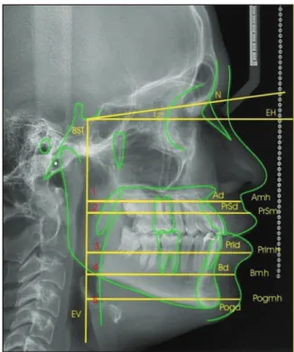
Two reference lines werefirst traced on the pre- and postop-erative radiographies: a horizontal axis (HA) line and a vertical axis (VA) line. The HA line was defined as the one traced 12 degrees below the Base of the sella-Nasion line starting from the base of the sella (►Fig. 1). This reference line traced in this manner becomes more parallel to the natural horizontal plane. The VA line was defined as the line perpendicular to the HA line passing through the base of the sella (►Fig. 1).
Points located on hard and soft tissues were established for the execution of the measurements. The points located on the hard tissues werefirst marked. The points on soft tissues that composed the horizontal lines were traced starting from VA, passing through the cephalometric point on hard tissue and continuing to the limit of the corresponding soft tissue (►Fig. 2).
Cephalometric Analysis
The following cephalometric points were marked (►Fig. 2):
N (nasion point): most anterior point of the frontonasal suture where the lines of the glabella profile meet those of the nasal bones
Ah (hard A point): the deepest point of the anterior curvature of the maxilla between the anterior nasal spine and the alveolar process
As (soft A point): prolongation of point A to its correspond-ing point on soft tissue
PrSh (superior hard prostion point): point located on the junction of the alveolar process with the crown of the upper incisors
PrSs (superior soft prostion point): prolongation of the PrSh to its corresponding point on soft tissue
PrIh (inferior hard prostion point): point located on the junction of the alveolar process and the crown of the lower incisors
PrIs (inferior soft prostion point): point on soft tissue corresponding to the prolongation of PrIh to its corre-sponding point on soft tissue
Bh (hard B point): deepest point of the anterior concavity of the mandible
Bs (soft B point): point on soft tissue corresponding to the prolongation of point B to its corresponding point on soft tissue
Pogh (hard pogonion point): most anterior point of the contour of the mandibular symphysis
Pogs (soft pogonion point): point on soft tissue corre-sponding to the prolongation of the Pog point to its corresponding point on soft tissue
Horizontal Lines
The following horizontal lines were marked:
BST-N: line traced from the BST point to the N point Line 1: line starting perpendicular to the vertical axis, passing through Ah and continuing to As
Line 2: line PrSh and continuing to PrSs
Line 3: line starting perpendicular to the vertical axis, passing through PrIh and continuing to PrIs
Fig. 1 Horizontal axis obtained with 12 degrees below the BST-N line and vertical axis perpendicular to the horizontal axis.
Abbreviations: BST, base of the sella turcica; HA, horizontal axis; N, nasion point; VA, vertical axis.
Line 4: line starting perpendicular to the vertical axis, passing through Bh and continuing to Bs
Line 5: line starting perpendicular to the vertical axis, passing through Pogh and continuing to Pogs
Le Fort I osteotomy was the surgical technique used for maxillary advancement.
Statistical Analysis
The Pearson correlation coefficient was used to determine the relationship between the changes in soft tissue and those of bone tissue. The calculation was used to test the hypothesis of a direct relationship between movement of bone tissue and points on soft tissue, yielding a correlation between the magnitudes analyzed. We used the Pearson correlation coef-ficient (r), which determines the variation of the modification of soft tissue in relation to hard tissue with the following meaning:Rof 0 to 0.49, no correlation between the measure-ments analyzed; R from 0.5 to 1.0, there is a correlation between hard and soft tissue.
We also used the intercept (I), which is the linear coeffi -cient of the model, Ahj.R2determines the percent variability of the data applied by the linear model coefficient of deter-mination. The level of significance was set atp<0.05.
Complex statistical calculations were performed and graphs and dispersal diagrams were generated to visualize the relationship of dislocation at points of bone and soft tissue.
Results
The results of the present study demonstrated that there was a strong correlation (r¼0.747) in the horizontal analysis between Ah and As in maxillary advancement, with a mean 0.859% variation in As for each 1% variation in the Ah point (►Fig. 3).
►Fig. 4demonstrates that a mean 0.698% variation in PrSs occurred for each 1% variation in the PrSh point due to maxillary advancement surgery.
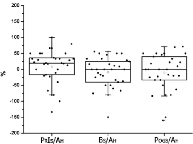
Analysis of the effects of maxillary advancement surgery on the soft tissues of the mandible using the horizontal relation of PrIs (line 3), Bs (line 4), and Pogs (line 5) with Ah demonstrated that the three soft points had zero or close to zero median and mean (►Fig. 5).
Discussion
The result of orthognathic surgeries in terms of facial es-thetics depends on the effects of the changes in soft tissues
Fig. 3 Horizontal relationship between Ah and As (line 1) in maxillary advancement. It can be seen that a strong correlation (r¼0.747) exists between Ah and As in the horizontal analysis The graph demonstrates that a mean 0.859% variation in As occurs for each 1% variation of the Ah point (y¼0.859x).
Abbreviations: Ah, hard A point; As, soft A point.
Fig. 4 Horizontal relationship between PrSh and PrSs (Line 2) in maxillary advancement. It can be seen that a strong correlation (r¼0.662) exists between PrSh and PrSs in the horizontal analysis. The graph demonstrates that a mean 0.698% variation in PrSs occurs for each 1% variation of the PrSh point (y¼0.698x).
Abbreviations: PrSh, superior hard prostion point; PrSs, superior soft prostion point.
that accompany the repositioning of bone structures. Thus, in surgical planning it is extremely important to know the correct relation between bone movements and their effects on soft tissues. The more precise this information, the greater the surgeon’s safety in planning the orthognathic surgery, not only for functional correction but also for the best esthetic result, so strongly desired by all patients. The identification of the aesthetic factors and the prediction of thefinal profile of the facial soft tissues play important roles in planning the orthognathic treatment.5However, it can be seen that the literature is quite divergent regarding the quantitation of the changes in soft tissues due to the bone movements performed in orthognathic surgeries.
In the present study, the evaluation of maxillary advance-ment surgery revealed that the amount of bone advanceadvance-ment of point Ah is reflected in 86% advancement of point As (►Fig. 3). This means that for each 1-mm advancement of the maxilla, the soft tissue of the upper lip in the subnasal region will advance by 0.86 mm. This movement shows a strong correlation between hard tissue and changes in soft tissue (r¼0.75). A survey of the literature showed that several authors who evaluated the changes in soft tissues due to maxillary advancement in point A detected 60 to 83% variation in such changes in the various studies.6–9In view of such variation in results, it is difficult to define the result that best reflects the reality. The findings of the present study were close to those reported by Brooks et al.10 A possible explanation for such variability is the methodology used in each study, as well as variations in race, amplitude of move-ments, or even sex of the patients studied. Some studies have used a distinct methodology based on angular measure-ments. In the present study, we used linear measurements considered to be more appropriate and applied a horizontal line with a well-defined bone point tofind its corresponding soft point. Tissue thickness was measured with considerable precision at the same soft and hard points before and after surgery. Thus we believe that we can safely state that, in maxillary advancement, the Ah point produces on average an 86% advancement of its corresponding soft tissue. We con-sider thisfinding to be important because maxillary move-ments are frequently used in daily clinical practice and, depending on the dimension of this movement, it is possible to predict the position of the upper lip. Thus the reestablish-ment of a harmonic profile can be predicted, as well as an appropriate upper lip–lower lip relationship with a better final esthetic result.
In the region of the upper lip vermilion, PrSs point, in maxillary advancement surgery, we observed that the hori-zontal advancement of soft tissue was 0.70 mm for each 1 mm of bone advancement of the PrSh point (i.e., 70% of the amount of bone advancement;►Fig. 4). This means that the upper lip in the vermilion region has a 30% smaller thickness after surgery, with a strong correlation between bone movement and changes in soft tissue (r¼0.662). Other studies about the modification of this point due to maxillary advancement have reported values ranging from 50 to 75%.6,11–15The results of the present study agree with those reported by Arnett et al,16but in any case, there is general
agreement that the upper lip does not accompany the mag-nitude of bone movement. Another interesting aspect is that although the soft tissues of the As point advance by 86%, the PrSs point advances by 70%. On this basis, advancement of the upper lip does not occur in a homogeneous manner, with its greater amplitude being in its portion close to the base of the nose, being progressively reduced up to the vermilion by16%.
Skeletal maneuvers not only can change the soft tissue morphology in the immediate vicinity, but also that in distant regions owing to the 3D composite anatomy of the craniofa-cial region. These changes affect the overall esthetic balance of the face. Thus, maxillofacial surgeons must pay due atten-tion to alteraatten-tions in the facial form before performing orthognathic surgery.17
Does the maxillary advancement surgery provoke changes also in soft tissues of the lower lip region?
Kim et al have confirmed that conventional Le Fort I osteotomy affected areas remote from the osteotomy line.18 However, the effect was not very pronounced compared with the nearer areas.
In the present study, analysis of the horizontal changes at points PrIs, Bs, and Pogs (►Fig. 5) in maxillary advance-ment surgery revealed that, on average or as the median, the thickness of the soft tissues did not change. However, a 25% variation of more or less may occur in PrIs, Bs, and Pogs. A possible explanation for such variation is that when the maxilla is advanced, the mandible is repositioned in class I occlusion, possibly producing small clockwise or counter-clockwise movements. These movements may provoke adjustments of soft tissues in the lower lip and chin region. We noted that the tissues underwent an equal and discrete adjustment of25% or suffered no change. Greater vari-ability occurred in the region of the mentolabial sulcus, possibly causing a slight modification in the shape of the sulcus, turning it less markedly. This type of analysis and the findings obtained here were not detected in the literature.
Conclusion
Based on the methodology employed and the results ob-tained, the soft tissues of the upper lip, lower lip, and chin regions were found to have different responses depending on the osseous movement applied to the maxilla. In general, maxillary advancement produces an 80% projection of the upper lip and a discrete change or no change in the tissues of the lower lip and chin.
References
1 Finlay LM. Craniometry and cephalometry: a history prior to the
advent of radiography. Angle Orthod 1980;50(4):312–321
2 Legan HL, Burstone CJ. Soft tissue cephalometric analysis for
orthognathic surgery. J Oral Surg 1980;38(10):744–751
4 Arnett GW, Jelic JS, Kim J, et al. Soft tissue cephalometric analysis:
diagnosis and treatment planning of dentofacial deformity. Am J Orthod Dentofacial Orthop 1999;116(3):239–253
5 Aydil B, Özer N, Marşan G. Facial soft tissue changes after maxillary
impaction and mandibular advancement in high angle class II cases. Int J Med Sci 2012;9(4):316–321
6 Lines PA, Steinhauser EW. Soft tissue changes in relationship to
movement of hard structures in ortognathic surgery: a prelimi-nary report. J Oral Surg 1974;32:891–896
7 Carlotti AE Jr, Aschaffenburg PH, Schendel SA. Facial changes
associated with surgical advancement of the lip and maxilla. J Oral Maxillofac Surg 1986;44(8):593–596
8 Medeiros PJ, Quintão CCA, Menezes LM. Avaliação da estabilidade
do perfil facial após tratamento cirúrgico. Ortodontia Gaúcha 1999;3:5–23
9 Vigorito JW. Traçado predictivo: proposta de um método em caso
de maloclusões dentárias com indicações para cirurgia ortogná-tica. Ortodontia 2000;33:63–73
10 Brooks BW, Buschang PH, Bates JD, Adams TB, English JD.
Predict-ing upper lip response to 4-piece maxillary LeFort I osteotomy. Am J Orthod Dentofacial Orthop 2001;120(2):124–133
11 Mansour S, Burstone C, Legan H. An evaluation of soft-tissue
changes resulting from Le Fort I maxillary surgery. Am J Orthod 1983;84(1):37–47
12 Epker B, Stella JP, Fish L. Dentofacial Deformities: An Integrated
Orthodontic Surgical Approach. St. Louis, MO: Mosby; 1985: 3–70
13 Hernandez-Orsini R, Jacobson A, Sarver DM, Bartolucci A.
Short-term and long-Short-term soft tissue profile changes after mandibular advancements using rigidfixation techniques. Int J Adult Orthod Orthognath Surg 1989;4:209–218
14 Jensen AC, Sinclair PM, Wolford LM. Soft tissue changes associated with double jaw surgery. Am J Orthod Dentofacial Orthop 1992; 101(3):266–275
15 Gregoret J. Ortodontia e cirurgia ortognática, diagnóstico e pla-nejamento. Barcelona, Spain: Livraria Santos; 1999
16 Arnett GW, McLaughlin RP. Planejamento facial e dentário para
ortodontistas e cirurgiões bucomaxilofaciais. São Paulo, Brazil: Artes Médicas; 2004
17 Vasudavan S, Jayaratne YSN, Padwa BL. Nasolabial soft tissue
changes after Le Fort I advancement. J Oral Maxillofac Surg 2012;70(4):e270–e277
18 Kim YI, Park SB, Son WS, Hwang DS. Midfacial soft-tissue changes