O R I G I N A L A R T I C L E UDC: 616.31::616.716.716.4-007-08 DOI: 10.2298/VSP141210051M
Radiographic cephalometry analysis of condylar position after
bimaxillary osteotomy in patients with mandibular prognathism
Rendgen-kefalometrijska analiza pozicije kondila nakon bimaksilarne
osteotomije mandibularnog prognatizma
Nikola D. Miković*, Miloš M. Lazarević*, Zoran Tati憇, Sanja Krejović-Trivić†, Milan Petrović*, Aleksandar Trivić§
*Clinic of Maxillofacial Surgery, Faculty of Dentistry, University of Belgrade, Belgrade, Serbia; †Clinic of Oral Surgery and Implantology, Military Medical Academy, Belgrade, Serbia;‡Facultyof Medicine of the MilitaryMedical Academy, University of Defence,
Belgrade, Serbia; §Clinic of Otorhinolaryngology and Maxillofacial Surgery, Clinical Center of Serbia, Faculty of Medicine, University of Belgrade, Belgrade, Serbia
Abstract
Background/Aim. Postoperative condylar position is a substantial concern in surgical correction of mandibular prognathism. Orthognathic surgery may change condylar position and this is considered a contributing factor for ear-ly skeletal relapse and the induction of temporomandibular disorders. The purpose of this study was to evaluate changes in condylar position, and to correlate angular skele-tal measurements following bimaxillary surgery. Methods.
On profile teleradiographs of 21 patients with mandibular angular and linear parametres, the changes in condylar posi-tion, were measured during preoperative orthodontic treat-ment and 6 months after the surgical treattreat-ment. Results. A statistically significant difference in values between the groups was found. The most distal point on the head of condyle point (DI) moved backward for 1.38 mm (p = 0.02), and the point of center of collum mandibulae point (DC) moved backward for 1.52 mm (p = 0.007). The amount of upward movement of the point DI was 1.62 mm (p = 0.04). Conclusion. In the patients with mandibular prognathism, the condyles tend to migrate upward and for-ward six months after bimaxillary surgery.
Key words:
prognathism; surgery, oral; postoperative period; cephalometry; temporomandibular joint; centric relation.
Apstrakt
Uvod/Cilj. Postoperativna pozicija kondila je značajna za hiruršku korekciju mandibularnog prognatizma. Ortognat-ska hirurgija može da promeni poziciju kondila, a to može biti jedan od faktora koji doprinosi ranom skeletnom recidi-vu i pojavi temporomandibularnih disfunkcija. Zbog toga je cilj ove studije bio da proceni promene pozicije kondila kao i da ne korelišu promene pozicije kondila sa angularnim ske-letnim promenama nakon bimaksilarne hirurgije. Metode.
Na telerendgenskim snimcima 21 bolesnika sa mandibular-nim prognatizmom mereni su angularni i linearni parametri koji opisuju promene u položaju kondila, pre ortodontske pripreme i šest meseci nakon hirurške korekcije. Rezultati.
Ustanovljena je statistička značajnost razlika u vrednosti pa-rametara između grupa. Tačka DI – najdistalnija tačka na glavi kondila, pomerila se unazad 1,38 mm (p = 0,02), a tač -ka DC – tačka koja označava centar collum mandibulae, pomerila se, takođe, unazad za 1,52 mm (p = 0,007). Vred-nost pomeranja tačke DI naviše bila je 1,62 mm (p = 0,04).
Zaključak. Kod bolesnika sa mandibularnim prognatiz-mom, kondili su težili da migriraju unapred i naviše šest me-seci nakon bimaksilarne operacije.
Ključne reči:
prognatizam; hirurgija, maksilofacijalna; postoperativni period; kefalometrija;
temporomandibularni zglob; centrički odnos.
Introduction
Mandibular prognathism (MP) or skeletal Class III maloc-clusion with a prognathic mandible has long been viewed as one of the most severe maxillofacial deformities 1. The treatment of
Vol. 73, No. 4 VOJNOSANITETSKI PREGLED Page 319 sagittal split osteotomy (BSSO). Another popular technique,
mostly used for maxillary reposition, is Le Fort I osteotomy. In some severe cases of MP both mandibular and maxillary osteotomy are needed, and that form of correction is commonly known as bimaxillary surgery.
One of the goals of bimaxillary surgery is maintaining skeletal and occlusal stability. Occlusal stability, which is one of the most important factors in the prevention of pos-toperative relapse in orthognathic surgery, results from go-od dental occlusion and a normal postoperative condylar position 3. Condylar processus is a part of the mandibular ramus and a part of the temporomandibular joint (TMJ), specific to the human body in its morphology, position and function 4. This makes it particularly important, both in functional and in anatomical terms, because of its shape and position depending on the position of the mandible, the function of the TMJ and facial appearance 5. Good dental occlusion depends on normal temporomandibular joint; that is, dental malocclusion or abnormal interdigitation with normal condylar position can be controlled postoperatively by orthodontic treatment, but an abnormal condylar positi-on can not be corrected postoperatively and eventually dis-rupts postoperative occlusal stability 3. Therefore, postope-rative condylar position is a substantial concern in the sur-gical correction of a mandibular prognathism. Orthognathic surgery may change condylar position and this is conside-red a contributing factor for early skeletal relapse 6–9 and the induction of temporomandibular disorders (TMDs) 10–13. Positional changes in the condyle have been hard to recognize and accurately measure following orthognathic surgery 14, 15. Displacement of the condyle can be expected as a result of four variables: anterior-posterior, vertical, medial-lateral, and along the long axis of the condyle 16.
The purpose of this study was to evaluate changes in condylar position, and to correlate angular skeletal measu-rements following bimaxillary surgery in patients with mandibular prognathism.
Methods
The study included 21 patients (13 males, 8 females; ages between 18–25 years). Clinical examinations and sta-ndardized lateral cephalometric radiographs were conduc-ted at the Belgrade University Faculty of Dentistry. The study was approved by the Ethics Committee at the Faculty of Dentistry in Belgrade. Informed consent was obtained from each patient. All the patients were diagnosed with mandibular prognathism on the basis of the following crite-ria: the angle of mandibular prognathism (SNB) ≥ 80°; the angle of sagittal intermaxillary relationship (ANB) ≤ 0°; reverse overlap of the frontal teeth and relationship of the first permanent molars in Class III, and had ended the growth and development of orofacial system. The patients with mandibular prognathism as a result of severe facial asymmetry, deformity secondary to trauma, syndromes, pa-tients with systemic disease, degenerative joint disease, and with signs and symptoms of temporomandibular dysfunction were not included in the study.
The presurgical protocol included preoperative ortho-dontic treatment, model surgery, cephalometric and photo-cephalometric analysis. The preoperative orthodontic trea-tment lasted from 18 to 24 months.
The surgery began with soft tissue incision and initial osteotomy of the ramus of the mandible as in BSSO, but with no definitive separation of bone fragments. The wound was filled with gauze soaked in saline and then the complete Le Fort I osteotomy was done. Using the interoc-clusal splint and maxillo-mandibular fixation, the maxilla was positioned in a certain position and fixed with mono-cortical screws (at least four) and L-shaped plates. After fixing the maxilla, the maxillo-mandibular fixation was re-moved, so the separation of mandibular bone fragments was completed. A separated central fragment of mandible was placed in the correct occlusion with the maxilla, the intermaxillary fixation was restored, and bone fragments of the mandible were fixed with monocortical screws and pla-tes. Monocortical screws were located on the buccal surfa-ce of the mandible, three of them on each side of the osteotomy line. Rigid intermaxillary fixation was maintai-ned for 6 to 8 weeks and after that period of time, the elas-tic fixation was maintained for 4 weeks. Postoperative ort-hodontic treatment started 6 to 8 weeks after the surgery.
Standardized lateral cephalometric radiographs were obtained at the following 2 stages in all the patients: before the preoperative orthodontic treatment (T1) and 6 months after the surgical treatment (T2).
The machine used to obtain lateral cephalometric ra-diographs was Ortoceph (Simens, Germany). The scanning settings of the machine were: 65–80 kVp tube voltage, 20 mA tube current, and 1–1.5 second scan time. All the pati-ents sat in an upright position with the teeth in centric oc-clusion. The patients’ Frankfort horizontal (FH) plane was parallel to the floor.
Cephalometric radiographs were scanned by a scanner EPSON 1600 PRO (Japan) into jpg format. In that way all the radiographs were converted into digital form. The software Ax Ceph version 2.3 (Audax, Slovenia) was used for computerized cephalometric analysis. Cephalometric analysis was carried out by one examiner and included the reference points and lines shown in Figures 1 and 2. Analyses were performed twice by the same examiner, on different days. Statistically significant differences did not appear between these two analyses.
Fig. 1 –Reference points included in the analysis.
S (sella) – The point representing the geometric center of the sella turcica.; N (nasion) – The most anterior (midline) point of the frontonasal suture; A (subspinale) – The deepest point in the bony concavity in the midline below the anterior nasal spine; Or (orbitalis)- The point representing the lowest point on the inferior orbital rim; Po (porion) – The most superior point of the external auditory meatus; Sna (spina nasalis anterior) – The most prominent point of maxilla; Snp (spina nasalis posterior) – The most distal point of the conjunction of palatinal bone and pterygomaxillar fissure; B (supramentalis) – The innermost point on the contour of the mandible between the incisor tooth and the bony chin; Me (menton) – The lowest point of the mandibular symphysis; Go (gonion) – the midpoint of the mandibular angle between the ramus and mandibular body; Cd (condylion) – the most posterosuperior point on head of the condyle; Ar (articularis) – The point midway between the two posterior borders of the left and the right mandibular rami at the intersection with the basilar portion of the occipital bone; DI – The most distal point on the head of the condyle; PI – The most anterior point on the head of the condyle; DC – The center point of the collum mandibulae on the Ba-N line; Xi – The point located at the geographic center of the ramus; Ba (basion) – The point of the anterior margin of the foramen magnum – The midpoint of the curvature between upper and the lower surfaces of the basilar portion of the occipital bone.
Fig. 2 – Reference planes included in the analysis.
N-S – The main plane of the anterior cranial base; Go-Me – The main plane of the mandible body; Sna-Snp – The main plane of the maxilla; Cd-DC-Xi – the centerline of the mandibular rami; X axis (Or-Po Frankfort horizontal (FH)) – The horizontal plane of the
head; Y axis – The vertical plane which is normal to the X osis and goes from the point S.
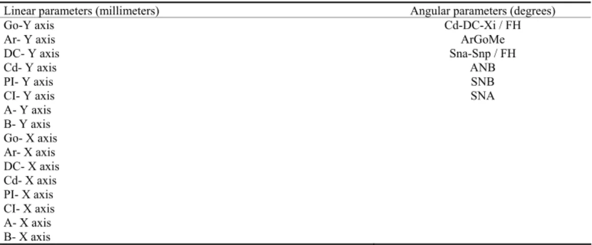
Table 1 Linear and angular parameters included in the analysis
Linear parameters (millimeters) Angular parameters (degrees)
Go-Y axis Cd-DC-Xi / FH
Ar- Y axis ArGoMe
DC- Y axis Sna-Snp / FH
Cd- Y axis ANB
PI- Y axis SNB
CI- Y axis SNA
A- Y axis B- Y axis Go- X axis Ar- X axis DC- X axis Cd- X axis PI- X axis CI- X axis A- X axis B- X axis
Vol. 73, No. 4 VOJNOSANITETSKI PREGLED Page 321
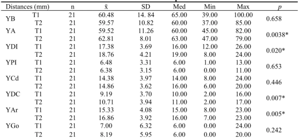
Table 2
Distances between the reference points and Y axis
Distances (mm) n ґ SD Med Min Max p
T1 21 60.48 14. 84 65.00 39.00 100.00 YB
T2 21 59.57 10.82 60.00 37.00 85.00 0.658
T1 21 59.52 11.26 60.00 45.00 82.00
YA
T2 21 62.81 8.01 63.00 47.00 79.00 0.0038*
T1 21 17.38 3.69 16.00 12.00 26.00
YDI
T2 21 18.76 4.21 19.00 8.00 24.00 0.020*
T1 21 6.48 3.31 6.00 1.00 13.00
YPI
T2 21 6.38 3.15 6.00 0.00 11.00 0.653
T1 21 14.38 3.97 14.00 8.00 24.00
YCd
T2 21 14.86 3.62 16.00 6.00 20.00 0.446
T1 21 9.19 3.70 10.00 2.00 16.00
YDC
T2 21 10.71 3.94 11.00 2.00 17.00 0.007*
T1 21 15.33 4.08 15.00 8.00 23.00
YAr
T2 21 16.86 3.92 16.00 7.00 23.00 0.005*
T1 21 7.00 6.32 6.00 0.00 24.00
YGo
T2 21 8.19 5.95 6.00 0.00 20.00 0.242
*p < 0.05 (2-tailed); ґ – mean; SD – standard deviation; Med – median;
Min-Max – minimal-maximal value; T1 – Standardized lateral cephalometric radiographs obtained before preoperative orthodontic treatment; T2 – Standardized lateral cephalometric radiographs obtained 6 months after the surgical treatment.
See abbreviations in Addendum.
Using the software, after insertion the digital (jpg) format of lateral cephalogram, calibration was set up. The calibration is used to convert pixels of the inmages into milimetres. A me-tal ruler on a cephalostat which is visible on radiography was used for calibration (Figure 3).
Fig. 3 –Calibration of the digital image usig the software “Ax Ceph”.
Then, the location of reference points and lines were de-fined. To analyze linear (anterior-posterior and vertical) mo-vement of the condyle, in every cephalometric radiograph the coordinate system with X and Y axis (as described in Figure 2) was inserted. After that, the distance between the points Go, Ar, DC, Cd, PI, CI, A, B and Y axis was measured to determi-ne horizontal skeletal changes postoperatively. The distance between the points Go, Ar, DC, Cd, PI, CI, A, B and X axis was measured to determine vertical skeletal changes postoperatively. Angles SNA, SNB, and ANB were used to describe skeletal changes after the intervention. The angle Ar-Go-Me and angle Cd-DC-Xi/FH were used to analyze rotation of the condyle after the intervention. Angle Sna-Snp/FH was
used to describe rotation of the maxilla after bimaxilary surgery (see Abbreviations in addendum).
Data analysis was not preformed until the last patient had been examined for the last time to prevent bias from the examiner’s awareness of any trends in the basic data.
Statistical analyses were performed with SPSS version 15 (SPSS, Chicago, Ill). For the assessment of the differences between angular and linear parameters before (T1) and after (T2) the surgery, Students paired t-test was used. Pearsons cor-relation was used to correlate changes in condilar position with angular skeletal changes following bimaxillary surgery. The differences were considered significant at p < 0,05.
Results
Horizontal skeletal changes: the mean setback of the man-dible 6 months postoperatively (T2-T1) was 0.91 mm at point B but the differences were not statistically significant (p = 0.658). Point Go showed tendency to go forward (1,19 mm) but also was not statistically significant (p = 0.242). On the other hand, maxilla was on average moved forward 3.29 mm at point A (p = 0.0038) (Table 2). Horizontal changes in condylar position: six months after the surgery the position of point DI and DC chan-ged significantly. Point DI moved backward 1.38 mm (p = 0.02), likewise, point DC moved backward for 1.52 mm (p = 0.007). The movement of the points PI and Cd were not statistically significant, but it was noted that point PI showed the tendency to move forward by 0.1 mm (Table 2).
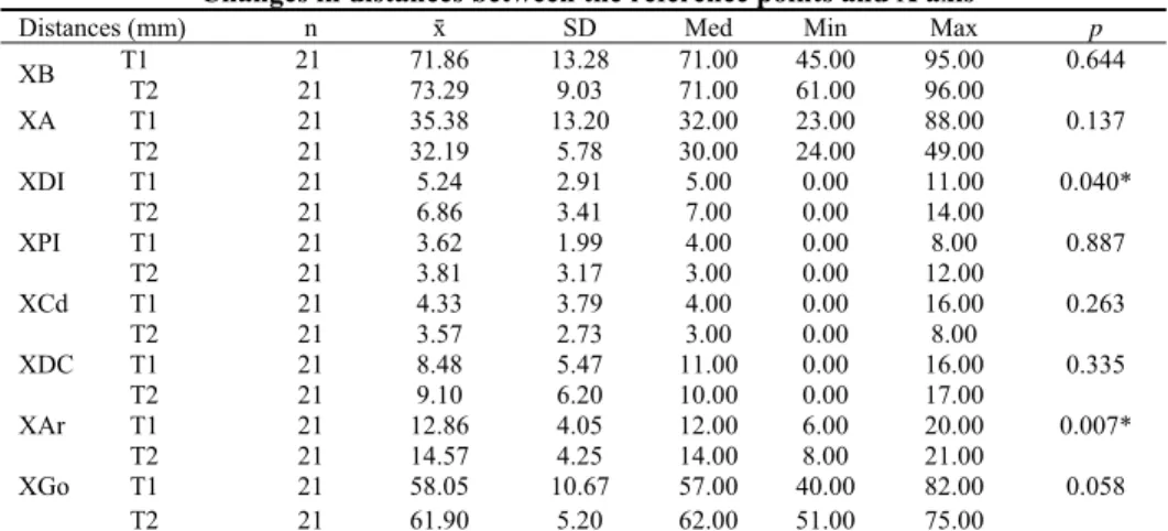
Vertical skeletal changes: the results showed the tendency of downward movement of the mandible at points B and Go (1.43 mm, p = 0.644; 3.85 mm p = 0,058, respectively). Maxilla was moved upward at point A for 3.18 mm but showed no stati-stical significance (Table 3).
Vertical changes in condylar position: the position of
Table 3 Changes in distances between the reference points and X axis
Distances (mm) n ґ SD Med Min Max p
T1 21 71.86 13.28 71.00 45.00 95.00 XB
T2 21 73.29 9.03 71.00 61.00 96.00
0.644
T1 21 35.38 13.20 32.00 23.00 88.00
XA
T2 21 32.19 5.78 30.00 24.00 49.00
0.137
T1 21 5.24 2.91 5.00 0.00 11.00
XDI
T2 21 6.86 3.41 7.00 0.00 14.00
0.040*
T1 21 3.62 1.99 4.00 0.00 8.00
XPI
T2 21 3.81 3.17 3.00 0.00 12.00
0.887
T1 21 4.33 3.79 4.00 0.00 16.00
XCd
T2 21 3.57 2.73 3.00 0.00 8.00
0.263
T1 21 8.48 5.47 11.00 0.00 16.00
XDC
T2 21 9.10 6.20 10.00 0.00 17.00
0.335
T1 21 12.86 4.05 12.00 6.00 20.00
XAr
T2 21 14.57 4.25 14.00 8.00 21.00
0.007*
T1 21 58.05 10.67 57.00 40.00 82.00
XGo
T2 21 61.90 5.20 62.00 51.00 75.00
0.058
*p < 0,05 (2-tailed); ); ґ – mean; SD – standard deviation; Med – median; Min-Max – minimal-maximal value; T1 – Standardized lateral cephalometric radiographs obtained before preoperative orthodontic treatment; T2 – Standardized lateral cephalometric radiographs obtained 6 months after the surgical treatment. See abbrevations in Addendum.
Table 4 Changes in angular parametres
Angle (°) n ґ SD Med Min Max p
T1 21 81.86 5.51 82.00 72.00 96.00 SNA
T2 21 83.62 5.59 84.00 74.00 94.00 0.049*
T1 21 86.57 6.03 88.00 77.00 103.00 SNB
T2 21 84.62 5.04 84.00 75.00 92.00 0.040*
T1 21 -4.71 2.41 -4.00 -10.00 0.00 ANB
T2 21 -0.95 2.62 -1.0 -6.00 3.00 < 0.001**
T1 21 139.48 7.94 137.00 126.00 155.00
ArGoMe
T2 21 134.38 8.55 134.00 119.00 154.00
0.009*
T1 21 61.90 6.20 62.00 43.00 70.00 Cd-DC-Xi/FH
T2 21 63.76 6.36 63.00 52.00 77.00
0.277
T1 21 4.10 3.27 4.00 0.00 9.00
SnaSnpFH
T2 21 5.19 3.37 5.00 1.00 11.00 0.128
*p < 0.05 (2-tailed); ** p < 0.001 (2-tailed); ); ґ – mean; SD – standard deviation; Med – median;
Min-Max – minimal-maximal value; T1 – Standardized lateral cephalometric radiographs obtained before the preoperative orthodontic treatment; T2 – Standardized lateral cephalometric radiographs obtained 6 months after the surgical treatment. See abbrevations in Addendum.
showed the trend to move upward (0.19 mm and 0.62 mm, respectively). On the contrary, point Cd showed the tendency to move downward (0.76 mm; p = 0.263) (Table 3).
The results suggest that point Ar was moved significantly from both X and Y axis. Point Ar moved downward (mean dif-ference T2-T1 was 1.71 mm; p = 0.007) and forward (T2-T1 was 1.53 mm; p = 0.005) (Tables 2 and 3).
SNA, SNB and ANB angle significantly changed postoperatively. SNA and ANB angle increased in dimensions (T2-T1) for 1.76° and 3.76° respectively (p = 0.049 and p < 0.001). On the other hand, SNB angle decreased for 1.95° (p = 0.04). Angles which predicted the rotation of the condyle – Ar-GoMe changed significantly (p = 0.009) for 5.1°, but Cd-DC-Xi/FH did not (p = 0.277). The rotation of maxilla (SnaSnp/FH angle) did not change significantly six months after the surgery (p = 0.128) (Table 4).
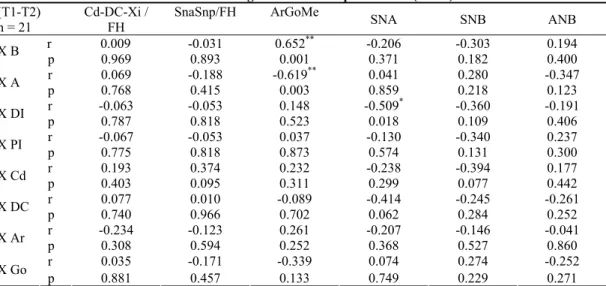
The study showed a positive correlation between the distance XB and the ArGoMe angle. The XA distance also correlated negatively with ArGoMe angle. The distance between the point DI and the X axis showed negative corre-lation with SNA angle (Table 5).
A positive correlation between the distance YB and the angle SNB, YA and SNB was noted. The distance between the Y axis and the point DI showed a positive cor-relation with the angle ArGoMe (Table 6).
Discussion
Vol. 73, No. 4 VOJNOSANITETSKI PREGLED Page 323
Table 5 Correlation of angular and linear parametres (Xaxis)
(T1-T2) n = 21
Cd-DC-Xi / FH
SnaSnp/FH ArGoMe
SNA SNB ANB
r 0.009 -0.031 0.652** -0.206 -0.303 0.194
X B
p 0.969 0.893 0.001 0.371 0.182 0.400
r 0.069 -0.188 -0.619** 0.041 0.280 -0.347
X A
p 0.768 0.415 0.003 0.859 0.218 0.123
r -0.063 -0.053 0.148 -0.509* -0.360 -0.191
X DI
p 0.787 0.818 0.523 0.018 0.109 0.406
r -0.067 -0.053 0.037 -0.130 -0.340 0.237
X PI
p 0.775 0.818 0.873 0.574 0.131 0.300
r 0.193 0.374 0.232 -0.238 -0.394 0.177
X Cd
p 0.403 0.095 0.311 0.299 0.077 0.442
r 0.077 0.010 -0.089 -0.414 -0.245 -0.261 X DC
p 0.740 0.966 0.702 0.062 0.284 0.252
r -0.234 -0.123 0.261 -0.207 -0.146 -0.041
X Ar
p 0.308 0.594 0.252 0.368 0.527 0.860
r 0.035 -0.171 -0.339 0.074 0.274 -0.252
X Go
p 0.881 0.457 0.133 0.749 0.229 0.271
*Correlation is significant at the level p < 0.05 (2-tailed); **Correlation is significant at the level p < 0.01 (2-tailed); (T1-T2) – The difference in dimensions in angles/distances before the preoperative orthodontic treatment and six months after the correction of mandibular prognathism.
See abbrevations in Addendum.
Table 6 Correlation of angular and linear parametres (Y axis)
(T1-T2) n = 21
Cd-DC-Xi / FH
SnaSnp/FH ArGoMe SNA SNB ANB
r -0.303 -0.005 -0.027 0.379 0.680** -0.360
Y B
p 0.181 0.983 0.906 0.090 0.001 0.109
r -0.174 -0.053 -0.218 0.422 0.499* -0.112
Y A
p 0.450 0.819 0.343 0.057 0.021 0.630
r -0.099 0.078 0.498* -0.235 -0.061 -0.194
Y DI
p 0.668 0.738 0.022 0.306 0.793 0.398 r 0.204 -0.393 -0.370 -0.308 -0.234 -0.090 Y PI
p 0.374 0.078 0.099 0.175 0.308 0.700 r -0.328 -0.123 0.178 -0.067 0.071 -0.163 Y Cd
p 0.147 0.595 0.440 0.774 0.759 0.481 r -0.294 -0.225 0.425 -0.179 -0.236 0.131 Y DC
p 0.195 0.327 0.055 0.437 0.303 0.571 r 0.112 0.206 0.422 -0.415 -0.276 -0.129 Y Ar
p 0.629 0.370 0.057 0.061 0.226 0.577 r 0.175 0.009 -0.275 0.193 0.189 0.081 Y Go
p 0.447 0.969 0.227 0.401 0.413 0.727 *Correlation is significant at level p < 0.05 (2-tailed); **Correlation is significant at level p < 0.01 (2-tailed);
(T1-T2) – difference in dimensions in angles/distances before preoperative orthodontic treatment and six months after the correction of mandibular prognathism.
See abbrevations in Addendum.
these reasons, in this study the patients’ jaw fragments were connected with rigid fixation.
Many researchers, using various radiographic methods, studied the movement of the condyle in patients after orthognat-hic surgery 20–22. However, there are still few studies that deal with bimaxillary orthognathic surgery mandibular prognat-hism 23, 24. In this study, four points on the condyle – DI, PI, DC and Cd were used and based on the distance of these points with the X and Y axis the anteroposterior and vertical changes in po-sition of the condyle before the preoperative orthodontic prepa-ration and 6 months after the bimaxillary surgical correction were established. The results of this study indicate the condyle tend to move forward and upward. The anterior condyle move-ment is similar with the study which Ueki et al. 25 conducted. They also reported that there was anterior and inferior
move-ment of the condyle after BSSO and intraoral vertical ramus osteotomy, but there was no statistically significant difference between these different techniques. The possible reason for mo-ving the condyle forward and downward is anatomical feature of the front part of the glenoid fossa 3.
forward rotation of the condyle. Contrary to the results of Hu et al. 26, a study by Harris et. al. 27 showed medial, posterior and superior movement of the condyle after BSSO, and also medial rotation of the condyle.
The results showed that the amount of the mandibular and maxillar movement postoperatively did not correlate statistically with condylar displacement as did the results of Harris et al. 27 and Lee and Park 3. Interestingly, only changes in ArGoMe angle correlated with the changes in the distance Y-DI, and changes in the angle SNA correlated with changes in the distance X-DI.
Conclusion
This study shows that the position of the condyle after bimaxillary orthognathic surgery is altered. In our group of pati-ents, six months after surgery, the condyles tend to migrate upward and forward. Only the most distal point on the head of the condyle (point DI) correlated with the gonial and SNA an-gle. Although this study yielded significant results over a period of six months, it was performed in a limited number of patients due to strict inclusion criteria. Further research on changes in condylar position is needed with a longer observation period.
R E F E R E N C E S
1. Graber LW. Chin cup therapy for mandibular prognathism. Am J
Orthod 1977; 72(1): 23−41.
2. Trauner R, Obwegeser H. The surgical correction of mandibular
prognathism and retrognathia with consideration of genioplasty. Oral Surg Oral Med Oral Pathol Oral Radiol Endod 1957; 10(7): 677−89.
3. Lee W, Park JU. Three-dimensional evaluation of positional
change of the condyle after mandibular setback by means of bilat-eral sagittal split ramus osteotomy. Oral Surg Oral Med Oral Pa-thol Oral Radiol Endod 2002; 94(3): 305−9.
4. Cutović T, Pavlović J, Kozomara R. Radiographic cephalometry
analy-sis of dimensions of condylar processus in persons with mandibu-lar prognathism. Vojnosanit Pregl 2008; 65(7): 513−9. (Serbian)
5. Ricketts RM, Roth RH, Chacones SJ, Schlhof RJ, Engel GA.
Orthodon-tic diagnosis and planing. Denver: Rocky Mountain Data Systems; 1982.
6. Joss CU, Vassalli IM. Stability after bilateral sagittal split osteotomy advancement surgery with rigid internal fixation: a systematic re-view. J Oral Maxillofac Surg 2009; 67(2): 301−13.
7. Epker BN, Wessberg GA. Mechanisms of early skeletal release
fol-lowing surgical advancement of the mandible. Br J Oral Surg 1982; 20(3): 175−82.
8. Will LA, Joondeph DR, Hohl TH, West RA. Condylar position
fol-lowing mandibular advancement: its relationship to relapse. J Oral Maxillofac Surg 1984; 42(9): 578−88.
9. Van SJ, Tiner BD, Keeling SD, Clark GM, Bays R, Rugh J. Condylar
position with rigid fixation versus wire osteosynthesis of a sagittal split advancement. J Oral Maxillofac Surg 1999; 57(1): 31−4.
10. Ellis E, Hinton RJ. Histologic examination of the
temporoman-dibular joint after mantemporoman-dibular advancement with and without rigid fixation: an experimental investigation in adult Macaca mulatta. J Oral Maxillofac Surg 1991; 49(12): 1316−27.
11. Lisniewska-Machorowska B, Cannon J, Williams S, Bantleon H.
Evalua-tion of force systems from a "free-end" force system. Am J Or-thod Dentofacial Orthop 2008; 133(6): 791−10.
12. Mladenović I, Jović N, Čutović T, Mladenović G, Kozomara R. Tem-poromandibular disorders after orthognathic surgery in patients with mandibular prognathism with depression as a risk factor. Acta Odontol Scand 2013; 71(1): 57−64.
13. Mladenović I, Dodić S, Stošić S, Petrović D, Cutović T, Kozomara R. TMD in class III patients referred for orthognathic surgery: psychologi-cal and dentition-related aspects. J Craniomaxillofac Surg 2014; 42(8): 1604−9.
14. Kawamata A, Fujishita M, Nagahara K, Kanematu N, Niwa K, Langlais
RP. Three-dimensional computed tomography evaluation of post-surgical condylar displacement after mandibular osteotomy. Oral Surg Oral Med Oral Pathol Oral Radiol Endod 1998; 85(4): 371−6.
15. Bettega G, Cinquin P, Lebeau J, Raphaël B. Computer-assisted
or-thognathic surgery: Clinical evaluation of a mandibular condyle re-positioning system. J Oral Maxillofac Surg 2002; 60(1): 27−34.
16. Kundert M, Hadjianghelou O. Condylar displacement after sagittal
splitting of the mandibular rami. A short-term radiographic study. J Maxillofac Surg 1980; 8(4): 278−87.
17. Ueki K, Degerliyurt K, Hashiba Y, Marukawa K, Nakagawa K,
Ya-mamoto E. Horizontal changes in the condylar head after sagittal
split ramus osteotomy with bent plate fixation. Oral Surg Oral Med Oral Pathol Oral Radiol Endod 2008; 106(5): 656−61.
18. Ueki K, Nakagawa K, Takatsuka S, Yamamoto E. Plate fixation
af-ter mandibular osteotomy. Int J Oral Maxillofac Surg 2001; 30(6): 490−6.
19. Nishimura A, Sakurada S, Iwase M, Nagumo M. Positional changes
in the mandibular condyle and amount of mouth opening after sagittal split ramus osteotomy with rigid or nonrigid osteosyn-thesis. J Oral Maxillofac Surg 1997; 55(7): 672−6.
20. Fang B, Shen GF, Yang C, Wu Y, Feng YM, Mao LX, et al.
Changes in condylar and joint disc positions after bilateral sagit-tal split ramus osteotomy for correction of mandibular prog-nathism. Int J Oral Maxillofac Surg 2009; 38(7): 726−30.
21. Kim YI, Jung YH, Cho BH, Kim JR, Kim SS, Son WS, et al. The
as-sessment of the short- and long-term changes in the condylar position following sagittal split ramus osteotomy (SSRO) with rigid fixation. J Oral Rehabil 2010; 37(4): 262−70.
22. Chen S, Lei J, Wang X, Fu K, Farzad P, Yi B. Short- and long-term
changes of condylar position after bilateral sagittal split ramus osteotomy for mandibular advancement in combination with Le Fort I osteotomy evaluated by cone-beam computed tomogra-phy. J Oral Maxillofac Surg 2013; 71(11): 1956−66.
23. Draenert FG, Erbe C, Zenglein V, Kämmerer PW, Wriedt S, Al
Na-was B. 3D analysis of condylar position after sagittal split
osteot-omy of the mandible in mono- and bimaxillary orthognathic surgery - a methodology study in 18 patients. J Orofac Orthop 2010; 71(6): 421−9.
24. Kim Y, Lee Y, Chun Y, Kang N, Kim S, Kim M. Condylar
posi-tional changes up to 12 months after bimaxillary surgery for skeletal class III malocclusions. J Oral Maxillofac Surg 2014; 72(1): 145−56.
25. Ueki K, Marukawa K, Nakagawa K, Yamamoto E. Condylar and
temporomandibular joint disc positions after mandibular osteot-omy for prognathism. J Oral Maxillofac Surg 2002; 60(12): 1424−32.
26. Hu J, Wang D, Zou S. Effects of mandibular setback on the
tem-poromandibular joint: a comparison of oblique and sagittal split ramus osteotomy. J Oral Maxillofac Surg 2000; 58(4): 375−80.
27. Harris MD, Van SJ, Alder M. Factors influencing condylar
posi-tion after the bilateral sagittal split osteotomy fixed with bicorti-cal screws. J Oral Maxillofac Surg 1999; 57(6): 650−4.
Vol. 73, No. 4 VOJNOSANITETSKI PREGLED Page 325
Addendum
Abbreviations: