Pituitary macroadenoma presenting
as a nasal tumor: case report
Macroadenoma hipoisário, apresentando-se como um tumor nasal: relato de caso
Nivaldo Adolfo Silva Junior
I, Fabiano Reis
II, Larissa Kaori Miura
III, Guilherme Henrique Vieira
IV, Luciano Souza Queiroz
V,
Heraldo Mendes Garmes
VI, Cristina Laguna Benetti-Pinto
VIIDepartments of Radiology, Pathology, Endocrinology and Gynecology, Faculdade de Ciências Médicas, Universidade Estadual de
Campinas (Unicamp), Campinas, São Paulo, Brazil
ABSTRACT
CONTEXT: Pituitary macroadenomas are rare intracranial tumors. In a few cases, they may present aggres-sive behavior and invade the sphenoid sinus and nasal cavity, causing unusual symptoms. In this paper, we report an atypical case of pituitary adenoma presenting as a nasal mass.
CASE REPORT: The patient was a 44-year-old woman who had had amenorrhea and galactorrhea for ten months, with associated nasal obstruction, macroglossia and acromegaly. Both growth hormone and pro-lactin levels were increased. Magnetic resonance imaging showed a large mass originating from the lower surface of the pituitary gland, associated with sella turcica erosion and tumor extension through the sphe-noid sinus and nasal cavity. Histopathological analysis demonstrated a chromophobe pituitary adenoma with densely packed rounded epithelial cells, with some atypias and rare mitotic igures. There was no evidence of metastases.
CONCLUSION: Macroadenoma invading the nasal cavity is a rare condition and few similar cases have been reported in the literature. This study contributes towards showing that tumor extension to the sphe-noid sinus and nasopharynx needs to be considered and investigated in order to make an early diagnosis when atypical symptoms like nasal obstruction are present.
RESUMO
CONTEXTO: Macroadenomas hipoisários são tumores intracraniais raros. Em alguns casos, podem apre-sentar comportamento agressivo e invadir o seio esfenoidal e a cavidade nasal, causando sintomas não usuais. Neste relato de caso, descrevemos um caso atípico de adenoma hipoisário manifestando-se como uma massa nasal.
RELATO DE CASO: A paciente de 44 anos, do sexo feminino, apresentava amenorreia e galactorreia por 10 meses associando-se a obstrução nasal, macroglossia e acromegalia. Os níveis do hormônio de cresci-mento e de prolactina apresentaram-se aumentados. Ressonância magnética mostrou uma grande massa originada da superfície inferior da glândula hipoisária associada com erosão da sela túrcica e extensão do tumor através do seio esfenoidal e cavidade nasal. Análise histopatológica demonstrou adenoma hi-poisário cromófobo com células epiteliais arrendondadas densamente agrupadas com algumas atipias e escassas iguras de mitose. Não houve evidências de metástase.
CONCLUSÃO: O macroadenoma invasivo para a cavidade nasal é uma condição rara e há poucos relatos similares descritos na literatura. Este trabalho contribui para mostrar que, na presença de sintomas atípicos como a obstrução nasal, a extensão para o seio esfenoidal e para a nasofaringe deve ser considerada e investigada para um diagnóstico precoce.
IMD, PhD. Attending Physician, Department
of Radiology, Faculdade de Ciências Médicas, Universidade Estadual de Campinas (Unicamp), Campinas, São Paulo, Brazil.
IIMD, PhD. Professor, Department of Radiology,
Faculdade de Ciências Médicas, Universidade Estadual de Campinas (Unicamp), Campinas, São Paulo, Brazil.
IIIMD. Resident, Department of Radiology,
Faculdade de Ciências Médicas, Universidade Estadual de Campinas (Unicamp), Campinas, São Paulo, Brazil.
IVMD. Faculdade de Ciências Médicas,
Universidade Estadual de Campinas (Unicamp), Campinas, São Paulo, Brazil.
VMD, PhD. Professor, Department of Pathology,
Faculdade de Ciências Médicas, Universidade Estadual de Campinas (Unicamp), Campinas, São Paulo, Brazil.
VIMD, PhD. Assistant Physician, Department of
Endocrinology, Faculdade de Ciências Médicas, Universidade Estadual de Campinas (Unicamp), Campinas, São Paulo, Brazil.
VIIMD, PhD. Professor, Department of Obstetrics
and Gynecology, Faculdade de Ciências Médicas, Universidade Estadual de Campinas (Unicamp), Campinas, São Paulo, Brazil.
KEY WORDS: Pituitary neoplasms. Nasal cavity.
Growth hormone-secreting pituitary adenoma. Amenorrhea.
Prolactin
PALAVRAS-CHAVE: Neoplasias hipoisárias. Cavidade nasal.
Adenoma hipoisário secretor de hormônio do crescimento.
INTRODUCTION
Pituitary macroadenomas are rare intracranial tumors. hey may occur in diferent settings of presentation and measure more than 10 mm in diameter. Usually, they exhibit endocrine symptoms and local mass efects that may include compression of the optical chiasm and consequent visual disturbance. In some cases, they become invasive, going beyond the sella turcica to adjacent structures such as the internal carotid and cavernous sinus. In rare cases, they extend to the sphenoid sinus and
nasopharynx.1-3 When this occurs, some unusual symptoms may
appear, which may or may not be accompanied by the expected indings, like epistaxis, nasal obstruction, painful sinuses and
purulent rhinorrhea.4-8
he reason why certain adenomas behave in a locally aggressive fashion is unknown and, although most cases are benign, these tumors can sometimes show malignant charac-teristics and so can be deined as carcinomas in the presence of
craniospinal and/or systemic metastases.9,10 In the present case,
scintigraphy and computed tomography ruled out the diag-nosis of a malignant tumor. he incidence of pituitary carci-noma is relatively low in patients with invasive macroadecarci-nomas
and, in many cases, the metastases may be silent.6 Since this is
an important diferential diagnose for other tumors involving the skull base, correct investigation is indispensable, given that pituitary neoplasms have a much more favorable prognosis and
low metastatic potential.4,10
Furthermore, improvements in diagnostic methods such as magnetic resonance imaging techniques and pathological exam-inations including immunohistochemical and ultrastructural studies provide the possibilities of precisely delimiting the extent
of the mass and describing the composition and kind of tumor.11
In this paper, we report a rare case of pituitary macroadenoma that was found to present growth extending through the sella turcica to invade the sphenoid sinus and nasopharynx. Although
this case represents an unusual kind of presentation, we show that invasive adenomas may manifest primarily as a nasal mass.
CASE REPORT
A 44-year-old woman who had had amenorrhea and galactor-rhea for 10 months, had been noting progressive growth of her face and extremities over the past two and a half years. Over the last six months, she had complained of severe nasal obstruction with respiratory distress and dysphagia due to macroglossia. Ater referral to our service, which forms part of a tertiary-level uni-versity hospital belonging to Universidade Estadual de Campinas (UNICAMP), she started to be followed up regarding endocrinol-ogy, neurosurgery and ophthalmology.
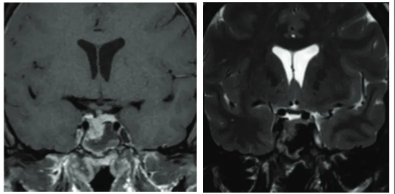
On physical examination, acromegaly was evident. he blood hormone levels were: growth hormone (GH) 34.20 ng/ml (n = 0.06-5.0 ng/ml), insulin-like growth factor-1 (IGF-1) 726 ng/ml and prolactin (PRL) ranging from 70 to 82.83 ng/ml in three samples (n = 4.79 – 23.3 ng/ml). ACTH, TSH, FSH and LH levels were all normal. Magnetic resonance imaging (MRI) depicted a large mass (3.2 x 4.1 x 3.2 cm) originat-ing from the lower surface of the pituitary gland, which was asso-ciated with sella turcica erosion and tumor extension through the
sphenoid sinus into the nasal cavity (Figure 1). he sella itself was
of normal dimensions and, because the tumor had expanded on the lower side, the optical chiasm was not afected, as was veriied through a normal visual ield examination.
Initially, considering the high levels of GH and that, besides surgery, adjuvant therapy would probably be necessary in order to reduce the tumor, pharmacological treatment with octreo-tide 30 mg (a somatostatin analogue that inhibits GH and IGF-1) was administered every 28 days. However, ater three months, the blood hormone levels were seen to remain elevated and the symptoms became severe. herefore, endoscopic sur-gery was indicated.
B
A
C
he surgical procedure was started by setting the speculum at the nostril, so as to reach the tumor through the transsphenoidal route. Because of the volume of the intranasal mass, the procedure was halted due to technical diiculties and only a biopsy was performed.
Two months later, it was decided to perform transcranial sur-gery. hrough bifrontal craniotomy, the anterior cranial fossa was accessed and, ater drilling the sphenoid surface, the mass was aspirated and removed.
Histopathological analysis on the biopsy and the frag-ments from the hypophysectomy gave rise to a diagnosis of iniltrating pituitary macroadenoma with nuclear atypia. Immunohistochemistry on the samples showed that they were
positive for GH and PRL (Figure 2). A visual estimate of marked
nucleus proportions showed that 3-5% were positive for Ki-67, with several typical mitoses, thus characterizing an atypical pitu-itary tumor. Since the diagnosis of carcinoma is conirmed only if metastases are present, whole-body bone scintigraphy and abdominal, pelvic and chest computed tomography were
per-formed. here was no evidence of metastases.12
Two months ater the surgery, the patient reported that her menstrual cycles had become regular and that her galactorrhea had ceased. hree months later, she noticed that her shoe size
had decreased by two sizes. Although her GH and PRL hor-mone levels showed progressive reduction over this period (GH = 3.41 ng/ml; PRL= 12.44 ng/ml), IGF-1 maintained high levels (IGF-1 = 455 ng/ml). A postoperative control MRI examination
showed evidence of tumor residues (Figure 3). he patient
con-tinues to be asymptomatic, with continuing pharmacological treatment with octreotide and endocrinological follow-up.
DISCUSSION
Even though pituitary macroadenoma is a rare condition, it exhib-its a variety of presentations and manifestations of symptoms when invading the sphenoid sinus and nasal cavity. In the present case, the appearance of some typical signs such as amenorrhea, galactor-rhea and growth of the face, tongue and extremities (resulting from increased GH and prolactin levels) contributed towards the inves-tigation and aided in making the diagnosis.
However, in many cases reported in the literature, less exu-berance of symptoms was reported. he patients showed nasal obstruction, sometimes with epistaxis and purulent rhinorrhea. In addition, the lack of endocrinal symptoms and the normal levels of pituitary hormones made it more diicult to suspect an adenoma, with a consequent risk of diagnostic delay. In almost
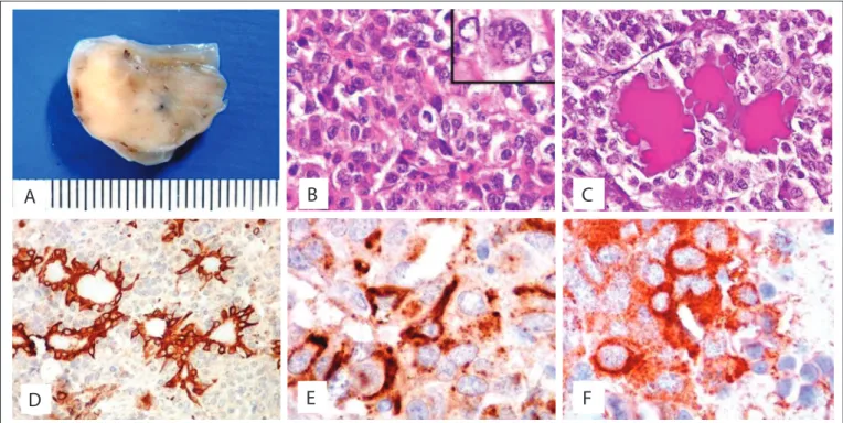
Figure 2. Polyp-like specimen excised from left nasal cavity. External surface was smooth and shiny, cut surface solid and irm, and translucent in some areas (A). Chromophobe pituitary adenoma showing densely packed rounded epithelial cells with some atypia and scattered mitotic igures (one at the center) (hematoxylin-eosin, HE; 100 X); inset: binucleated cell with prominent nucleoli and rare cytoplasm (400 X) (B). Glands of nasal mucosa surrounded by tumor cells, in which mucus is highlighted in magenta in the dilated gland lumina (periodic-acid Schif, PAS; 100 X) (C). Immunohistochemical analysis for 34Βe12 (an antibody for high molecular weight keratins), demonstrating mucosal glands trapped amid negative adenoma cells (100 X) (D). Immunohistochemical analysis (400 X) of the pituitary adenoma cells in the cytoplasm, which are focally positive for growth hormone (GH) (E) and prolactin (F).
A
B
C
E
all of the cases, the diagnosis was only made ater removing the
obstructing tissue for microscopic examination.6,7
Invasive pituitary neoplasm may occur either as an exten-sion from an intrasellar leexten-sion or as an ectopic tumor. Kikuchi et al. and other authors described similar cases of a large pituitary
adenoma consequent to an ectopic focus in the nasal cavity.13
In the present case, the histological analysis and the imaging examinations reinforced the evidence showing that the pituitary macroadenoma extended inside the sella turcica. Magnetic reso-nance imaging showed that the mass originated from the right lower surface of the pituitary gland, with associated sella turcica erosion and tumor extension through the sphenoid sinus into the
nasopharyx.14 However, when the tumor originates from an
ecto-pic focus, with a normal pituitary gland, it becomes more diicult to restrict the diferential diagnoses.
Although pituitary adenomas are considered to be benign tumors, they can show malignant characteristics when the presentations are
Table 1. Search strategies performed on June 25, 2013, and the results from each database
Electronic databases Key words Results
Found Related
Medline via PubMed (pituitary adenomas) AND (nasal cavity) AND (obstruction) 12 5 Lilacs via Bireme (pituitary neoplasms) OR (neoplasias hipoisarias) OR (neoplasias hipoisárias) AND (sphenoid sinus) OR
(seno esfenoidal) OR (seio esfenoidal) AND (nasal obstruction) OR (obstrucción nasal) OR (obstrução nasal) 18 0 Embase via Elsevier (pituitary adenomas) AND (nasal cavity) AND (obstruction) 0 0
atypical, such as in the case reported here. Invasive pituitary tumors are relatively rare, as are pituitary carcinomas. In cases of the lat-ter condition, a pituitary tumor that is either not contiguous with the primary sellar tumor and/or a pituitary tumor that has
metasta-sized to distant sites from the pituitary gland needs to be present.12
his shows that for correct diagnosis in cases of tumors in the nasal cavity, it is very important to exclude other intracranial tumors, such as craniopharyngioma, chordoma, chondrosarcoma and
meningi-oma.9 his diagnosis is usually made by means of histological analysis,
using the endocrine growth pattern comprising tumor cells arranged in packets, ribbons or rosettes, with delicate neuroendocrine markers and pituitary hormones.
It seems that invasive pituitary macroadenomas do not
fol-low any standard. As shown in Table 1, few cases have been
described in the literature and there is still much to study about these tumors, given that the reason why some of them behave in
a more locally aggressive manner remains unknown.10 he case
presented here is important, because it shows that even when the presentation is atypical, with only a few symptoms like nasal obstruction, the diagnostic hypothesis of macroadenoma needs to be taken into consideration whether or not its usual clinical signs are present, especially when caused by variation in endo-crinal hormone levels.
CONCLUSION
Invasive macroadenoma to the nasal cavity is a rare condition with few cases reported in the literature and the reason why some are more locally aggressive is still unknown. he description of this case of atypical presentation and its peculiarities shows that exten-sion of the tumor to the sphenoid sinus and nasopharynx needs to be taken into consideration when the usual hormonal manifes-tations of adenomas present associations with nasal symptoms. In this manner, early diagnosis and intervention may be facilitated.
REFERENCES
1. Cole IE, Keene M. Nasal obstruction in pituitary tumours. J Laryngol Otol. 1981;95(2):183-9.
2. Daita G, Makino K, Goto S, Ueno K, Takamura H. [Nasopharyngeal extension of a large chromophobe adenoma of the pituitary (author’s transl)]. No Shinkei Geka. 1976;4(7):699-706.
3. Cook RJ, Besser M. A 16 year old boy with poor vision and nasal obstruction. Aust N Z J Surg. 1987;57(7):485-8.
4. Luk IS, Chan JK, Chow SM, Leung S. Pituitary adenoma presenting as sinonasal tumor: pitfalls in diagnosis. Hum Pathol. 1996;27(6):605-9. 5. Beriat GK, Doğan C, Akmansu SH, Karadağ D, Doğan H. A rare cause of
nasal obstruction: giant invasive nonfunctioning pituitary adenoma. Kulak Burun Bogaz Ihtis Derg. 2010;20(6):309-13.
6. Godey B, Morandi X, Le Gall F, et al. Pituitary adenomas with infra-sellar extension into the nasopharynx. J Laryngol Otol. 1999;113(12):1109-11.
7. Dent JA, Rickhuss PK. Invasive pituitary adenoma presenting with nasal obstruction. J Laryngol Otol. 1989;103(6):605-9.
8. Summers GW. Nasal obstruction caused by a pituitary chromophobe adenoma. Laryngoscope. 1976;86(11):1718-21.
9. Inagawa H, Ishizawa K, Mitsuhashi T, et al. Giant invasive pituitary adenoma extending into the sphenoid sinus and nasopharynx: report of a case with intraoperative cytologic diagnosis. Acta Cytol. 2005;49(4):452-6.
10. Margo CE, Gonzalvo AA. Extracranial manifestation of invasive pituitary adenoma. South Med J. 1987;80(3):381-3.
11. Jankiewicz-Wika J, Pawlikowski M, Zawirski M, Stepień H. [Pituitary adenoma penetrating the sphenoidal sinus and nasal cavity: a case report]. Neurol Neurochir Pol. 2001;35(4):727-32.
12. Kaltsas GA, Nomikos P, Kontogeorgos G, Buchfelder M, Grossman AB. Clinical review: Diagnosis and management of pituitary carcinomas. J Clin Endocrinol Metab. 2005;90(5):3089-99.
13. Kikuchi K, Kowada M, Sasaki J, Sageshima M. Large pituitary adenoma of the sphenoid sinus and the nasopharynx: report of a case with ultrastructural evaluations. Surg Neurol. 1994;42(4):330-4.
14. Davis PC, Hofman JC Jr, Spencer T, Tindall GT, Braun IF. MR imaging of pituitary adenoma: CT, clinical, and surgical correlation. AJR Am J Roentgenol. 1987;148(4):797-802.
Sources of funding: None
Conlict of interest: None
Date of irst submission: March 21, 2013
Last received: December 16, 2013
Accepted: January 29, 2014
Address for correspondence: Fabiano Reis