Nóbrega, BB et al.
Pulmonary sarcoidosis: high-resolution computed tomography findings
Pulmonary sarcoidosis: high-resolution computed
tomography findings*
BRUNO BARCELOS DA NÓBREGA, GUSTAVO DE SOUZA PORTES MEIRELLES, GILBERO SZARF, DANY JASINOWODOLINSKI, JORGE ISSAMU KAVAKAMA
J Bras Pneumol 2005; 31(3): 254-60.
*Study conducted at the Universidade Federal de São Paulo - Escola Paulista de Medicina (UNIFESP- EPM), Centro de Medicina Diagnóstica Fleury Institute of Radiology at the Hospital das Clínicas of the Faculdade de Medicina da Universidade de São Paulo (INRAD-HCFMUSP)
Correspondence to: Bruno Barcelos da Nóbrega. Alameda Ribeirão Preto, 551 apto.14, Bela Vista. CEP 01331-001, São Paulo, SP. Phone: 55 11 288-4801. E-mail: [email protected]
Submitted: 4 August 2004. Accepted, after review: 29 October 2004.
Key words: Sarcoidosis. Lung. High-resolution computed tomography.
Sarcoidosis is a systemic disease of unknown etiology, characterized by noncaseating granulomas. Although it may affect any organ, morbidity and mortality are most commonly related to pulmonary involvement, which is found in 80-90% of patients. This study illustrates the principal manifestations of sarcoidosis seen in high-resolution computed tomography scans, including typical as well as atypical forms.
INTRODUCTION
Sarcoidosis is a multisystemic disease of u n k n o w n e t i o l o g y t h a t i s v a r i a b l e i n i t s p r e s e n t a t i o n , p r o g r e s s i o n a n d p r o g n o s i s . Pulmonary involvement is seen in up to 90% of patients, and 20-25% of those present permanent
functional impairment(1-4).
Pulmonary involvement tends to be bilateral and asymmetric, predominantly found in the upper lobes. Radiologic findings are atypical in 25% of the cases, and chest X-rays reveal normal results
in 5-10% of patients(5,6).
The objective of this study was to present, in a succinct and illustrative way, the main aspects of
pulmonary sarcoidosis revealed in high-resolution computed tomography scans.
TYPICAL FINDINGS
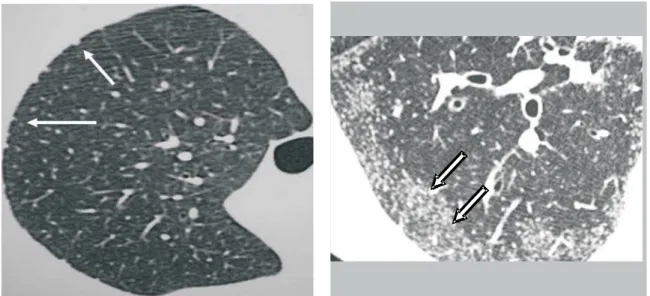
Nodules. The nodular pattern is the pattern most frequently seen in pulmonary sarcoidosis. The nodules are generally small and present perilymphatic distribution, involving the peribronchovascular cuffs, interlobular septa, subpleural region, centrilobular areas and the entire length of the fissures (Figures 1 to 3). Nodules may be large or cavitary, sometimes mimicking neoplasms, and are found in 15-25% of the cases (Figure 4)(1,4,7-10).
Figure 1. Micronodular form. Micronodules in the subpleural regions (arrows)
Figure 2 A and B. Micronodular pattern. Micronodules in the upper lobes, some of which are confluent (arrows)
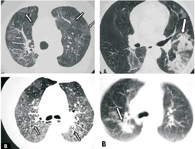
Figure 3. Micronodular form. Confluent peripheral micronodules (arrows)
Nóbrega, BB et al.
Pulmonary sarcoidosis: high-resolution computed tomography findings
Figure 5. Ground-glass pattern. A – Scattered bilateral ground-glass opacities (arrows). B –Ground-glass pattern accompanied by traction bronchiectasis (arrow)
Figure 6. Parenchymal opacities A – Poorly defined opacities in the left lung, accompanied by air bronchograms (arrow). B – Irregular consolidation in the right lung (arrow)
Figure 7. Parenchymal opacities with imprecise, sparse borders in the right upper lobe, surrounded by ground-glass pattern (arrows)
Ground-glass opacities. Ground-glass opacities are found in 14-83% of patients, tending to present scattered distribution. It may occur in isolation or be accompanied by signs of pulmonary fibrosis (Figure 5)(11-13).
Parenchymal opacities. Parenchymal opacities are less common than ground-glass opacities and are generally seen in the initial stage of the disease. They can be found throughout the peribronchovascular cuffs or on the lung periphery. Their borders are irregular, and this type of opacity is frequently accompanied by air bronchograms and nodules in the adjacent
parenchyma (Figures 6 and 7)(14).
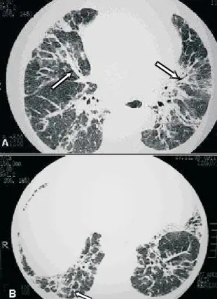
Figure 8. Reticular opacities. Pulmonary sarcoidosis with septal thickening of the upper lobes, forming polygonal arches (arrows), simulating lymphangitic carcinomatosis
Figure 9. Reticular opacities. A – Irregular thickening of peribronchovascular cuffs (arrows). B – thickening of posterior basal interlobular septa (arrow) accompanied by distortion of the lung architecture
Figure 10. Areas of air trapping in the lower lobes (arrows)
thickening is typically found along the peribronchovascular cuffs but can also be seen in other topographies (Figure 8). Irregular linear opacities adjacent to the bronchovascular cuffs have also been described and attributed to early
manifestations of pulmonary fibrosis (Figure 9)(7,9).
Air trapping. Air trapping during expiration is relatively frequent, due to the presence of peribronchial or submucosal granulomas or to peribronchiolar fibrosis, leading to small airway obliteration (Figure 10) (15).
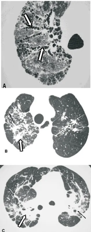
Fibrosis. Parenchymal abnormalities may evolve to fibrosis, which is accompanied by architectural distortion, volumetric loss, linear opacities, honeycombing, fibrotic masses, bronchiectasis and traction bronchiolectasis. Fibrosis is usually found in
the upper and middle lung fields (Figures 11 and 12)(7).
B r o n c h i e c t a s i s. B r o n c h i e c t a s i s i s a n uncommon finding in sarcoidosis and is frequently associated with fibrosis (traction bronchiectasis). Other less common etiologies are airway obstruction caused by granulomas and extrinsic
compression caused by lymph nodes (Figure 13)(6).
ATYPICAL FINDINGS
Pseudotumoral form. In some cases, coalescence of interstitial granulomas results in large conglomerates with the formation of masses (pseudotumors not accompanied by air bronchograms) that compress
adjacent air spaces (Figures 14 and 15)(16).
Cysts. The etiology of cysts is uncertain. Of all the possible causes, peripheral air imprisonment, alveolar distention caused by endobronchial component, the destruction of the alveolar parenchyma, and retractions and collapses of the surrounding parenchyma have been reported (Figure 16)(7).
Nóbrega, BB et al.
Pulmonary sarcoidosis: high-resolution computed tomography findings
Figure 11. Fibrosis. A – Traction bronchiectasis (arrows), architectural distortion and ground-glass pattern, showing the classical aspect of fibrosis. B – Irregular parenchymal opacities accompanied by architectural distortion (arrows). C – Fibrotic conglomerate forming masses with retractile aspect (arrows)
Figure 12. Fibrosis. Posterior subpleural honeycombing, predominantly in the lower lobes (arrows)
Figure 13A. Bronchiectasis (arrows) associated with peripheral micronodules. B – Irregular fibrotic mass with traction bronchiectasis (arrows)
first be ruled out. True cavities, as well as apical cysts,
may be the focus of saprophytic colonizations(17,18).
Bronchial changes (thickening/stenosis).
Lenique et al. reported thickening of the bronchial walls, caused by the deposition of granulomas along the peribronchovascular interstice, in 65% of patients (Figure 17)(19).
Lobar atelectasis. The occlusion of a lobar bronchus and consequent atelectasis may be caused by intraluminal disease or extrinsic compression. There are no radiological signs that help distinguish between lobar atelectasis caused by sarcoidosis and
that resulting from other causes(17).
Unilateral pulmonary parenchymal lesions. It has been reported in the literature that unilateral parenchymal changes are caused mainly by localized interstitial disease, accompanied by an alveolar pattern, or by localized interstitial disease, accompanied by a reticulonodular pattern or presenting as a pulmonary coin lesion(7,17).
A
Figure 14. Pseudotumoral form. Mass with irregular borders in the right upper lobe (arrow). Ground-glass areas characterized in the left lower lobe and adjacent to the mass (arrows)
Figure 15. Pseudotumoral form. A – High-resolution c o m p u t e d t o m o g r a p h y w i t h l u n g w i n d o w s h o w i n g spiculated mass in the posterior basal segment of the l e f t l o w e r l o b e ( a r r o w ) . B – D e n s i t y m e a s u r e m e n t during the pre-contrast phase (40 UH). C – Scan three minutes after intravenous administration of iodinated contrast material, highlighting the lesion (maximum density of 76 UH). Swensen’s protocol positive(16). A n a t o m o p a t h o l o g i c a l e x a m i n a t i o n o f t h e s u r g i c a l sample confirmed sarcoidosis
Fairy-ring sign. The "fairy-ring sign" is caused by multiple peripheral granulomatous nodules bordering areas of preserved lung
parenchyma (Figure 18)(20)..
Figura 16. Sarcoidose com cistos bilaterais associados a fístula broncopleural com formação de hidropneumotórax à direita (setas)
Figure 17. Bronchial involvement. Unilateral presentation, characterized by concentric thickening of the bronchial walls (arrows), with nodules with lymphatic distribution in the right lower lobe
Nóbrega, BB et al.
Pulmonary sarcoidosis: high-resolution computed tomography findings
CONCLUSIONS
Because of the broad spectrum of presentations, pulmonary sarcoidosis may mimic other interstitial pulmonary diseases. High-resolution computed tomography is the most sensitive and specific imaging method for the evaluation of this disease. In those cases in which atypical manifestations predominate, the knowledge of the various tomographic patterns helps us limit the differential diagnosis and select the best location for an eventual biopsy.
REFERENCES
1. Traill ZC, Maskell GF, Gleesson FV. High-resolution CT f i n d i n g s o f p u l m o n a r y s a r c o i d o s i s . A J R 1 9 9 7 ; 168:1557-60.
2. Remy-Jardin M, Giroud F, Remy J, Wattine L, Wallaert B, Duhamel A. Pulmonary sarcoidosis: role of CT in the evaluation of disease activity and functional impairment in prognosis assessement. Radiology 1994;191:675-80. 3. Miller BH, Rosado-de-Christenson ML, Mcadans HP, Jishback NF. Thoracic sarcoidosis: radiologic-pathologic correlation. Radiographics 1995;15:421-37.
4. Müller NL, Kulling P, Miller RR. The CT findings of pulmonary sarcoidosis: analysis of 25 patients. AJR 1989;152:1179-82.
5. Webb WR, Müller NL, Naidich DP. Doenças caracterizadas principalmente por opacidades nodulares e reticulonodulares. In: Webb WR, Müller NL, Naidich DP. TC de alta resolução do pulmão. 3a
ed. Rio de Janeiro: Guanabara Koogan; 2002:245-336.
6. Hamper VM, Fishman EK, Khouri NF, Jonhs CT, Wong K P, S i e g e l m a n S S . Ty p i c a l a n d a t y p i c a l C T manifestations of pulmonary sarcoidosis. J Comput Assist Tomogr 1986, 10:929-36
7. Chiles C. Imaging features of thoracic sarcoidosis. Semin Roentgenol. 2002;37:82-93.
8. Nishimura K, Itoh H, Kitaichi M, Nagai S, Izumi T. Pulmonary sarcoidosis: correlation of CT and histopathologic findings. Radiology 1993;189:105-9. 9. Brauner MW, Lenoir S, Grenier P, Cluzel P, Batteste JP, Valleyre D. Pulmonary sarcoidosis: CT assessment of lesion reversibility. Radiology 1992;182:349-54. 10. Nakatsu M, Hatabu H, Morikawa K , Vematsu H, Ohio Y,
Nishimura K, et al. Large coalescent parenchymal nodules in pulmonary sarcoidosis: “Sarcoid galaxy” sign. AJR 2002;178:1389-93.
11 . Brauner MW, Grenier P, Monpoint D, Lenoir S, Gremoux H. Pulmonary sarcoidosis: evaluation with high-resolution CT. Radiology 1989;174:467-71.
1 2 . Neto ALF, Marchiori E, Capone D, Mogami R. Aspectos de tomografia computadorizada de alta resolução na sarcoidose. Radiol Bras 1996: 29:325-30.
13. Lynch DA, Webb WR, Gainsu G, Itulbarg M, Golden J. Computed tomography in pulmonary sarcoidosis. J Comput Assist Tomogr 1989; 13:405-10.
1 4 . Jokoh T, Ikezoe J, Takeuchi N, Kohuo N, Tomiyama N, Akira M, et al. CT findings in pseudoalveolar sarcoidosis. J Comput assist Tomogr 1992;16:904-7
1 5 . Hansell DM, Milne DG, Wisher ML, Wells AV. Pulmonary sarcoidosis: morphologic associations of airflow o b s t r u c t i o n a t t h i n s e c t i o n C T. R a d i o l o g y 1998;209:697-704
1 6 . Swensen SJ, Viggiano RW, Midthun DE, Müller NL, S h e r r i c k A , Y a m a s h i t a K , e t a l . L u n g N o d u l e Enhancement at CT: Multicenter Study1 Radiology 2000; 214: 73-80.
1 7 . Rockoff SD, Rohatgi PK. Unusual manifestation of thoracic sarcoidosis. AJR 1985;144:513-28.
1 8 . Ichikawa Y, Fujimoto K, Shiraishi T, Oizumi K. Primary cavitary sarcoidosis: high resolution CT findings. AJR 1994;163:745
1 9 . Lenique F, Brauner MW, Grenier P, Bettesti JP, Loixau A, Valeyre D. CT assessment of bronchi in sarcoidosis: endoscopic and pathologic correlations. Radiology 1995;194:419-23.