261
Jornal Brasileiro de Pneumologia 31(3) - Mai/Jun de 2005
Tuberculous spondylitis in teenager*
MARTA MACIEL LYRA CABRAL, BRUNO CANTO C. DE A. AZEVEDO, LÍLIAN MARIA LAPA MONTENEGRO, ROSANA DE ALBUQUERQUE MONTENEGRO, ANDREA SANTOS LIMA, HAIANA CHARIFKER SCHINDLER
*Study carried out at the Hospital das Clínicas - Universidade Federal de Pernambuco (HC - UFPE, Federal University of Pernambuco Hospital das Clínicas) and at the Centro de Pesquisa Aggeu Magalhães/Fundação Oswaldo Cruz (CPqAM/FIOCRUZ, Aggeu Magalhães Research Center/Oswaldo Cruz Foundation).
Correspondence to: Haiana Charifker Schindler. Av. Moraes Rego, s/n, Cidade Universitária. Cx. Postal 7472, CEP: 50670-420, Recife, PE, Tel: 55 81 2101-2560 E-mail: [email protected]
Submitted: 25 May 2004. Accepted, after review: 1 September 2004. J Bras Pneumol 2005; 31(3): 261-4.
Key words: Tuberculosis. Spondylitis. PCR. Diagnostic.
This article presents a case report of osteoarticular tuberculosis affecting lumbar sacral column non-typical attack. The diagnosis remains a medical challenge because the symptoms and bone lesions are not specific and can be mislead with other morbidity such as inflammatory, circulatory, metabolic, traumatic, congenital and tumoral diseases. The disease is degenerative and the prognosis not satisfactory. Besides the clinical aspects and laboratory, imaging results, including computed tomography and magnetic ressonance, are discussed. A PCR system targeting the IS 6110 of M. tuberculosis was positive, strongly suggesting the presence of this pathogen. This assay would be particularly indicated when quick and sensitive diagnosis of tuberculosis is required.
INTRODUCTION
In 1779, the English surgeon Sir Percival Pott described the spinal deformity symptom triad, abscess and paraplegia, relating the last to spinal deformity. In his honor, vertebral tuberculosis (vertebral TB) was renamed Pott's disease, also known as tuberculous spondylitis(1).
Tuberculous spondylitis is an entity that causes a characteristic bone deformity. Until 1944, when streptomycin was discovered by the American researcher, Waksman, there was no treatment for the disease. Koch's bacillus (Mycobacterium tuberculosis) may be found in the osteoarticular system at any age. However, in developing countries, it is more common in children (up to age 10) and in adolescents(2).
Osteoarticular TB is generally secondary to pulmonary TB and almost always occurs, through lymphohematogenous dissemination, at least one year after the primary infection. Occasionally, it may occur contiguously due to the proximity of the spinal column to the affected pleura. Occasionally, there may be local TB development, complicated by closed
fractures, or there may be direct penetration of the bacillus into open fractures(3).
Osteoarticular TB accounts for at least 10% of the extrapulmonary forms of the disease, and the spinal column is the site most commonly affected(4).
In addition to general symptoms (fever, weight loss, anorexia, adynamia and others), pain, antalgic posture, gait disturbances and restriction of movement should be investigated. The onset of kyphosis occurs early in Pott's disease and is the leading cause of spinal deformity in childhood.
Diagnosis of paucibacillary TB, as well as of the extrapulmonary forms, continues to be a challenge because the symptoms, in addition to frequently having an insidious onset, are quite often mistakenly attributed to other diseases(5).
In view of the difficulty in diagnosing TB in certain forms and situations, molecular-based approaches, which are becoming increasingly sensitive and specific, have been developed. Among these, the polymerase chain reaction (PCR) method stands out. This method allows the amplification of
262
Cabral, MML, et al.
Espondilite tuberculosa em adolescente
the M. tuberculosis DNA in different biological samples collected from a patient suspected of having the disease. Theoretically, a copy of the target DNA can be detected in a single bacterial cell (6-8).Currently, these methods are being used only at
the research level.
Herein, we report the case of an adolescent presenting heterogeneous alterations of the lumbar spine followed by involvement of the sacral vertebra. Several nonspecific treatments were administered and, after eight months, based on clinical and laboratory testing aspects, imaging (including computed tomography and magnetic resonance), histopathological examination and PCR, a treatment regimen for tuberculous Spondylitis was initiated.
CASE REPORT
A fourteen-year-old mulatto female patient (Brazilian, born in João Pessoa, in the state of Pernambuco, and a student) sought treatment at the outpatient clinic of the Federal University of Pernambuco Hospital das Clínicas after experiencing lumbar pain, whose frequency and intensity were worsening, for approximately eight months, together with difficulty in flexing the dorsolumbar spine and difficulty in walking. Her condition did not improve with the use of painkillers or anti-inflammatories, or both.
Initially, she had sought treatment for mild lumbar pain at a health clinic in João Pessoa, where the physician suggested that her condition was of psychosomatic origin.
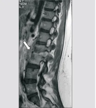
As her condition worsened, leading to difficulty in moving about, she was advised to seek medical assistance at a neurosurgery outpatient clinic, where she was submitted to lumbosacral spine computed tomography, magnetic resonance imaging, chest X-ray, biopsy of the affected site and blood count (including determination of erythrocyte sedimentation rate). Computed tomography revealed irregularity and reduction of the articular space between L2 and L3, concomitant with sclerosis of the corresponding vertebral bodies, as well as fracture in L2. The vertebral disk between L5 and S1 presented a slight protrusion, without causing significant intrarachidian repercussions. The findings suggested the possibility of an inflammatory or infectious process, or both, involving the L2 and L3 vertebral bodies, as well as the corresponding intersomatic disk (Figure 1). Magnetic resonance revealed reduction of the L2/L3
disk space and cold abscess in the corresponding area, which reinforced the possibility of a spondylodiscitis-like process (Figures 2 and 3).
The chest X-ray was normal and a needle biopsy of the site of the vertebral lesions revealed irregular fragments, together with fibroconnective, cartilage and bone tissue with reparative alterations, as well as fibrosis and foci of dystrophic calcifications. The culture was negative for pyogenic bacteria as well as for mycobacteria. The blood work only revealed an increased rate of erythrocyte sedimentation. On that occasion, an antibiotic (ciprofloxacin) and an anti-inflammatory (rofecoxib) were started.
After having taken the medication for ten days, without any signs of improvement, the patient was referred to our clinic for a definitive diagnosis.
The patient reported a history of episodes of bronchial asthma and other forms of allergy (rhinitis and allergy to dipyrone and acetylsalicylic acid), as well as two episodes of nonspecific pneumonia (at the age of two and at the age of four). The patient had received the Bacillus Calmette-Guérin vaccine, as evidenced by a scar on her right arm. Upon physical examination, the patient presented general aspect of suffering, hypoactivity, and eupnea but no fever. The patient weighed 75.3 kg (p = 95 according to the National Center for Health Statistics), and her blood pressure was 120 x 80 mmHg. Further evaluation revealed lower back pain, a limp, inability to flex the dorsolumbar spine and pain upon palpation of the paravertebral gutters. We did not find any external signs of spinal column inflammation.
Although epidemiology was negative for TB, we requested a tuberculin test, together with repetitions of the same exams performed in João Pessoa, in order to complement the diagnostic investigation of the case. The tuberculin test result (induration) was 18 mm and the molecular blood test, performed through nested PCR using the 6110 insertion sequence (IS6110) of M. tuberculosis, was positive. The PCR was performed at the Centro de Pesquisas Aggeu Magalhães/Fundação Oswaldo Cruz (Aggeu Magalhães Research Center/ Oswaldo Cruz Foundation), and the patient was included in the protocol of a paucibacillary TB study that is currently underway.
263
Jornal Brasileiro de Pneumologia 31(3) - Mai/Jun de 2005
findings, we decided to start the triple-treatment regimen for bone TB.
After two months of treatment, the patient presented clinical improvement and was re-evaluated monthly as an outpatient. The treatment continued for nine months, and there was a significant evolution of the general profile. In the last month of the treatment regimen, the patient no longer presented pain, and her motor skills were fully restored.
DISCUSSION
The most severe form of skeletal TB is tuberculous spondylitis. The lesion usually affects the anterior portion of the vertebral body, near the intervertebral disk. The M. tuberculosis infection may reach the cortex, destroy the intervertebral disk and fragment the adjacent vertebral body. A case of tuberculous spondylitis with lumbar (L2, L3 and L5) and sacral (S1) involvement has been reported.
Despite the resurgence of TB and its increasing incidence, cases that involve the lowest portion of the spinal column are still rare, affecting less than 5% of TB patients(9-13).
Various studies have shown that tuberculous spondylitis may involve the lower portion of the spinal column and the lumbosacral junction. In 1991, Mansberg reported the case of an L5 fracture concomitant with L5/S1 intervertebral disk herniation, caused by M. tuberculosis infection in the body of L5 vertebra(11).
In 1998, Rajasekaram published the findings of a 15-year follow-up of 53 patients. In most of the cases there was L3 and L4 involvement, and there was dissemination to the lower levels of the lumbosacral junction in 12 cases. That resulted in rectification of lumbar lordosis in 35 patients and kyphosis in 14 patients(13).
Diagnosis of intrapulmonary TB continues to be a challenge because the symptoms, usually insidious, can be mistakenly attributed to other causes. Spinal TB may be concomitant with pulmonary involvement, and occurs in less than 1% of patients. It is even rarer when there is no pulmonary alteration(14-17).The most common
site of spinal TB infection is low thoracic and lumbar vertebrae, regardless of being primary or secondary to pulmonary TB(5). Infection of the lumbosacral junction
is much rarer and occurs in only 2% to 3% of all cases of spinal TB(9-13).
Magnetic resonance is the most sensitive and most
Figure 1. Magnetic resonance imaging (MRI) showing sclerosis of vertebral bodies
Figure 2. Magnetic resonance imaging (MRI) showing fusion of L1-L2 vertebral bodies
264
Cabral, MML, et al.
Espondilite tuberculosa em adolescente
specific method for differentiating the nature of the vertebral collapses, in addition to showing early alterations(9,18,19) that aid in diagnosing tuberculous
spondylitis(20). However, it may produce similar imaging
results in cases of vertebral neoplasia and infection by pyogenic bacteria(9, 21).
In pyogenic infections, reduction of the intervertebral space is atypical(20,22). Nevertheless, for our patient, the
imaging revealed intervertebral reduction concomitant with involvement of consecutive vertebrae, both of which are considered characteristic of tuberculous spondylitis(9).
In order to diagnose vertebral TB, special attention should be given to the joint analysis of several factors such as detailed clinical and epidemiological history, analysis of imaging results obtained through computed tomography and magnetic resonance, histopathological examination findings, tuberculin test and, if possible, molecular tests using the PCR method. Nested PCR is one variant of the method and is currently used only at the research level. It has presented higher sensitivity and specificity, especially in paucibacillary forms (extrapulmonary TB and TB in children), thereby allowing better diagnostic clarification, even prior to culture results. Blood, as a biological sample, has been tested, with encouraging results, in patients who present negative sputum smears and cannot expectorate, as is the case for children under the age of ten, as well as in patients who present extrapulmonary forms(23-25).
Therefore, further studies are necessary in order to validate these new diagnostic approaches, thereby providing us with more efficient tools for early detection of the M. tuberculosis.
ACKNOWLEDGMENTS
The authors would like to thank Dr. Emanuel Sarinho, head of the Department of Pediatric Allergology of the Federal University of Pernambuco Hospital das Clínicas, for having referred this case for diagnostic investigation.
REFERENCES
1. Sternbach G. Percivall Pott: tuberculous spondylitis. J Emerg Med 1996; 14:72-83.
2. Rosenbaum S. Tuberculose osteoarticular. In: Sant’Anna CC. Tuberculose na infância e na adolescência. 1° ed. São Paulo: Atheneu 2002; 137-52.
3. Stuart D. Local osteo-articular tuberculosis complicating closed fracture. Report of two cases. J Bone Joint Surg Br 1976; 58:248-9.
4. Kadah H, Naughton B. Pott´s paraplegia: a complication of tuberculous spondylitis. J Am Geriatr Soc 1992; 40:710-2. 5. Wellons JC, Zomorodi AR, Villaviciencio AT, Woods CW, Lawson WT,Eastwood JD. Sacral tuberculosis: a case report and review of the literature. Surg Neurol 2004;61:136-9. 6. Bennedsen JV, Thompsen GE, Pfyffer G, Funke K, Feldmann
A. Utility of PCR in diagnosing pulmonary tuberculosis. J Clin Microbiol 1996; 34:1407-11.
7. Chang C, Yuen K, Chan K, Yam W, Yim K. Single-tube nested PCR in the diagnosis of tuberculosis. J Clin Pathol 1996;49:290-4.
8. Schindler HC, Montenegro L, Montenegro R, Carvalho AB, Abath FHC, Jaureguiberry G. Development and optimization of PCR-based malaria diagnostic methods and their comparasion with QBC assay. Am Soc Trop Med Hyg 2001; 65:355-61.
9. Ahmadi J, Bajaj A, Destian S, Segall HD, Zee CS. Spinal tuberculosis: atypical observations at MR imaging. Radiology 1993, 189 pp. 489–93.
10. Dayras JC, Lorilloux J, Hugonet M, Benichou P. Tuberculose osseuse a localisation sacree. Ann Pèdiatr Paris 1985; 32:289–93.
11. Mansberg VJ, Rowe U, Walker C. Atypical case of Pott’s disease. Australas Radiol 1991; 35:191–6.
12. Pun WK, Chow SP, Luk KDK. Tuberculosis of the lumbosacral junction, long-term follow-up of 26 cases. J Bone Joint Surg 1990; 72:675–8.
13. Rajasekaran S, Shanmugasundaram TK, Prabhakar R, Dheenadhayalan J, Shetty AP, Shetty DK. Tuberculous lesions of the lumbosacral lesion, a 15-year follow-up of patients treated by ambulant chemotherapy 1998; 23:1163–7. 14. Davidson PT, Horwitz I. Skeletal tuberculosis. Am J Med
1970; 48:72–84.
15. Gorse GJ, Pais MJ, Kusske JA, Cesario TC, Tuberculous spondylitis, a report of six cases and a review of the literature. Medicine 1983; 62:178–93.
16. Oman B, Robertson JM, Nelson RJ, Chiu LC. Pott’s disease, a resurgent challenge to the thoracic surgeon. Chest 1989; 95:145–50.
17. Rezai AR, Lee M, Cooper PR, Errico TJ, Koslow M. Modern management of spinal tuberculosis. Neurosurgery 1995; 36:87–98.
18. http://www.fleury.com.br/htmls/cdrom/capitulo2.htm. 19. Daher S, Pimenta Júnior WE, Souza Júnior ZA, Machado
MM, Rosa ACF, Teixeira KS, et al. Qual o seu diagnóstico? Radiol Bras 2004; 37.
20. Rahman N. Atypical forms of spinal tuberculosis. J Bone Joint Surg Br 1980;62:162–5.
21. McGahan JP, Dublin AB. Evaluation of spinal infections by plain radiographs, computed tomography, intrathecal metrizamide, and CT-guided biopsy. Diagn Imag Clin Med 1985;54:11–20.
22. Gupta RK, Agarwal P, Rastogi HS, Kumar RV, Phadke N, Krishnani N. Problems in distinguishing spinal tuberculosis from neoplasia on MM. Neuroradiology 1996;38:S97–S104. 23. Huggett JF, McHugh TD, Zumla A. Tuberculosis: amplification-based clinical diagnostic techniques. The J Int of Biochem & Cell Biol 2003; 35: 1407-12.
24. Folgueira L, Delgado R, Palenque E, Aguado JM, Nobrega AR. Rapid diagnostic of Mycobacterium tuberculosis bacteremia by PCR. J Clin Micro. 1996; 34: 512-5. 25. Delacourt C, Poveda JD, Chureau C, Beudon N, Mahut B, Blic J,