Utilization of microsatellites for
the analysis of genomic alterations
in colorectal cancers in Brazil
1Departamento de Bioquímica, Instituto de Ciências Biológicas,
Universidade Federal de Minas Gerais, 30161-970 Belo Horizonte, MG, Brasil Departamentos de 2Cirurgia and 3Anatomia Patológica, Faculdade de Medicina,
Universidade Federal de Minas Gerais, 30130-100 Belo Horizonte, MG, Brasil
4Núcleo de Genética Médica de Minas Gerais (GENE),
30130-909 Belo Horizonte, MG, Brasil A.K. Fuzikawa1,
L.A. Haddad1,
J.R. da-Cunha-Melo2,
G. Brasileiro-Filho3
and S.D.J. Pena1,4
Abstract
Two different pathogenetic mechanisms are proposed for colorectal cancers. One, the so-called “classic pathway”, is the most common and depends on multiple additive mutational events (germline and/or somatic) in tumor suppressor genes and oncogenes, frequently involv-ing chromosomal deletions in key genomic regions. Methodologically this pathway is recognizable by the phenomenon of loss of heterozy-gosity. On the other hand, the “mutator pathway” depends on early mutational loss of the mismatch repair system (germline and/or so-matic) leading to accelerated accumulation of gene mutations in critical target genes and progression to malignancy. Methodologically this second pathway is recognizable by the phenomenon of microsat-ellite instability. The distinction between these pathways seems to be more than academic since there is evidence that the tumors emerging from the mutator pathway have a better prognosis. We report here a very simple methodology based on a set of tri-, tetra- and pentanucle-otide repeat microsatellites allowing the simultaneous study of micro-satellite instability and loss of heterozygosity which could allocate 70% of the colorectal tumors to the classic or the mutator pathway. The ease of execution of the methodology makes it suitable for routine clinical typing.
Correspondence
S.D.J. Pena
Departamento de Bioquímica e Imunologia, ICB, UFMG Caixa Postal 486
30161-970 Belo Horizonte, MG Brasil
Fax: 55 (031) 227-3792 E-mail: [email protected]
Research supported by CNPq and FAPEMIG.
Received February 28, 1997 Accepted June 3, 1997
Key words
•Polymerase chain reaction •Colorectal carcinoma •Microsatellite •p53
•DCC
•Microsatellite instability
Introduction
The characterization of chromosomal and molecular alterations in many types of neoplasias has led to the current paradigm that cancer is a genomic disease (1). In colorectal carcinomas, which are among the most prevalent human cancers worldwide (2), a series of studies have established that additive mutations in c-Ki-ras, an oncogene,
together with the inactivation of the tumor suppressor genes p53 (located on chromo-some 17p), DCC (deleted in colon cancer, located on chromosome 18q) and APC
(adenomatous polyposis coli, located on chro-mosome 5q),occur in most patients and are implicated in the stepwise transformation of the normal mucosa into a malignant tumor (3). Inherited germline mutations in APC
of the necessary mutational steps and thus are associated with a strong familial predis-position to colorectal cancer (4). It is impor-tant to note that, while mutations in onco-genes are generally single dominant events, the inactivation of tumor suppressors is de-pendent on the functional loss of both copies of the relevant genes. While the first of the two losses occurs most frequently by gene mutations, the second one is more often a chromosomal event, generally a deletion (5-7). Since the deletion generally involves si-multaneous loss of genetic loci near the tu-mor suppressor − and occasionally loss of the whole chromosome or chromosome arm
− these events are strongly associated with loss of heterozygosity (LOH) of hypervaria-ble polymorphisms (minisatellites and microsatellites) located in the deleted re-gion.
Recently, studies of an autosomal domi-nant form of colorectal cancer known as “hereditary non-polyposis colorectal cancer” (HNPCC) have shown that this tumor is caused by germline mutations in human DNA repair genes, principally hMSH and hMLH1
but also hPMS1 and hPMSH2 (reviewed in Refs. 8-10). Mutations in these genes lead to a defect in the correction of mismatches occurring during DNA replication, with the subsequent accumulation of mutations throughout the genome, including tumor sup-pressor genes and oncogenes, which can cause malignant transformation (9,11). Mis-match repair defects occur also in a substan-tial proportion (15-25%) of sporadic colo-rectal cancers (9).
Microsatellites are genomic sequences consisting of 2-6-bp motifs repeated in mul-tiple tandem copies (reviewed in Ref. 12). Because of their repetitive nature, these se-quences are prone to frequent changes in the repeat number by replication slippage (13). Since these mutations are almost always cor-rected by the DNA repair system, microsat-ellites, although polymorphic in populations, are somatically stable. In the case of tumors
with defects in mismatch repair, however, new mutant alleles are frequently observed in microsatellites, a phenomenon termed “microsatellite instability” or “replication error phenomenon (RER)” (8). It is ques-tionable if microsatellite instability has any direct relationship with carcinogenesis, but at any rate it serves as a marker for the presence of mismatch repair deficiency in the tumors (8,9).
In summary, two different pathways of colorectal carcinogenesis can be recognized. One, called the “classic pathway”, is the most common and depends on multiple ad-ditive mutational events (germline and/or somatic) in tumor suppressor genes and on-cogenes, frequently involving chromosomal deletions in key genomic regions (3,7). Meth-odologically this pathway is recognizable by the LOH phenomenon. On the other hand, the “mutator pathway” depends on early mutational loss of the mismatch repair sys-tem (germline and/or somatic) leading to accelerated accumulation of gene mutations in critical target genes and progression to malignancy (8-10). Methodologically this second pathway is recognizable by the phe-nomenon of microsatellite instability. The distinction between these pathways seems to be more than academic. There is mounting and convincing evidence that the tumors emerging from the mutator pathway have lower proliferative activity and overall bet-ter prognosis than those emerging from the classical pathway (14-16). Thus, laboratory analysis of colorectal tumors aimed at estab-lishing the pathogenetic pathway may soon be incorporated into medical practice. There-fore, it will be necessary to develop simple methods applicable to the routine clinical laboratory.
are abundant and sensitive, but cumbersome to type because alleles can only be easily resolved on sequencing gels and generally require isotopic labeling for visualization of the PCR products. In contrast, as we have shown elsewhere, microsatellites with re-peats of more than 3 bp can be much more simply typed in short non-denaturing gels with non-isotopic silver staining (17-19). We report here a very simple methodology based on a set of tri-, tetra- and pentanucleotide repeat microsatellites which allows the si-multaneous study of microsatellite instabil-ity and loss of heterozygosinstabil-ity and thus helps to allocate a given colorectal tumor to the classic or mutator pathway. The ease of ex-ecution of the methodology makes it suitable for routine clinical typing. Moreover, the present study provides the first evaluation of the relative importance of the two pathways in the pathogenesis of colorectal carcinoma in Brazil.
Material and Methods
Patients
A sequential sample of 20 colorectal ad-enocarcinomas was obtained at the Hospital das Clínicas, Federal University of Minas Gerais, and the Colo-Proctology Service of the Santa Casa de Misericórdia, Belo Hori-zonte, Brazil. There was no selection on the basis of age, location of the tumor, histo-pathological classification or family history. However, all tumors were subsequently found to be isolated cases in the family. All 20 samples consisted of paired fragments of normal colonic mucosa and tumoral tissue from the same patient and were subjected to microscopic analysis prior to DNA extrac-tion to ensure the absence of neoplastic infil-tration in the mucosa and the predominance (>80%) of neoplastic cells in the tumor samples. DNA was extracted from the tumor samples by an alkaline extraction procedure (20). To establish a population baseline, all
tri-, tetra- and pentanucleotide loci were stud-ied in DNA extracted from the blood of at least 100 unrelated individuals randomly chosen among those presenting for paternity tests at the Núcleo de Genética Médica de Minas Gerais, Belo Horizonte.
Microsatellite analysis
We used microsatellites to detect loss of heterozygosity in the p53, DCC and APC
loci. For p53 we used the pentanucleotide repeat p53ALU (21,22) (AAAAT repeats) which is located in the first intron of the p53
gene; for DCC we used the closely linked tetranucleotide repeat D18S51 (AAAG re-peats; 23) and for APC we used the linked CA-repeat microsatellite D5S299 (24). For the detection of microsatellite instability we utilized the previous three microsatellites plus the trinucleotide repeats D13S308E (17) and D2S196E (19) (containing CAT and ACA repeats, respectively) and the tetra-nucleotide repeat D12S67 (25) containing GATA repeats. PCR reactions were carried out in 20-µl volumes with 10-100 ng of genomic DNA as a template. The primers used are listed in Table 1. For resolution of the alleles of the D5S299 CA-repeat locus, the products were denatured and resolved on a sequencing gel (50 cm x 30 cm x 0.1 cm); after electrophoresis, a strip of the area ex-pected to contain the products was cut and stained with silver salts for visualization (18). For the tri-, tetra- and pentanucleotide mi-crosatellites, non-denaturing electrophore-sis was performed on short gels (17 cm x 10 cm x 0.15 cm) followed by silver staining (18).
Results
Loss of heterozygosity
(30%) were non-informative (homozygous) for p53ALU; in the 14 heterozygous samples LOH was observed in eight cases (57%). Loss of heterozygosity in DCC was analyzed using microsatellite D18S51, which is lo-cated on 18q21.3, in the proximity of the
DCC gene on 18q21.1. At this locus, four samples were non-informative (20%) and in the 16 heterozygous samples LOH was ob-served in six cases (37.5%). For D5S299 17 patients were informative, but LOH was not observed in any of them.
Microsatellite instability
All 20 adenocarcinomas were success-fully amplified using the six microsatellites (Table 2). The presence of extra bands in the amplification of tumoral DNA that were not present in the amplification of DNA from the normal mucosa defined microsatellite instability (Figure 1B) and was observed in five tumors. This frequency of 25% agrees well with those reported in previous studies, which range from 15% to 28% for sporadic colorectal tumors (8,9). Two of these five samples showed instability in two microsat-ellites, one in three loci and the remaining
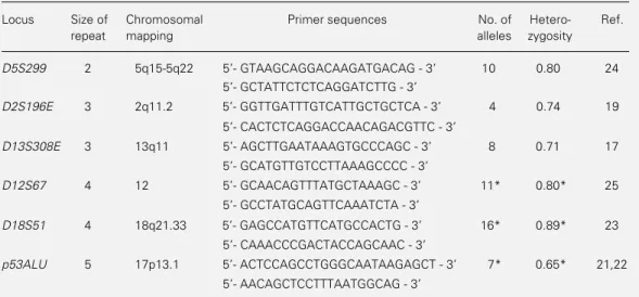
Table 1 - Details of the microsatellites studied and some of their population parameters.
*Observations made in the present study based on a random sample of N Brazilians (N = 408 for D12S67; N = 311 for D18S51; N = 100 for p53ALU).
Locus Size of Chromosomal Primer sequences No. of Hetero- Ref.
repeat mapping alleles zygosity
D5S299 2 5q15-5q22 5- GTAAGCAGGACAAGATGACAG - 3 10 0.80 24 5- GCTATTCTCTCAGGATCTTG - 3
D2S196E 3 2q11.2 5- GGTTGATTTGTCATTGCTGCTCA - 3 4 0.74 19 5- CACTCTCAGGACCAACAGACGTTC - 3
D13S308E 3 13q11 5- AGCTTGAATAAAGTGCCCAGC - 3 8 0.71 17 5- GCATGTTGTCCTTAAAGCCCC - 3
D12S67 4 12 5- GCAACAGTTTATGCTAAAGC - 3 11* 0.80* 25 5- GCCTATGCAGTTCAAATCTA - 3
D18S51 4 18q21.33 5- GAGCCATGTTCATGCCACTG - 3 16* 0.89* 23 5- CAAACCCGACTACCAGCAAC - 3
p53ALU 5 17p13.1 5- ACTCCAGCCTGGGCAATAAGAGCT - 3 7* 0.65* 21,22 5- AACAGCTCCTTTAATGGCAG - 3
present in the normal mucosa (Figure 1A). Obviously, homozygous patients could not be informative for LOH. For this reason, we established the level of heterozygosity of
p53ALU and D18S51 in the Brazilian popu-lation. The location of the pentanucleotide repeat polymorphism p53ALU in the first intron of the p53 gene (21,22) is ideal for studies of loss of heterozygosity. We typed this polymorphism in 100 randomly chosen individuals (Table 1) and found that 65 of them were heterozygous. This means that in 35% of cases the polymorphism will unfor-tunately not be informative. However, it still appears to be the best microsatellite marker for this purpose. In contrast, D18S51, which is not located as ideally but still closely linked to the DCC gene (23), exhibited a much higher heterozygosity of 89% among 311 individuals. The D5S299 polymorphism is closely linked to the APC gene and has an expected heterozygosity of 0.70 in Europe-ans (24). This polymorphism has the disad-vantage of being a dinucleotide repeat and of needing more complex techniques for allele resolution.
two samples in four distinct loci.
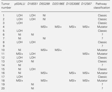
Pathogenetic classification of colorectal tumors
Of the 20 tumors studied, 14 (70%) could be assigned to the classic or mutator path-way of carcinogenesis (Table 2). Nine cases (45%) showed LOH at p53ALU and/or
D18S51 and absence of microsatellite insta-bility and could thus be assigned to the clas-sic pathway. Five cases (25%) showed mi-crosatellite instability and absence of LOH and could thus be allocated to the mutator pathway (including one case that presented both replication errors in two microsatellites and LOH in D18S51; see Discussion below). Six tumors could not be classified because they showed neither LOH nor microsatellite instability.
Discussion
In the present study we were able to assign 70% of the colorectal tumors to the classic or the mutator pathway (9). There are both theoretical and methodological reasons why it was not possible to establish the patho-genetic pathway of all colorectal tumors. First, in addition to the established so-called classic and mutator pathways, there may exist (and probably do exist) as yet unknown other route(s). Another reason for uncer-tainty is that microsatellite instability may be present in the tumor but may not be detected (10).
The fact that LOH is thought to indicate the classic pathway of tumorigenesis de-pends on probabilistic considerations. In normal states the chromosomal mutation rate is higher than the genic one. Thus, we expect to see frequent chromosomal deletions in tumors with an intact repair system undergo-ing the classic pathway of tumorigenesis. In the mutator pathway, gene mutation rates are elevated by hundred- or thousand-fold and thus much more likely to occur than the
Figure 1 - Examples of the detection with microsatellites of loss of heterozygosity and microsatellite instability in colorectal carcinomas. A, Silver-stained polyacryla-mide gel containing D18S51 alleles amplified from the normal mucosa (N) and tumor (T) of four patients (identified by numbers) with colorectal adenocarcinoma. The tumor from patient 2 presents loss of heterozygosity (arrowhead). Patient 6 is homozygous and consequently is not informative. B, Silver-stained polyacrylamide gel containing D12S67 alleles amplified from the normal mucosa (N) and tumor (T) of four patients (identified by numbers) with colorectal adenocarcinoma. The tumors from patients 11, 16 and 18 present microsatellite instability identified by the appearance of new mutant alleles (arrowheads).
Patient
5 6 7 2
N T N T N T N T
7 11 16 18
N T N T N T N T
Patient
A
B
chromosomal accidents that lead to LOH. These predictions were largely observed ex-perimentally (11). However, there is no com-pelling reason why LOH may not occur in tumors with microsatellite instability (11), as shown by the one instance of a tumor with both microsatellite instability and LOH at
D18S51 in the present study.
would still remain the same. Hence, for clini-cal studies we can apparently limit ourselves to the five other microsatellites, consider-ably simplifying the problem.
The great majority of studies focusing on
Table 2 - Microsatellite findings in each of the 20 colorectal tumors studied.
LOH = Loss of heterozygosity; NI = not informative (only indicated for the p53ALU,
D18S51 and D5S299); MSI+ = microsatellite instability present.
Tumor p53ALU D18S51 D5S299 D2S196E D13S308E D12S67 Pathway
number classification
1 LOH LOH NI Classic
2 LOH LOH NI Classic
3 LOH Classic
4 MSI+ MSI+ MSI+ MSI+ Mutator
5 LOH Classic
6 NI NI ?
7 NI NI ?
8 LOH LOH Classic
9 ?
10 NI MSI+ MSI+ Mutator
11 MSI+ LOH MSI+ Mutator
12 LOH LOH Classic
13 LOH NI Classic
14 NI ?
15 NI LOH Classic
16 NI MSI+ MSI+ MSI+ Mutator
17 LOH Classic
18 MSI+ MSI+ MSI+ MSI+ Mutator
19 NI ?
20 NI ?
colorectal cancer have come from First World countries where this tumor is the second or third most common cancer and has shown a rising incidence (2). In Third World coun-tries, colorectal cancer seems to be less com-mon and it was important to ascertain whether the relative importance of the two recog-nized pathogenetic mechanisms was the same in both environments. The rates of DNA replication errors, as well as the rates of LOH linked to the tumor suppressor genes
p53 and DCC obtained in this study, were closely similar to the frequencies reported by others. We observed loss of heterozygos-ity at p53 in 8 of 14 informative tumors (57%), a value similar to published data ranging from 40 to 76% (26-28). We ob-served loss of heterozygosity at D18S51 in 6 of 16 informative tumors (38%), a value similar to published data ranging from 34 to 52% (27,28). On the other hand, we did not observe any LOH in 17 tumors informative for D5S299. However, when compared with the expected frequency (16-20%; Refs. 27-29) this is not significantly different (χ2 =
2.5; 0.2>P>0.1), suggesting that, at least at this data level, the epidemiology of colorectal cancers in Brazil does not differ significantly from that of Europe and the United States.
References
1. Cavenee WK & White RL (1995). The ge-netic basis of cancer. Scientific American, 272: 50-57.
2. Dunlop MG (1992). Screening for large bowel neoplasms in individuals with a family history of colorectal cancer. British Journal of Surgery, 79: 488-494. 3. Vogelstein B & Kinzler KW (1993). The
multistep nature of cancer. Trends in Ge-netics, 9: 138-141.
4. Groden J, Thliveris A, Samowitz W, Carlson M, Gelbert L, Albertsen H, Joslyn G, Stevens J, Spirio L, Robertson M, Sargeant L, Krapcho K, Wolff E, Burt R, Hughes JP, Warrington J, McPherson J, Wasmuth J, Le Paslier D, Abderrahim H, Cohen D, Leppert M & White R (1991). Identification and characterization of the familial adenomatous polyposis coli gene.
Cell, 66: 589-600.
5. Miyaki M, Konishi M, Kikuchi-Yanoshita R, Enomoto M, Igari T, Tanaka K, Muraoka M, Takahashi H, Amada Y, Fukayama M, Maeda Y, Iwama T, Mishima Y, Mori T & Koike M (1994). Characteristics of somatic mutation of the adenomatous polyposis coli gene in colorectal tumors. Cancer Re-search, 54: 3011-3020.
6. Nigro JM, Baker SJ, Preisinger AC, Jessup JM, Hostetter R, Cleary K, Bigner SH, Davidson N, Baylin S, Devilee P, Glover T, Collins FS, Weston A, Modali R, Harris CC & Vogelstein B (1989). Muta-tions in the p53 gene occur in diverse human tumour types. Nature, 342: 705-708.
7. Vogelstein B, Fearon ER, Hamilton SR, Kern SE, Preisinger AC, Leppert M, Nakamura Y, White R, Smits AMM & Bos JL (1988). Genetic alterations during colorectal tumor development. New Eng-land Journal of Medicine, 319: 525-532. 8. de la Chapelle A & Peltomäki P (1995).
Genetics of hereditary colon cancer. An-nual Review of Genetics, 29: 329-348. 9. Eshleman JR & Markowitz SD (1996).
Mismatch repair defects in human carci-nogenesis. Human Molecular Genetics, 5: 1489-1494.
11. Reitmair AH, Cai JC, Bjerknes M, Redston M, Cheng M, Pind MT, Hay K, Mitri K, Bapat BV, Mak TW & Gallinger S (1996). MSH2 deficiency contributes to acceler-ated APC-mediacceler-ated intestinal tumorigen-esis. Cancer Research, 56: 2922-2926. 12. Jeffreys AJ & Pena SDJ (1993). A brief
introduction to human DNA fingerprint-ing. In: Pena SDJ, Chakraborty R, Epplen JT & Jeffreys AJ (Editors), DNA Finger-printing: State of the Science. Birkhäuser Verlag, Basel, 1-20.
13. Dover G (1995). Slippery DNA runs on and on and on Nature Genetics, 10: 254-256.
14. Bocker T, Schlegel J, Kullmann F, Stumm G, Zirngibl H, Epplen JT & Ruschoff J (1996). Genomic instability in colorectal carcinomas: comparison of different evaluation methods and their biological significance. Journal of Pathology, 179: 15-19.
15. Bubb VJ, Curtis LJ, Cunningham C, Dunlop MG, Carothers AD, Morris RG, White S, Bird CC & Wyllie AH (1996). Microsatellite instability and the role of hMSH2 in sporadic colorectal cancer. On-cogene, 12: 2641-2649.
16. Lothe RA, Peltomäki P, Meling GE, Aaaltonen LA, Nyström-Lahti M, Pylkkanen L, Heimdal K, Andersen TI, Moller P, Rognum TO, Fossa SD, Haldorsen T, Langmark F, Brogger A & de la Chapelle A (1993). Genomic instability in colorectal cancer; relationship, clinico-pathological variables and family history. Cancer Research, 53: 5849-5852. 17. Haddad LA & Pena SDJ (1993). CAT
re-peat polymorphism in a human expressed sequence tag (EST00444) (D13S308). Hu-man Molecular Genetics, 2: 1748.
18. Santos FR, Pena SDJ & Epplen JT (1993). Genetic and population study of an Y-linked tetranucleotide repeat DNA poly-morphism with a simple non-isotopic technique. Human Genetics, 90: 655-656. 19. Haddad LO, Fuzikawa A & Pena SDJ (1997). Simultaneous detection of size and sequence variation in the polymor-phic microsatellite D2S196E (EST 00493).
Human Genetics, 99: 796-800.
20. Vago AR, Macedo AM, Oliveira RP, Andrade LO, Chiari E, Galvão LMC, Reis DA, Pereira MES, Simpson AJG, Tostes S & Pena SDJ (1996). kDNA signatures of
Trypanosoma cruzi strains obtained di-rectly from infected tissues. American Journal of Pathology, 149: 2153-2159. 21. Futreal PA, Barrett JC & Wiseman RW
(1991). An Alu polymorphism intragenic to the TP53 gene. Nucleic Acids Re-search, 19: 6977.
22. Hahn M, Serth J, Fislage R, Wolfes H, Allhoff E, Jonas U & Pingoud A (1993). Polymerase chain reaction detection of a highly polymorphic VNTR segment in in-tron 1 of the human p53 gene. Clinical Chemistry, 39: 549-550.
23. Urquhart A, Oldroyd NJ, Kimpton CP & Gill P (1995). Highly discriminating heptaplex short tandem repeat PCR sys-tem for forensic identification. Biotech-niques, 18: 116-121.
24. Van Leeuwen C, Tops C, Breukel C, van der Klift H, Fodde R & Khan PM (1991). CA repeat polymorphism at the D5S299 locus linked to adenomatous polyposis coli (APC). Nucleic Acids Research, 19: 5805.
25. Roewer L, Arnemann J, Spurr NK, Grzeschik K-H & Epplen JT (1992). Simple repeat sequences on the human Y chro-mosome are equally polymorphic as their autosomal counterparts. Human Genet-ics, 89: 389-394.
26. Lothe A, Nakamura Y, Woodward S, Gedde-Dahl T & White R (1988). VNTR (variable number of tandem repeats) markers show loss of chromosome 17p sequences in human colorectal carcino-mas. Cytogenetics and Cell Genetics, 48: 167-169.
27. Law DJ, Olschwang S, Monpezat JP, Lefrancois D, Jagelman D, Petrelli NJ, Thomas G & Feinberg AP (1988). Con-certed nonsyntenic allelic loss in human colorectal carcinoma. Science, 241: 961-965.
28. Iacopetta B, Di Grandi S, Dix B, Haig C, Soong R & House A (1994). Loss of het-erozygosity of tumour suppressor gene loci in human colorectal carcinoma. Euro-pean Journal of Cancer, 30A: 664-670. 29. Solomon E, Voss R, Hall V, Bodmer WF,