The Le bane se m utatio n as an
im po rtant cause o f fam ilial
hype rcho le ste ro le m ia in Brazil
Departamento de Clínica Médica, Faculdade de Medicina de Ribeirão Preto,
Universidade de São Paulo, Ribeirão Preto, SP, Brasil F.L. Alberto, M.S. Figueiredo,
M.A. Zago, A.G. Araújo and J.E. Dos-Santos
Abstract
Familial hypercholesterolemia (FH) is a common autosomal disorder that affects about one in 500 individuals in most Western populations and is caused by a defect in the low-density-lipoprotein receptor (LDLr) gene. In this report we determined the molecular basis of FH in 59 patients from 31 unrelated Brazilian families. All patients were screened for the Lebanese mutation, gross abnormalities of the LDLr gene, and the point mutation in the codon 3500 of the apolipoprotein B-100 gene. None of the 59 patients presented the apoB-3500 muta-tion, suggesting that familial defective ApoB-100 (FDB) is not a major cause of inherited hypercholesterolemia in Brazil. A novel 4-kb deletion in the LDLr gene, spanning from intron 12 to intron 14, was characterized in one family. Both 5' and 3' breakpoint regions were located within Alu repetitive sequences, which are probably involved in the crossing over that generated this rearrangement. The Lebanese mutation was detected in 9 of the 31 families, always associated with Arab ancestry. Two different LDLr gene haplotypes were demon-strated in association with the Lebanese mutation. Our results suggest the importance of the Lebanese mutation as a cause of FH in Brazil and by analogy the same feature may be expected in other countries with a large Arab population, such as North American and Western Euro-pean countries.
Co rre spo nde nce
J.E. Dos-Santos
Departamento de Clínica Médica FMRP, USP
14049-900 Ribeirão Preto, SP Brasil
Fax: + 55-16-633-1144 E-mail: jedsanto@ fmrp.usp.br
Research supported by FAPESP, CNPq and FINEP.
Received August 13, 1998 Accepted March 17, 1999
Ke y wo rds
·Familial hypercholester-olemia
·LDL receptor gene
·Lebanese mutation
·Haplotype analysis
·apoB-100 gene
·Alu sequences
Intro ductio n
Familial hypercholesterolemia (FH) is a common autosomal disorder caused by a defect in the low-density-lipoprotein recep-tor (LDLr) gene. It affects about one in 500 individuals in most Western populations and is characterized by elevated serum LDL cho-lesterol levels, tendon xanthomas and pre-mature coronary artery disease (1). The anal-ysis of the LDLr gene in FH patients has led
tic) and control sera. LDL cholesterol was calculated using the Friedwald equation (8). In Table 1, we summarize the clinical fea-tures of the families investigated here.
D NA analysis
High molecular weight genomic DNA was isolated from peripheral blood leuko-cytes by phenol/chloroform extraction (9).
Southern blotting/hybridization assay.
Five to 7 µg of DNA were digested with
XbaI, KpnI and BamHI restriction endonu-cleases, separated on 1% agarose gels, and blotted onto nylon membranes (GeneScreen Plus, DuPont, Wilmington, DE) (9). The filters were hybridized to fragments of hu-man LDLr cDNA (obtained from clone pLDLR3, purchased from American Type Culture Collection, Rockville, MD) at 42o
C for 16-48 h and washed at 65oC for 60 min. The probes were labelled with [a-32
P]dCTP by random oligonucleotide priming (9). The membranes were exposed to x-ray films (X-Omat AR, Kodak) with intensifying screens for 1 to 7 days. In order to characterize the deletion observed in family 2, Bam HI-di-gested DNA samples were hybridized to the oligonucleotides A (5'-ATGACAGGGGTCT CCTATACCAGGAGAAGGTGTTGGA GTG-3') and B (5'-CGATCCGACGTCAC CGGTGGGTCCTCAGTAGGTGGCAGTC CG-3') as specific probes for exons 13 and 15, respectively. Hybridization conditions were as described above, except that the filters were washed at 55o
C for 30 min.
Haplotype analysis. For LDLr gene hap-lotype determination, the following poly-morphic sites were investigated: StuI (exon 8), AvaII (exon 13), ApaLI (intron 15), PvuII (intron 15) and NcoI (exon 18) (10). The
ApaLI, PvuII, and NcoI polymorphisms were analyzed by Southern blotting (as described above) using a 2.1-kb BamHI fragment (ex-ons 10-18) of the LDLr cDNA as probe. StuI and AvaII polymorphisms were analyzed by PCR amplification (see below) of a DNA The Brazilian population is composed of
various ethnic groups, mainly Caucasians of European descent, Blacks, Amerindians, and, to a much lesser extent, Asians and Arabs (4). In a preliminary study we identified the Lebanese allele in 5 out of 10 unselected Brazilian families with FH, always in asso-ciation with Arab ancestry, suggesting an important contribution of this ethnic group to the prevalence of FH in Brazil (5).
In the present study, we extended our search for the Lebanese allele and screened for other structural abnormalities of the LDLr gene in Brazilian FH patients. In addition, the LDLr gene haplotypes associated with FH were determined in selected cases. Fi-nally, we also examined the role of the point mutation in the codon 3500 of the apolipo-protein B-100 gene as another cause of hy-percholesterolemia in these patients.
Patie nts and Me tho ds
Patie nts
Diagnos-segment containing the polymorphic site fol-lowed by digestion with the appropriate en-zyme. When necessary, non-affected mem-bers of the families were also studied in order to identify the LDLr gene haplotype in association with the abnormal gene.
Polymerase chain reaction (PCR). The Lebanese allele was detected by the selec-tive amplification of exon 14 of the LDLr gene followed by HinfI digestion, as previ-ously described (5). To characterize the
de-letion identified in family 2, a segment con-taining the deletion breakpoints was ampli-fied using the primers P1 (5'-CTTGGAGGA TGAAAAGGCTGG-3') and P2 (5'-TGACC TTTAGCCTGACGGTGGATG-3'), which are complementary to exons 12 and 15, re-spectively. The polymorphic site AvaII was investigated using the primers P3 (5'-CTTCC TTCCTTGCTGCCTGT-3') and P4 (5'-TG TGAGGCAGCTCCTCATGT-3'). The prim-ers P5 (5'-CCAAGCCTCTTTCTCTCTCTC
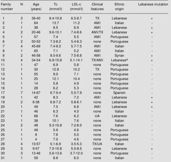
Table 1 - Clinical and laboratory features, ethnic background and detection of the Lebanese mutation in the patients included in this study.
Age (in years) at diagnosis; N: number of analyzed individuals per family; Tc: total cholesterol; LDLc: low -density lipoprotein cholesterol. TX: tendon xanthomata; AM I: acute myocardial infarction; UA: unstable angina. Families 1 to 10 w ere reported previously (Ref. 5); + and - denote the presence (alw ays in heterozy-gosity) or absence of the Lebanese mutation; aone patient w ith clinical and laboratory data compatible w ith homozygous FH, and all other patients w ere diagnosed as heterozygotes for FH. Cholesterol mg/dl = mmol/l x 38.6.Triglyceride mg/dl = mmol/l x 88.5.
Family N Age Tc LDL-c Clinical Ethnic Lebanese mutation number (years) (mmol/l) (mmol/l) features origin
1 3 36-40 8.4-10.8 6.5-8.7 TX Lebanese +
2 1 64 13.7 11.2 AM I Italian
-3 1 38 8.5 6.9 AM I Lebanese +
4 2 20-46 9.6-10.1 7.4-8.6 AM I/TX Lebanese +
5 1 57 7.4 5.5 AM I Portuguese
-6 2 50-52 7.3-8.2 5.4-6.3 none Portuguese
-7 4 45-69 7.4-9.3 5.7-7.5 AM I Italian
-8 1 65 7.1 5.2 AM I Italian
-9 2 46-58 8.5-9.6 7.5-8.8 AM I Syrian +
10 4 34-54 6.9-15.8 5.1-14.1 TX/AM I Lebanesea +
11 1 47 6.9 5.8 none Portuguese
-12 1 30 12.8 10.2 TX Portuguese
-13 1 55 9.0 7.1 none Portuguese
-14 1 25 12.1 10.4 none Portuguese
-15 1 14 5.8 4.9 none Portuguese
-16 1 28 6.2 5.3 none Portuguese
-17 7 14-67 6.7-9.4 5.5-7.9 none Spanish
-18 1 42 8.3 7.2 AM I Lebanese +
19 2 6-38 6.8-7.2 5.8-6.1 none Lebanese +
20 1 49 7.5 6.8 AM I Lebanese +
21 1 46 6.3 4.0 none Italian
-22 1 68 7.6 6.2 UA Lebanese
-23 1 38 10.1 7.6 none Italian
-24 2 48 9.3-10.8 7.2-8.0 none Italian
-25 1 49 5.9 4.6 none Portuguese
-26 1 9 7.6 5.0 none Portuguese
-27 1 4 7.0 4.6 none Portuguese
-28 4 13-57 5.1-6.9 3.5-5.3 TX/UA Italian
-29 3 9-57 7.0-10.8 5.5-8.5 none Lebanese +
30 3 13-46 5.6-13.6 3.7-12.0 none Portuguese
-TTCCA-3') and P6 (5'-CCACCCGCCGCCT TCCCGTGCT-3') were used to analyze the polymorphic site StuI. All PCR experiments were performed using a 50-µl total reaction volume containing 100 ng of genomic DNA, 10 mM Tris-HCl, 1.5 mM MgCl2, 0.01% BSA, 200 µM of each dNTP, 0.25 µM of each primer, and 1 U of Taq DNA poly-merase; 35 cycles of amplification were car-ried out (typically, 94o
C for 30 s, 55o C for 50 s and 72o
C for 1 min) in a Perkin Elmer Cetus thermal cycler (Norwalk, CT). All oligonucleotides were synthesized with an Applied Biosystems 391 oligonucleotide syn-thesizer, based on the cDNA sequence pre-viously reported (11).
Detection of apoB-3500 mutation. To detect the apoB-3500 mutation, a single primer-template mismatch approach with subsequent digestion with the enzyme MspI was performed as described (12).
Re sults
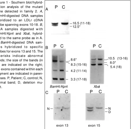
For only one of the 31 families included in this study was it possible to identify a gross deletion as the molecular defect asso-ciated with FH. DNA samples from the af-fected member of family 2 (Table 1) di-gested with BamHI and hybridized to a cDNA probe that covers exons 10-18 revealed an abnormal band of 12.5 kb in addition to a reduction in intensity of the expected 16.5-kb band (Figure 1A). XbaI-digested DNA hybridized to this probe revealed an abnor-mal band of 8.0 kb and a reduction in inten-sity of the normal 10.5-kb band, which con-tains exons 13-16 (Figure 1B). BamHI/KpnI double-digested DNA hybridized to the same probe showed a reduced intensity for the normal 4.2-kb band (exons 11-14) and an abnormal band of 8.6 kb (Figure 1B). These results indicate the occurrence of a partial deletion of about 4 kb encompassing exon 14 in a heterozygous patient. By probing
BamHI-digested DNA samples with oligo-P C
10.5 (13-16) 8.0* 5.4 (18)
P C P C
16.5 (11-18) 12.5*
8.6* 8.3 (15-16)
4.2 (11-14)
3.3 (17-18)
BamHI/KpnI Xbal
N D N
Figure 1 - Southern blot/hybrid-ization analysis of the mutant gene detected in family 2. A,
BamHI-digested DNA samples hybridized to an LDLr cDNA probe spanning exons 10-18. B, DNA sam ples digest ed w it h
BamHI/KpnI and XbaI, hybrid-ized to the same probe as in A.
C, BamHI-digested DNA sam-ples hybridized t o specif ic probes for exons 13 and 15. The ast erisks indicat e abnorm al bands; the size of the bands (in kb) are indicated on the right; the exons contained w ithin each segment are indicated in paren-theses. P, Patient; C, control; N, normal band; D, deletion mu-tant.
exon 13 exon 15
normal gene 4.2 2.5
0.6 0.4
M 1 2 3
S P E X P E S P P P P
15 13 14
12
mutant gene
S P E P P P
15 12
0.5 kb Figure 2 - PCR amplification
across the deletion junctions of the mutation characterized in family 2. Top, PCR products separated on 1% agarose gel. Lane M , molecular w eight mark-er; lanes 1 and 2, tw o affected members of the family; lane 3, unaffected control sample. Bot-tom, Restriction map of the de-letion mutant (the normal map is show n for comparison). Ar-row s, Alu sequences; horizontal bars, deletion breakpoint re-gions; solid vertical bars, exons; horizontal line, introns; S, SphI; P, PstI; E, EcoRV; X, XbaI.
A
B
nucleotides A (complementary to exon 13) and B (complementary to exon 15) we were able to locate the 5' and 3' deletion break-points within introns 12 and 14, respectively (Figure 1C). Further analysis was performed by selective amplification of the region span-ning the deletion breakpoints using the prim-ers P1 and P2 (complementary to exons 12 and 15, respectively), which are located 2.5 kb apart from each other in the mutant gene (Figure 2). These primers are separated by a segment of 6.5 kb in the wild type gene, precluding its amplification under the condi-tions described here (lane 3, Figure 2). The 2.5-kb amplified mutant segment was then subjected to a series of digestions with the enzymes XbaI, PstI, EcoRV, and SphI, to provide a restriction map (Figure 2). This strategy enabled us to determine both 5' and 3' breakpoint regions (within ± 900 bp of certainty), both within Alu repetitive se-quences (Figure 2).
The Lebanese mutation was detected in 9 of the 31 families, always as a heterozygote and in association with Arab ancestry (Table 1). Haplotype analysis revealed a common single haplotype in association with the Lebanese mutation in 8 families: +++ (+ and -indicate the presence and absence of the sites StuI, AvaII, ApaLI, PvuII, and NcoI, respectively). This particular haplotype has been reported in association with the Leba-nese mutation in all of the few patients that have been evaluated for this purpose (10). In one family (family 20, Table 1) a distinct haplotype (+----) was observed. None of the patients presented the apoB-3500 mutation (data not shown).
D iscussio n
The molecular basis of FH in the Brazil-ian population is poorly characterized. In the present study, we also searched for the muta-tion in the codon 3500 of the apolipoprotein B-100 gene because the two diseases, FH
and familial defective apolipoprotein B-100 (FDB), have similar clinical features and prevalence (12,13). None of the 59 patients investigated here presented the apoB-3500 mutation. These results agree with the study of Schuster et al. (14) in a German popula-tion, who found only 3% of FDB patients among those previously diagnosed as FH. Therefore, we assume that the vast majority, if not all, of our patients are in fact affected by FH.
dia-betes mellitus (22), lipoprotein lipase defi-ciency (23,24), C3 defidefi-ciency (25), some types of familial thrombophilia (16), Lesch Nyhan syndrome (26), adenosine deaminase deficiency (27), and others (25).
In a previous paper we pointed out the possible contribution of the Lebanese allele to the prevalence of FH in Brazil, far in excess of the proportion of individuals of Arab ancestry in the Brazilian population (5). Now we confirm that result by the anal-ysis of a larger number of FH families. We predict that this observation will contribute to the early diagnosis of FH in cases pre-sumed to be of Arab ancestry. Indeed, ante-natal diagnosis could be performed in se-lected cases.
Arab ancestry was present in all patients bearing the Lebanese mutation in our popu-lation. Haplotype analysis revealed the same single haplotype in 8 of 9 FH families bear-ing the Lebanese mutation: ++--. This par-ticular haplotype has been previously de-scribed in association with the Lebanese mutation (10). Surprisingly, a different hap-lotype was determined in one family: +----, which shows different results for polymor-phic sites located at 5' (AvaII) and 3' (ApaLI) of the Lebanese mutation. The occurrence of two different haplotypes in association with the same mutation may be explained by sev-eral genetic mechanisms: a) modification of the ancestor haplotype by at least two re-combination events, b) gene conversion, and c) occurrence of a recurrent mutation in chromosomes bearing different haplotypes (28). Extensive haplotype studies in FH pa-tients from Middle East populations would be necessary to elucidate this point.
The Lebanese mutation was detected in approximately 30% of our patients. Our se-ries contained 59 FH individuals from 31 families. This is the largest series of patients with FH reported in the country and includes patients mainly from the Southern region of
Brazil. If there are more than 50 different single point mutations, which can lead to familial hypercholesterolemia, the fact that almost 30% of the mutations detected in our population are Lebanese indicates the im-portance of the Lebanese mutation in the population studied. Thus, it seems also rea-sonable to conclude that the Lebanese muta-tion is an important cause of FH in Brazil. This finding was originally unexpected be-cause of the negligible contribution of Arabs to the composition of the Brazilian popula-tion (4). However, we should consider that, although constituting a small group, the Ar-abs present a much higher prevalence of FH than the groups that primarily compose the Brazilian population (Caucasians of Euro-pean descent, Blacks and Amerindians), which explains our observation. It is also tempting to speculate that the same feature may be occurring in other countries with mixed populations (and an even larger Arab population), such as North American and Western European countries (e.g., France, Germany, and England). Supporting this as-sumption, Cavanaugh et al. (29) reported the detection of the Lebanese mutation in 2 out of 15 (13%) Australian patients with FH, always in association with Arab descent. The authors interpreted this finding as the result of the recent immigration of Arabs to the Australian continent. To our knowledge, no other study was conducted to search for the Lebanese mutation in FH patients out-side the Middle East. It is therefore recom-mended that this approach be extended to other countries.
Ackno wle dgm e nts
Re fe re nce s
1. Goldstein JL & Brow n M S (1989). Familial hypercholesterolaemia. In: Scriver CR, Beaudet AL, Sly WS & Valle D (Editors),
The M etabolic Basis of Inherited Disease.
6th edn. M cGraw -Hill, New York, 1215-1250.
2. Hobbs HH, Brow n M S & Goldstein JL (1992). M olecular genetics of the LDL re-ceptor gene in familial hypercholesterole-mia. Human M utation, 1: 445-466. 3. Hobbs HH, Russel DW, Brow n M S &
Goldstein JL (1990). The LDL receptor locus in familial hypercholesterolaemia: molecular analysis of a membrane pro-tein. Annual Review of Genetics, 24: 133-170.
4. Daune D (Editor) (1991). Britannica Book of the Year. Encyclopaedia Britannica, Chi-cago, 560-561.
5. Figueiredo M S, Santos JE, Alberto FL & Zago M A (1992). High frequency of the Lebanese allele of the LDLr gene among Brazilian patients w ith familial hypercho-lesterolaemia. Journal of M edical Genet-ics, 29: 813-815.
6. Allain CC, Poon LS, Chon CSG, Richmond U & Fu PC (1974). Enzymatic determina-tion of total serum cholesterol. Clinical Chemistry, 20: 470-475.
7. Fossati P & Principe L (1982). Serum triac-ylglycerols determined colorimetrically w ith an enzyme that produces hydrogen peroxide. Clinical Chemistry, 28: 2077-2080.
8. Friedw ald W, Levy R & Fredrickson D (1972). Estimation of the concentration of low -density lipoprotein cholesterol in plasma w ithout use of the preparative ul-tracentrifuge. Clinical Chemistry, 18: 499-502.
9. Sambrook J, Fritsch EF & M aniatis T (Edi-tors) (1989). M olecular Cloning: A Labora-tory M anual. 2nd edn. Cold Spring Harbor Laboratory Press, New York.
10. Berkman N, Weir BS, Pressman-Schw artz S, Ayeleth R & Leitersdorf E (1992). Hap-lotype analysis at the low -density lipopro-tein receptor locus: application to the study of familial hypercholesterolemia in Israel. Human Genetics, 88: 405-410. 11. Yam am ot o T, Davis CG, Brow n M S,
Schneider W J, Linet t e Casey M , Goldstein JL & Russel DW (1984). The human LDL receptor: a cysteine-rich pro-tein w ith multiple alu sequences in its mRNA. Cell, 39: 27-38.
12. Hansen PS, Rudiger N, Tybjaerg-Hansen
A, Faegerman D & Gugersen N (1991). Detection of the apoB-3500 mutation (glu-tamine for arginine) by gene amplification and cleavage w ith M spI. Journal of Lipid Research, 32: 1229-1233.
13. Tybjaerg-Hansen A & Hum phries SE (1992). Familial defective apolipoprotein B-100: a single mutation that causes hypercholesterolaem ia and prem ature coronary artery disease. Atherosclerosis, 96: 91-107.
14. Schuster H, Rauh G, Gerl C, Keller C, Wol-fram G & Zöllner N (1991). Use of DNA haplotype analysis in diagnosis of familial hypercholest erolaem ia in 31 Germ an families. Journal of M edical Genetics, 28: 865-870.
15. Rüdiger NS, Hansen PS, Jørgensen M , Faergeman O, Bolund L & Gregersen N (1991). Repetitive sequences involved in the recombination leading to deletion of exon 5 of the low -density-lipoprotein re-ceptor gene in a patient w ith familial hy-percholesterolemia. European Journal of Biochemistry, 198: 107-111.
16. Olds RJ, Lane DA, Chow dhury V, De Stefano V, Leone G & Thein SL (1993). Complete nucleotide sequence of the an-tithrombin gene: evidence for homolo-gous recombination causing thrombo-philia. Biochemistry, 32: 4216-4224. 17. Rüdiger NS, Gregersen N &
Kielland-Brandt M C (1995). One short w ell con-served region of Alu-sequences is in-volved in human gene rearrangements and has homology w ith prokaryotic-chi.
Nucleic Acids Research, 23: 256-260. 18. Horsthemke B, Beisiegel U, Dunning A,
Havinga JR, Williamson R & Humphries S (1987). Unequal crossing-over betw een tw o alu-repetitive DNA sequences in the low -density-lipoprotein-receptor gene.
European Journal of Biochemistry, 164: 77-81.
19. M iyake Y, Tajim a S, Funahashi T & Yamamoto A (1989). Analysis of a recy-cling-impaired mutant of low density lipo-protein receptor in familial hypercholes-terolemia. Journal of Biological Chemis-try, 264: 16584-16590.
20. Lehrman M A, Russel DW, Goldstein JL & Brow n M S (1987). Alu-Alu recombination deletes splice acceptor sites and pro-duces secreted low density lipoprotein receptor in a subject w ith familial hyper-cholesterolem ia. Journal of Biological Chemistry, 262: 3354-3361.
21. Kigaw a K, Kihara K, M iyake Y, Funahashi T, Yamamura T & Yamamoto A (1993). Low -density lipoprotein receptor mutation that deletes exon 2 and 3 by Alu-Alu re-combination. Journal of Biochemistry, 113: 372-376.
22. Elbein SC (1992). Linkage disequilibrium among RFLPs at the insulin-receptor lo-cus despite intervening Alu repeat se-quences. American Journal of Human Ge-netics, 51: 1103-1110.
23. Benlian P, Etienne J, Gennes JL, Noé L, Brault D, Raisonnier A, Arnault F, Hamelin J, Foubert L, Cuat JC, Tse CT & Galibert F (1995). Homozygous deletion of exon 9 causes lipoprotein lipase deficiency: pos-sible intron-Alu recombination. Journal of Lipid Research, 36: 356-366.
24. Chuat JC, Raisonnier A, Etienne J & Galibert F (1992). The lipoprotein lipase-encoding human gene: sequence from intron-6 to intron-9 and presence in in-tron-7 of a 40-million-year-old Alu se-quence. Gene, 110: 257-261.
25. Botto M , Fong KY, So AK, Barlow R, Routier R, M orley BJ & W alport M J (1992). Homozygous hereditary C3 defi-ciency due to a partial gene deletion. Pro-ceedings of the National Academy of Sci-ences,USA, 89: 4957-4961.
26. M arcus S, Hellgren D, Lam bert B, Fällström SP & Wahlström J (1993). Du-plication in the hypoxanthine phosphori-bosyl-transferase gene caused by Alu-Alu recombination in a patient w ith Lesch Nyhan syndrome. Human Genetics, 90: 477-482.
27. Shovlin CL, Simmonds HA, Fairbanks LD, Deacock SJ, Hughes JM B, Lechler RI, W ebster ADB, Sun XM , W ebb JC & Soutar AK (1994). Adult onset immunode-ficiency caused by inherited adenosine deaminase deficiency. Journal of Immu-nology, 153: 2331-2339.
28. Antonarakis SE, Boehm CD, Sergeant GR, Theisen CE, Dover GJ & Kazazian Jr HH (1984). Origin of beta-S-globin gene in Blacks: contribution of recurrent mutation or gene conversion or both. Proceedings of the National Academy of Sciences, USA, 81: 853-856.
29. Cavanaugh JA, Easteal S, Simons L & Serjeanson S (1992). A rapid method for diagnosis of the Lebanese allele in the low -density lipoprotein receptor gene.