ABSTRACT
http://dx.doi.org/10.1590/1678-775720140259
Host response mechanisms in periodontal diseases
Nora SILVA1, Loreto ABUSLEME1, Denisse BRAVO1, Nicolás DUTZAN2, Jocelyn GARCIA-SESNICH2, Rolando
VERNAL2, Marcela HERNÁNDEZ3, Jorge GAMONAL2
1- Laboratory of Microbiology, Department of Pathology, Faculty of Dentistry, University of Chile, Santiago, Chile.
2- Laboratory of Periodontal Biology, Department of Conservative Dentistry, Faculty of Dentistry, University of Chile, Santiago, Chile. 3- Laboratory of Periodontal Biology, Department of Pathology, Faculty of Dentistry, University of Chile, Santiago, Chile.
Corresponding address: Jorge Gamonal - Facultad de Odontología - Universidad de Chile - Avenida Sergio Livingstone 943, Comuna de Independencia - Santiago - Chile - Phone: 0056-2-978 1700 - e-mail: [email protected]
Submitted: November 10, 2014 - Modiication: January 25, 2015 - Accepted: February 11, 2015
P
eriodontal diseases usually refer to common inlammatory disorders known as gingivitis and periodontitis, which are caused by a pathogenic microbiota in the subgingival bioilm, including Porphyromonas gingivalis, Aggregatibacter actinomycetemcomitans, Tannerella forsythia and Treponema denticola that trigger innate, inlammatory, and adaptive immune responses. These processes result in the destruction of the tissues surrounding and supporting the teeth, and eventually in tissue, bone and inally, tooth loss. The innate immune response constitutes a homeostatic system, which is the irst line of defense, and is able to recognize invading microorganisms as non-self, triggering immune responses to eliminate them. In addition to the innate immunity, adaptive immunity cells and characteristic cytokines have been described as important players in the periodontal disease pathogenesis scenario, with a special attention to CD4+ T-cells (T-helper cells). Interestingly, the T cell-mediated adaptive immunity development is highly dependent on innate immunity-associated antigen presenting cells, which after antigen capture undergo into a maturation process and migrate towards the lymph nodes, where they produce distinct patterns of cytokines that will contribute to the subsequent polarization and activation of speciic T CD4+ lymphocytes. Skeletal homeostasis depends on a dynamic balance between the activities of the bone-forming osteoblasts (OBLs) and bone-resorbing osteoclasts (OCLs). This balance is tightly controlled by various regulatory systems, such as the endocrine system, and is inluenced by the immune system, an osteoimmunological regulation depending on lymphocyte- and macrophage-derived cytokines. All these cytokines and inlammatory mediators are capable of acting alone or in concert, to stimulate periodontal breakdown and collagen destruction via tissue-derived matrix metalloproteinases, a characterization of the progression of periodontitis as a stage that presents a signiicantly host immune and inlammatory response to the microbial challenge that determine of susceptibility to develop the destructive/progressive periodontitis under the inluence of multiple behavioral, environmental and genetic factors.Keywords: Periodontitis. Periodontal diseases. Progressive periodontitis. Pathogenesis. Osteoimmunology. Metalloproteinases.
I - E T I O L O G Y O F P E R I O D O N T A L
DISEASE
Periodontal diseases usually refer to common inlammatory disorders known as gingivitis and periodontitis, which are caused by a pathogenic microbiota in the subgingival bioilm. The bacterial challenge induces the production of cytokines and chemokines by the gingival epithelium, resulting in the expression of adhesion molecules,
A hallmark of periodontitis is that is caused by complex subgingival microbial communities, which can comprise around 500 bacterial species171. The human body contains various and distinctive ecosystems providing a unique environment for colonizing microorganisms. Moreover, the oral cavity presents numerous surfaces for microbial colonization. For instance, the periodontal sulcus/ pocket is partially sheltered from the physical shear forces in the oral cavity and contains hard and non-shedding surfaces of the tooth root, along with the shedding surfaces of gingival mucosa in permanent contact with the gingival crevicular luid211.The epithelial surfaces are continuously replaced; however, several of the most pathogenic bacterial species are able to invade the gingival cells and tissues, remaining viable and evading the action of different immune cells237. Therefore, thesubgingival environment has distinct ecological determinants, and it can be considered as one of the most interesting oral niches for characterizing the intimate interplay between oral microbial communities and the immune host response.
Usually, a few bacterial species in the subgingival biofilm have been associated with diseased periodontal tissues and identified as putative pathogens. It was concluded that there is strong evidence that Porphyromonas gingivalis (P. gingivalis), Aggregatibacter actinomycetemcomitans (A. actinomycetemcomitans) and Tannerella forsythia (T. forsythia) are periodontal pathogens when their presence is in suficient numbers and in susceptible hosts31.
Although many efforts have been made to characterize the prevalence of the periodontal pathogens described above, there is little consensus due to the varied methodologies used. For that reason, our group investigated the differences in 9 bacterial species of the subgingival microbiota of chronic periodontitis populations in Chile, Spain and Colombia using the same clinical and microbiological methods90.In that study, the red complex species P. gingivalis and T. forsythia, were detected in high frequencies in most of the patients and the actives sites. P. gingivalis were the most frequently detected in Chilean patients (83.8%) and the less frequent in Colombians (65.9%), while the frequency of T. forsythia was lower in Chile (16.2%) than in Colombia (39%) and Spain (36.1%)90. Additionally, our group reported that active sites in Chileans with chronic periodontitis showed higher mean percentages for P. gingivalis of the total anaerobic than inactive sites, and no signiicant differences were found for T. forsythia204. Our findings are in agreement with the ones previously described by López, et al.137 (2004), who found a higher prevalence of P. gingivalis in Chilean than US subjects, using checkerboard
DNA-DNA hybridization. This study demonstrated that more than 90% of active sites are colonized by red complex bacteria, but the prevalence of T. forsythia was lower than P. gingivalis and similar in Chilean and in US subjects137 (Figure 1).
The high prevalence of P. gingivalis and T. forsythia in Chileans with chronic periodontitis was in accordance with studies performed in other Latin American populations. Using DNA-based methods, it was reported a high frequency of P. gingivalis and T. forsythia in Brazilian (68% and 45%, respectively) and Mexican population (100% and 97.4%, respectively)33, 252 (Figure 1). In addition, using microbiological cultures, Botero, et al.21 (2007) were able to detect P. gingivalis and T. forsythia in a high frequency in Colombians patients with chronic periodontitis (75.4% and 50%, respectively). Together these results indicate that P. gingivalis and T. forsythia are frequently detected in Latin American population with chronic periodontitis, however, the prevalence of P. gingivalis is higher in Chile compared to other Latin American countries (Figure 1).
Regarding A. actinomycetemcomitans prevalence in chronic periodontitis, our group reported that this pathogen was detected in a lower proportion than P. gingivalis and T. forsythia in Chilean population, using microbiological culture54 and DNA probes137. Also using microbiological cultures, lower proportions and frequency of A. actinomycetemcomitans were found in Chilean (19.4%), Colombian (17.1%) and Spaniard (16.7%) populations. However, the Chilean population presented a higher frequency of detection than US patients137 (Figure 1). In contrast, in Brazilian35 and Mexican256 patients with chronic periodontitis, a higher frequency of detection of A. actinomycetemcomitans has been reported, using PCR and checkerboard DNA-DNA hybridization, respectively. These findings are worth noting, since it has been demonstrated that the frequency detection of some periodontopathic microorganisms in chronic periodontitis subjects was signiicantly higher when detected by PCR than in microbiological cultures21.
On the other hand, a very high prevalence of P. gingivalis was also found in patients with aggressive periodontitis in Chileans55,135,136, Brazilians35, Mexicans256 and Colombians subjects21. Interestingly, concerning the microbiological proiles described in periodontitis, some studies have found differences in the periodontal microbiota between chronic and aggressive periodontitis21,35,55,256, however, more recent studies have reported only minor differences in the microbiological proiles of both clinical entities186.
aggressive periodontitis have a higher prevalence of P. gingivalis compared with the ones with chronic periodontitis (Figure 1). For example, our group reported that Chilean patients with aggressive periodontitis have 86.6%-100% of P. gingivalis
frequency of isolation, compared with 76.4% in chronic periodontitis55. Also using microbiological cultures, it was reported a frequency of 92.8% in Colombian subjects with aggressive periodontitis compared with 75.4% for the ones with chronic
Author Number of individuals and country Disease type (Sample description) Method of analysis
Prevalence of periodontopathic bacteria per individual
López, et al.135 (1995) n=15, Chileans Localized Juvenile Periodontitis (LJP; n=10), Generalized Juvenile Periodontitis (GJP; n=5) Microbiological Culture (7 bacterial species) P. gingivalis A. actinomycetemcomitans LJP 70.0% 40.0% GJP 80.0% 60.0% López, et al.136 (1996) n=24, Chileans Localized Juvenile Periodontitis (LJP; n=18), Generalized Juvenile Periodontitis (GJP; n=6) DNA probes (3 bacterial species) P. gingivalis A. actinomycetemcomitans LJP 94.4% 38.9% GJP 0% 0% Tinoco, et al.235
(1997) n=25, Brazilians Localized Aggressive periodontitis (LAgP) Microbiological Culture (1 bacterial species)
A. actinomycetemcomitans LAgP 76% López134 (2000) n=60, Chileans Progressive Periodontitis (PP) DNA probe (3 bacterial species) P. gingivalis A. actinomycetemcomitans PP 96% 11.6% López,
et al.137 (2004) n=26, Chileans Chronic Periodontitis (CP) Checkerboard DNA-DNA hybridization (40 bacterial species) P. gingivalis T. forsythia A. actinomycetemcomitans CP >9x10⁵ counts >6x10⁵ counts >3x10⁵ counts Gajardo,
et al.55 (2005) n=53, Chileans Chronic Periodontitis (CP; n=17), Localized (LAgP; n=30) and Generalized Aggressive Periodontitis (GAgP; n=6) Microbiological Culture (8 bacterial species) P. gingivalis A. actinomycetemcomitans CP 76.4% 35.2% LAgP 86.6% 16.6% GAgP 100% 33.3% Cortelli, et al.35 (2005) n=203, Brazilians Aggressive Periodontitis (AgP; n=25) Chronic Periodontitis (CP; n=178) PCR (5 bacterial species) P. gingivalis T. forsythia A. actinomycetemcomitans CP 68.0% 45.5% 41.6% AgP 80.0% 56.0% 72.0% Ximenez-Fyvie, et al.256 (2006) n=77, Mexicans Generalized Aggressive Periodontitis (GAgP; n=19) Chronic Periodontitis (CP; n=39) Periodontally Healthy (H; n=19) Checkerboard DNA-DNA hybridization (40 bacterial species) P. gingivalis T. forsythia A. actinomycetemcomitans CP 100% 97.4% 89.7% GAgP 100% 94.4% 94.7% H 89.5% 89.5% 73.7% Botero, et al.21 (2007) n=110, Colombians Chronic Periodontitis (CP; n=68), Aggressive Periodontitis (AgP; n=12) and Periodontally Healthy
(H; n=30)
Microbiological culture, biochemical tests and PCR
(11 bacterial species) P. gingivalis T. forsythia A. actinomycetemcomitans CP 75.4% 50.0% 15.4% AgP 92.8 % 51.4 % 10.0% H 10.0 % 6.7 % 6.7 % Herrera, et al.90 (2008) n=114, Colombians (co), Chileans (ch), Spaniards (sp) Chronic Periodontitis (Chileans; n=37, Colombians; n=41, Spaniards; n=36) Microbiological culture (9 bacterial species) P.gingivalis T. forsythia A. actinomycetemcomitans CP (ch) 83.4% 16.2% 19.4% CP (co) 65.9% 39.0% 17.1% CP (sp) 77.8% 36.1% 16.7% Rescala, et al.186
(2010) n=44, Brazilians Generalized Chronic periodontitis (GCP; n=20), Generalized Aggressive periodontitis (GAgP; n=14), Gingivitis (G;
n=10) Checkerboard DNA-DNA hybridization (40 bacterial species) P. gingivalis T. forsythia A. actinomycetemcomitans GCP >6x10⁵ counts >6x10⁵ counts <2x10⁵ counts GAgP >6x10⁵ counts >6x10⁵ counts <2x10⁵ counts G <2x10⁵ counts <1x10⁵ counts <2x10⁵ counts
periodontitis21. According to these results, using PCR, it has been reported that Brazilian patients with aggressive periodontitis had 80% of prevalence compared with 68% for chronic periodontitis patients, whereas similar prevalence was observed for T. forsythia in both pathologies35. In contrast, studies performed with checkerboard DNA-DNA hybridization method showed a similar amount of P. gingivalis and T. forsythia in Mexicans subjects256 as well as in Brazilians186 (Figure 1).
After red complex bacteria, the prevalence of A. actinomycetemcomitans is the most elevated in Chilean134,136 and Brazilian235 populations with aggressive periodontitis; however, it is quite different among populations. For example, Chilean subjects had a lower frequency of detection of this microorganism in aggressive (16.6% in localized aggressive periodontitis and 33.3% in generalized aggressive periodontitis) than in chronic periodontitis (35.2%)54. In addition, Botero, et al.21 (2007) found in Colombian patients a 10% of frequency of detection in aggressive and 15.4% in chronic periodontitis. In contrast, Ximenez-Fyvie, et al.256 (2006) describes in Mexican subjects a higher prevalence of A. actinomycetemcomitans (94.7%) in generalized aggressive periodontitis patients.
Regarding the studies described above, some of them have shown differences on the microbial proiles in subjects with the same clinical entities, when comparing populations of different countries. Therefore, the only presence of particular bacteria could not explain by itself the etiology of periodontal diseases. Consequently, many of the current studies are focusing their analysis on the bacterial subspecies of the main periodontopathogens (P. gingivalis, A. actinomycetemcomitans and T. forsythia), their serotypes and strains with speciic genetic characteristics151.
P. gingivalis, a Gram-negative, black pigmented, assacharolytic and strict anaerobic bacteria, has long been considered an important member of the periodontopathic microbiota involved in periodontal disease progression and bone and tissue destruction93. A large body of evidence supports that P. gingivalis produces an array of potential virulence factors164. Some of them are lipopolysaccharide, capsule, hemagglutinins, imbriae, serB protein and cysteine proteases called “gingipains”82,93,100,176,177,261. The pathological actions of gingipains includes aminoacid uptake from host proteins, imbrial maturation, macrophage CD14 degradation and cleavage of complement component C5131. Altogether, these effects facilitate a sustained colonization of P. gingivalis.
On the other hand, similar to other Gram-negative bacteria, this microorganism can synthesize an extracellular capsule, composed of negatively charged polysaccharides that enable
this bacterium to withstand phagocytosis50,198. Six different serotypes based on capsular (K) antigens have been described for P. gingivalis and designated K1- K6125,243. Recently, Vernal, et al.249 (2009) has demonstrated that each P. gingivalis K serotype induces a different type of immune response, suggesting a role of the capsule in the activation of dendritic cells. This virulence factor may be an important pathogenic determinant in the initiation, progression, and/or severity of the periodontitis.
In addition, another critical virulence factor is major imbriae. These are ilamentous components on the cell surface, and their subunit protein, fimbrillin (FimA), reportedly acts on bacterial interaction with host tissues by mediating bacterial adhesion and colonization in targeted sites4. P. gingivalis strains had been classified into six different variants (types I–V and type Ib) based on their different nucleotide sequences. Several studies have been performed to investigate the prevalence and distribution of these imA genotypes in subjects with different periodontal conditions in different geographical locations147-149,157. From those results, it can be inferred that the type II imA genotypes is the most prevalent in periodontitis patients, while the second most prevalent has been variably found to be type IV, type Ib, or type I, depending on the ethnic population studied. Conversely, type I and type III imA are more prevalent in non-periodontitis subjects5.
Furthermore, lipopolysaccharide represents a major component of P. gingivalis outer membrane and in contrast to most Gram-negative bacteria, lipid A from P. gingivalis lipopolysaccharide present heterogeneous molecules that induce a weak agonistic and antagonistic innate immune response42,104. These mechanisms are proposed to be essential in disrupting the oral homeostasis and oral health.
Regarding A. actinomycetemcomitans, is a Gram-negative bacterium that produces numerous factors that have been well-characterized, including adherence proteins, biofilm polysaccharides, lipopolysaccharide, and toxins51. Speciically, 2 protein toxins have been related to periodontal diseases mediated by A. actinomycetemcomitans: the cytolethal distending toxin (CDT)202 and leukotoxin (LtxA)116,126, which have been implicated in impaired of human lymphocyte function by perturbing cell cycle progression and the lymphocytic and monomyelocytic lineages lysis207, respectively. Both proteins are delivered to the host cell by outer membrane vesicles (OMVs)190 and participate in immune evasion mechanisms107.
recognized to be involved in the pathogenesis of periodontitis, which includes sialidase (SiaHI) and NanH, a cysteine protease with hemolysin activity (PrtH), a trypsin-like protease and an extracellular protein, BspA, mediating the attachment to ibronectin and ibrinogen, an apoptosis-inducing activity, alpha-D-glucosidase and N-acetyl-beta-glucosaminidase, a hemagglutinin, components of the bacterial S-layer, and methylglyoxal production81,122,200.
Taken together, these antecedents are showing that several virulence factors produced by these periodontal pathogens might be important in the immune response impairment, and permit to these bacteria to exert a signiicant impact on the oral homeostasis and the development of periodontal diseases.
I I - I N N A T E I M M U N I T Y I N
PERIODONTAL DISEASES
Periodontal disease is initiated by small subset of endogenous gram-negative periodontal bacteria, including Porphyromonas gingivalis, Aggregatibacter actinomycetemcomitans, Tannerella forsythia
and Treponema denticola, which trigger innate, inlammatory, and adaptive immune responses. These processes result in the destruction of the tissues surrounding and supporting the teeth, and eventually result in tissue, bone, and, inally, tooth loss5,113,130.
The innate immune response constitutes a homeostatic system, which is the irst line of defense and is able to recognize invading microorganisms as non-self, triggering immune responses to eliminate them. The effectors mechanisms of innate immune are improved by adaptive immune involving an eficient loop for microbial clearance, where triggering of proper innate mechanisms ensures an effective adaptive immune response, which potentiate these innate effectors functions against periodontopathic bacteria. The primary response to pathogens in the innate immune system is triggered by Pattern Recognition Receptors (PRRs) that bind Pathogen-Associated Molecular Patterns (PAMPs), found in a broad type of organisms. These receptor types include toll-like receptors, nucleotide-binding oligomerization domain (NOD) proteins, cluster of differentiation 14 (CD14), complement receptor-3, lectins and scavenger receptors3,9.
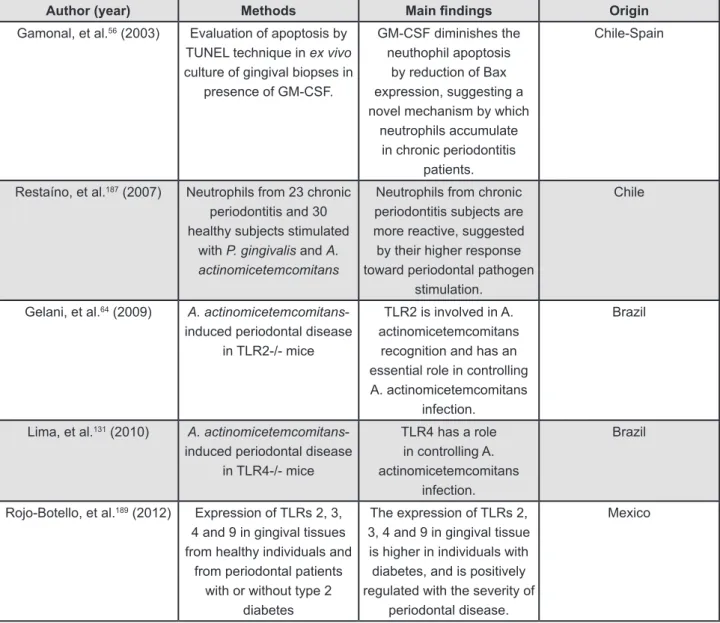
Latin-American research about innate immune response and periodontitis is mainly focused on the toll-like receptor and neutrophils. Such research is developed in Brazil, Chile and Mexico56,64,131,187,189 (Figure 2).
Toll-like receptors
Among the most important families of
pathogen-associated molecular patterns are the toll-like receptors (TLRs), which recognize, with selectivity, a large number of varied and complex pathogen-associated molecular patterns. Toll-like receptors are evolutionarily conserved proteins that also have a highly conserved intracellular TIR domain involved in protein-protein interaction and signaling activation. The extracellular domain, with Leucine-rich repeats (LRRs), is related to ligand recognition. Leucine-rich repeats motifs vary among toll-like receptors, although how they can discriminate among different pathogen-associated molecular patterns still remains unclear3. Among the 10 human toll-like receptors identiied so far, toll-like receptor-2 and toll-like receptor-4 are the most deined members. Toll-like receptor-2 is mostly involved in the recognition of a variety of different bacterial cell components, such as peptidoglycan and lipoproteins. Toll-like receptor-4 has been shown to speciically recognize lipopolysaccharide of Gram-negative bacteria and acts in cooperation with several protein components, such as lipopolysaccharide-binding protein and CD143,9,112,152. Research has established that toll-like receptors are expressed in periodontal tissues152,222. Since the gingiva is constantly exposed to microbes present in plaque bioilm, toll-like receptors signaling plays an important role in the innate immune response and maintenance of periodontal health. However, over-production of pro-inflammatory cytokines due to chronic stimulation of toll-like receptors, they may lead to tissue destruction13,254.
with type 2 diabetes, with regard to tissues from subjects without diabetes and with or without periodontitis189. These indings are consistent with the notion sustaining a correlation between toll-like receptors expression with disease severity, or with the degree of inlammation.
Using a model of experimental periodontitis, a Brazilian group has studied the mechanisms that modulate the outcome ofA. actinomycetemcomitans -induced periodontal disease in toll-like receptors 2-/-64 and toll-like receptors 4-/-131 mice. Toll-like receptors 2-/- mice developed more severe periodontitis
after infection, characterized by significantly higher bone loss. Also an intraperitoneal A. actinomycetemcomitans infection model was used to characterize the role of toll-like receptors 2 in the recruitment of macrophages and neutrophils. The
initiation of the inlammatory response in toll-like receptors 2-/- mice was reduced compared to wild type (WT) controls. Speciically, they observed that the inlux of neutrophils and macrophages into the peritoneal cavities was signiicantly decreased, and the levels of interleukin-1β, tumor necrosis factor-α, CXCL2, and CCL5 were signiicantly lower in toll-like receptors 2-/- mice64. These data highlight the
involvement of toll-like receptors 2 in recognizing
A. actinomycetemcomitans and its essential role in controlling A. actinomycetemcomitans infection. On the other hand toll-like receptors 4-/- mice
orally infected with A. actinomycetemcomitans
developed less severe periodontitis, when compared with wild type (WT) control mice, characterized by a signiicantly lower bone loss accompanied by a reduced inflammatory cell migration and
Author (year) Methods Main indings Origin
Gamonal, et al.56 (2003) Evaluation of apoptosis by TUNEL technique in ex vivo
culture of gingival biopses in presence of GM-CSF.
GM-CSF diminishes the neuthophil apoptosis
by reduction of Bax expression, suggesting a novel mechanism by which
neutrophils accumulate in chronic periodontitis
patients.
Chile-Spain
Restaíno, et al.187 (2007) Neutrophils from 23 chronic periodontitis and 30 healthy subjects stimulated
with P. gingivalis and A. actinomicetemcomitans
Neutrophils from chronic periodontitis subjects are more reactive, suggested by their higher response toward periodontal pathogen
stimulation.
Chile
Gelani, et al.64 (2009) A. actinomicetemcomitans -induced periodontal disease
in TLR2-/- mice
TLR2 is involved in A. actinomicetemcomitans
recognition and has an essential role in controlling A. actinomicetemcomitans
infection.
Brazil
Lima, et al.131 (2010) A. actinomicetemcomitans -induced periodontal disease
in TLR4-/- mice
TLR4 has a role in controlling A. actinomicetemcomitans
infection.
Brazil
Rojo-Botello, et al.189 (2012) Expression of TLRs 2, 3, 4 and 9 in gingival tissues from healthy individuals and
from periodontal patients with or without type 2
diabetes
The expression of TLRs 2, 3, 4 and 9 in gingival tissue is higher in individuals with diabetes, and is positively regulated with the severity of
periodontal disease.
Mexico
Figure 2- Evidence regarding innate immune response in periodontitis conducted by Latin-American groups GM-CF: Granulocyte monocyte-colony stimulating factor
TRL:Toll-like receptor
inlammatory cytokine production (
interleukin-1β and tumor necrosis factor-α) in periodontal tissues. Moreover, this absence of toll-like receptors 4 activation leads to impaired myeloperoxidese activity in the periodontum and possibly facilitates systemic bacteria dissemination and proliferation135. Regarding P. gingivalis, an interaction of imbriae with TLR2 has also been shown to induce cross talk of TLR2 with the coreceptors CR5, CXCL4, and CR3, resulting in distinct responses82. The plasticity and diversity in downstream signaling when TLR2 is activated is further underscored by the observation that P. gingivalis live cells induce a different pattern of TLR2-dependent cytokines than that induced by FimA or LPS preparations alone in mouse peritoneal macrophages265. It is likely that different TLR2-activating ligands vary in their abilities to engage TLR2 and speciic coreceptors. In terms of responses, TLR2 engagement has been shown to trigger production of both proinlammatory and anti-inlammatory cytokines.
The bacterial composition of plaque remains relatively stable despite regular exposure to minor environmental perturbations. The changes that affect the homeostasis leading to shifts for example in the frequent exposure of plaque to low pH; an increase low of gingival crevicular luid; and as microlora shifts from being mainly Gram-positive to being comprised of increased levels of obligately anaerobic, asaccharolytic Gram-negative organisms and the rate of acid production following sugar intake could be reduced by luoride, have been incorporated into a new hypothesis: the ecological plaque hypothesis, to explain the relationship between the plaque microlora and the host in health and disease, and to identify new strategies for disease prevention145.
The recent advancements in the periodontal research ield are consistent with a new model of pathogenesis, according to which periodontitis is initiated by a synergistic and dysbiotic microbial community rather than by a select bacterial complex, that is, polymicrobial synergy and dysbiosis model (PSD model)79,80.
Polymorphonuclear neutrophils
Polymorphonuclear neutrophils are one of the irst-responders of inlammatory cells to migrate toward the site of periodontal inflammation, following chemoattractants such as interleukin-8 secreted by oral epithelial cells, connective ibroblast and immune cells. In addition, polymorphonuclear neutrophils are recruited to the site of injury within minutes following injury, and are the hallmark of acute inlammation39.
Though the polymorphonuclear neutrophils are essentially protective cells, alterations in polymorphonuclear neutrophil function result
from periodontitis subjects. When the sample was embedded in complete medium containing granulocyte monocyte-colony stimulating factor, the total number of TUNEL-positive cells was reduced, therefore showing its delaying effect on the apoptotic process. The localized accumulation of functional polymorphonuclear neutrophils at sites of inlammation is pivotal in the host defense against infection, and the orderly elimination of these cells is equally important in the resolution of the inlammatory response56.
III- I N V O L V E M E N T O F T - C E L L
SUBSETS, CYTOKINES AND ORAL
VIRUSES IN PERIODONTAL DISEASES
PATHOGENESIS
In addition to the innate immunity, adaptive immunity cells and characteristic cytokines have been described as important players in the periodontal disease pathogenesis scenario, with a special attention to CD4+ T-cells (T-helper cells). Interestingly, T cell-mediated adaptive immunity development is highly dependent on innate immunity-associated antigen presenting cells, which after antigen capture undergo into a maturation process and migrate towards the lymph nodes, where they produce distinct patterns of cytokines that will contribute to the subsequent polarization and activation of speciic T CD4+ lymphocyte38.
CD4+ T-cells were subdivided initially into two subsets, designated T-helper 1 and T-helper 2, on the basis of their pattern of cytokine production155. As a general rule, T-helper 1 cytokines have been associated with infectious inflammatory bone destruction, while its classic antagonists T-helper 2 cytokines are described to minimize bone loss30,73,180,184. Nevertheless, the discovery of new T-helper subsets with prominent roles in the modulation of host responses determined the re-examination of T-helper 1/T-helper 2 dichotomy paradigm in chronic inflammatory diseases, including periodontal diseases21,73,103,205,252. Th17 cells emerged as a remarkable T subset, with inflammatory properties involved in a series of infectious, autoimmune and osteolytic processes44,106,224,226.
On the other hand, regulatory T cells (Tregs) comprise a FOXp3-dependent CD4+CD25+ T-helper subpopulation with suppressive effects on inlammatory osteolysis, thought to be mediated by cytokines such transforming growth factor-b, interleukin-10 and the CLTA418,106. Other regulatory T cell subsets, namely T-helper 3 and Tr1, are characterized by the specific production of transforming growth factor-b or interleukin-10, respectively16,17,103.
Also, additional T-helper subsets, such as
T-helper 9 and T-helper 22 cells, are described to play important roles in the modulation of inlammatory immune responses, interacting with the previously mentioned T-helper subpopulations103,154,252.
Therefore, we must consider that there are at least four types of T-helper cells, T-helper 1, T-helper 2, T-helper 17, and T-regulatory cells, which collectively play an important role in orchestrating adaptive immune responses to various infectious agents, and also a great inluence on the inlammatory and autoimmune diseases outcome. Upon TCR-mediated cell activation, and under a critical influence of cytokines that generate specific patterns of signaling and activate definite transcription factors, naïve CD4+ T cells can differentiate into characteristic polarized subsets. Speciic subsets of cytokines are involved in differentiation of each lineage: interleukin-12/interferon-γ for T-helper 1, interleukin-4/interleukin-2/interleukin-7 for T-helper 2, transforming growth factor-β/ interleukin-6/interleukin-21/interleukin-23 for T-helper 17, and transforming growth factor-β/ interleukin-2 for T-regulatory cells. The major effect of such polarizing cytokines is the generation of speciic transcription factors involved in T-helper cell differentiation: T-bet controls T-helper 1 fate, GATA3 is involved in T-helper 2 generation, RORγt for T-helper 17, and Foxp3 for T-regulatory cells.
Once differentiated, the polarized T-helper subsets express a signature pattern of chemokine receptors expression that determines the selective migration of CD4+ T helper subsets to the response foci, in which these cells will produce distinct and characteristic cytokines20,108,155. Indeed, the transcription factors involved in T-helper cell differentiation directly bind to the effector cytokine genes (Ifng, Il4/Il13, Il17a/Il17f; for T-helper 1, T-helper 2 and T-helper 17 cells respectively) at promoters and regulatory elements, resulting the prototypical production of cytokines that deines the T-helper subsets266,267. In this context, we describe in the sequence the major T-helper subsets and their potential role in the pathogenesis of periodontal diseases.
T-helper 1
degradation of the RANK adapter protein TRAF6 by the ubiquitin-proteasome system, resulting in the inhibition of the RANKL-signaling and its subsequent osteoclastogenic events227. These results can be interpreted in support of the hypothesis that T-helper 1 cells are associated with the stable lesions while T-helper 2 cells are associated with disease progression66. However, Garlet lab group57,58 demonstrated in vivo that the pro-inlammatory effect of interferon-γ mediates the upregulation of the levels of tumor necrosis factor-a and interleukin-1b, which in turn can upregulate RANKL, being the in vivo outcome opposite to the in vitro scenario.
Accordingly, as previously demonstrated by Gamonal and Garlet groups, interferon-γ is present at high levels in human and experimental periodontal lesions, being associated with progressive lesions or higher severity periodontal diseases forms46,61,94. Human and experimental data suggests that interferon-γ can contribute to periodontitis onset and progression by distinct mechanisms. With special interest in T-helper 1 cells, our group demonstrates in a series of studies that interferon-γ stimulates osteoclast formation and bone loss in vivo via antigen-driven T cell activation or through the chemoattraction of RANKL+ cells58,63,183. In fact, T-helper 1 cells (characterized as CD3+CCR5+CXCR3+ cells) are an important source of RANKL throughout experimental periodontitis183. Complementarily, interferon-γ can contribute to the migration of CD4/80+ cells, whose phenotype demonstrate monocyte/macrophage-like phenotype that can be interpreted as a potential osteoclast precursor’s subpopulation. Finally, interferon-γ seems to play an active role in the control of periodontal infection, possibly mediated by macrophages and neutrophils activation146,199 clearly evidenced by the severe impairment of protective immunity to A. actinomycetemcomitans infection, as demonstrated by the higher bacterial load in periodontium, increased acute phase response, and bacterial dissemination followed by death of interferon-γ KO mice56. The immune protection mediated by interferon-γ characteristically involves leukocyte recruitment and its subsequent activation at inlammatory foci199.
T-helper 2
T-helper 2 cells commitment and action is primarily dependent of interleukin-4, the prototypical T-helper 2 cytokine, and of the transcription factor GATA37,152,191. Interleukin-4 presents marked suppressive and anti-inlammatory properties mediated by its capacity to inhibit the transcription of pro-inlammatory cytokines and interferon-γ, then suppressing the polarization of T-helper 1 cells2,7,20,102. Also, in spite of some controversies, interleukin-4 can present anabolic
properties in distinct tissues, characterized by the ability to inhibit the production of tissue degrading factors such as matrix metalloproteinases (and other proteases) and the major osteoclastogenic factor RANKL, and concomitantly induce the upregulation of its respective inhibitors TIMPs and osteoprotegerin98,192,194. Therefore, interleukin-4 could contribute to the attenuation of both soft and mineralized tissue destruction, which could reinforce its potential protective nature in periodontitis pathogenesis67.
Indeed, the concentration of interleukin-4 in gingival crevicular luid was demonstrated to decrease from periodontal health to disease, suggesting that this cytokine could mediate the remission or improvement of periodontal lesions179. Experimental data support the protective role of T-helper 2 cells, since T-helper 2-biased immune response in mice resulted in minimal lesions after P. gingivalis challenge220, and the adoptive transfer T-helper 2 cells attenuates the severity of periodontal diseases47. In this context, it is important to remember that interleukin-4 cab induces the production of cytokines with similar or complementary suppressive properties, such as interleukin-10105,172. Interestingly, interleukin-10 was considered initially one of the T-helper 2-signature cytokines along interleukin-4, but more recent studies have been associating this cytokine more closely to T-regulatory cells. It is also important to mention that recent studies have been identiied an interesting plasticity among T-helper subsets, which may also account for some controversies in previous studies.
However, early studies demonstrated that B cells outnumber T cells in periodontal lesions, which in the view of the association of T-helper 2 responses with B cell function and humoral immunity development, results in the hypothesis that a T-helper 2 lesion was involved in the progression of chronic periodontitis65. Recent evidences, demonstrating that B cells can be a RANKL source, and that the majority of B cells in periodontal lesions are RANKL+110 are interpreted along with the T-helper 2 lesion hypothesis. While in this context the T-helper 2-B cell axis is considered detrimental, it is important to consider that the humoral immunity axis involving T-helper 2 and B cells is also thought to contribute to the host protection against periodontal pathogens75,168,169,200.
T-helper 17
factor-β and interleukin-6 signaling together cooperate to drive T-helper 17 commitment, while the cytokine interleukin-23 ampliies and stabilizes the T-helper 17 phenotype in chronic inlammatory reactions. Interestingly, T-helper 17 cells develop through cytokine signals distinct from, and antagonized by, products of the T-helper 1 and T-helper 2 lineages7,44,194. Once differentiated, RORγ can bind to multiple sites in the Il17a/Il17f locus, including the promoters of these cytokines, leading to the characteristic interleukin-17 production, which in turn leads to the induction of many proinlammatory factors such as tumor necrosis factor, interleukin-6, and interleukin-1β. In fact, these innate immunity cytokines have been associated with periodontitis, since the upregulation of such mediators expression/production by interleukin-17 clearly points to a destruction of such cytokines in periodontitis context. Accordingly, Gamonal groups presented the initial evidences that T-helper 17 cytokines are produced in periodontal lesions and are supposed to contribute to disease progression, which was conirmed latter by Garlet group and others12,24,162,163,223,247. Besides the classical phagocyte activating cytokines, recent evidences demonstrate that the T-helper 17/interleukin-17 axis, by itself or along with pro-inlammatory and T-helper 1 cytokines, mobilizes macrophages and neutrophils204,262, which could also contribute to exacerbate host responses in periodontal tissues. In accordance with this hypothesis, interleukin-17 was also recently described to increase toll-like receptors responsiveness in human gingival epithelial cells13, suggesting that this cytokine can play an active role in potentiating innate immunity mechanisms in periodontal environment. In addition to the upregulation of pro-inlammatory cytokines production, interleukin-17 was described as an inducer of RANKL production120,195,197. However, experimental studies in rodents demonstrate that the interleukin-17 deficient mice may present increased or decreased bone lesions in response to periodontal pathogens challenge, being the exact role of interleukin-17 in periodontitis still unclear166,262.
T-regulatory cells
Regulatory T cells were initially described as CD4+CD25+ T cells that speciically regulate the activation, proliferation, and effector function of activated conventional T cells, determining the outcome of several immunological settings ranging from infectious diseases to immunopathology and autoimmunity7,15,194,203. T-regulatory cells characteristically express as phenotypic markers the transcription factor forkhead box P3 (FOXp3), CD103, the glucocorticoid-inducible tumor necrosis factor receptor (GITR), the inhibitory molecule
cytotoxic T-lymphocyte-nassociated molecule 4 (CTLA-4) and cell surface transforming growth factor-β1, among other surface molecules203. However, recent studies demonstrate that FOXP3+ T cells are not a uniform population, but phenotypically and functionally diverse populations. Currently, there are two accepted major types of FOXP3+ T-regulatory cells: endogenous or natural Treg (nTreg), a stable subset derived from the thymus and thought to control autoreactivity, and adaptive or induced T-regulatory cells (aTreg or iTreg), a less stable subpopulation supposed to regulate responses upon antigenic exposure in the periphery15,150,191,193. Interestingly, T-regulatory cells from noninflamed peripheral tissues are considered to be in a resting state with little suppressor activity, but under certain inlammatory conditions, these cells are stimulated and undergo rapid reprogramming to acquire helper/effector functions. Foxp3 is the master regulator for both iT-regulatory cells and nT-regulatory cells, and its stimulation through TCR and transforming growth factor-βR signaling is essential to drive iT-regulatory cells development. Along with other transcription factors, such as Runx, Foxp3 positively regulates T-regulatory cells-speciic gene expression and negatively regulates genes not expressed in Tregs15,150,191.
with a decline in the disease progression rate, and its inhibition resulted in increased alveolar bone loss and inlammatory cell migration59. In addition to the attenuation of tissue destruction, T-regulatory cells-associated cytokines such as interleukin-10 and transforming growth factor-β are associated with tissue repair in different models29.
Additional T-helper subsets
While T-helper 1, T-helper 2, T-helper 17 and T-regulatory cells are the most recognized and studied T-helper subsets, recent studies suggest the existence of other CD4 lymphocytes subtypes with distinguished immunoregulatory properties. T-helper 9 cells characteristically produce interleukin-9, initially designated as a T-helper 2 cytokine that exerts pro- or anti-inflammatory activities by modulating T-regulatory cells and/or T-helper 17 cells development and function49,128,129,161. Additionally, the recently identiied T-helper 22 cells produces interleukin-22, which can exerts pro-inlammatory effects by a synergistic action with classic pro-inlammatory mediators such as tumor necrosis factor-α of interleukin-1749,252. Preliminary data from Garlet group demonstrate that both interleukin-9 and interleukin-22 are overexpressed in diseased periodontal tissues, reinforcing the complexity of cytokine networks in the inlamed periodontal environment. Here, we simultaneously investigated the expression of pro- and anti-inlammatory, T-helper 1, T-helper 2, T-helper 9, T-helper 17, T-helper 22 and T-regulatory cells cytokines/markers, and the major osteoclastogenesis regulators RANKL and osteoprotegerin, in human chronic periapical granulomas and their possible correlations with lesions activity pattern147 in order to obtain a more complete picture of the immunoregulatory scenario
in periapical lesions, which ultimately can contribute to the development and to the improvement of the diagnosis and treatment of these pathologies.
The T helper immunoregulatory network
Despite the reports regarding the expression of prototypical T-helper markers in diseased periodontal tissues, the related hypothesis regarding their role in the pathogenesis of periodontal diseases are often conlicting. In fact, since the production/expression of such factors is usually investigated individually or in small clusters, it does not allow the complete immunoregulatory scenario determination, where the potential synergic or antagonist action of cytokines should be considered. When interpreting in vivo data, the putative function of cytokines must be estimated in the view of a complex milieu, with presence of several other cytokines, which can modulate or be modulated by them in multiple ways until the establishment of an overall outcome. In addition, since the presence of speciic periodontopathogens is able to interfere with cytokine milieu, the in vivo scenario with multiple bacterial species turns this network even more complex (Figure 3). Interestingly, the simultaneous presence of T-helper 1 and T-helper 2 was previously reported in periodontal lesions, but the interferon-g and interleukin-4 levels of these cytokines were described to be inversely correlated in accordance with the mutual inhibitory activity of these T cell subsets62. More recently, studies demonstrate that periodontal lesions simultaneously express high levels of both interleukin-17 and interferon-g, suggesting a possible cooperative detrimental role for these cytokines46,223. Conversely, in other models, T-helper 1 and T-helper 17 mediators seem to be independently associated to the progression
of inlammatory bone lesions, suggesting that distinct pathways could result in a similar bone loss outcome26,236,244. The possible interactions among T-helper subsets would also involve potentially protective mechanisms, since a T-helper 2/T-regulatory cells crosstalk mediated by transforming growth factor-β and interleukin-4 may drive the polarization of T-helper 9 cells103, described to mediate immunosuppressive actions along interleukin-4 and interleukin-10 producing cells229,257.
Oral viruses in pathogenesis of periodontal disease
Periodontitis is considered to be a process that involves a multifactorial interaction among microbial, host and environmental modulating factors84. It is accepted that periodontitis is associated with speciic bacterial species and polymicrobial colonization of the teeth surfaces6, however, the amount of bacterial plaque per se does not completely explain the clinical and pathological features of periodontitis32. Periodontal diseases are inlammatory processes characterized by dense accumulation of immune cells, including polymorphonuclear neutrophils, T and B lymphocytes, plasma cells, mast cells, monocytes and macrophages25. The T cells play an important role in the immune response, regulating the polyclonal activation of the B cells and plasma cells in the periodontitis-affected sites. The interaction between host immune system and periodontopathogens is relevant to the pathogenesis of periodontitis53.
Our knowledge of viral infections has increased significantly in the past couple of decades. Herpesviruses may cause illness by mechanisms that are direct, indirect or immune-response linked, and illnesses range from subclinical or mild disease to encephalitis, pneumonia and other potentially lethal infections, and even to lymphoma, sarcoma and carcinoma. In the oral cavity, herpesviruses are involved in acute gingival infections, destructive periodontal disease, apical periodontitis, ulcerations of mucosa, odontogenic cyst, giant cell granuloma, autoimmune disease
and various types of neoplasm33. Contreras,
et al.33 (1999) suggested that the presence of herpesviruses in the periodontal sites could play a role on the pathogenesis of human periodontitis. Herpesviruses are ubiquitous and after primary infection can persist latently in the host in several types of cells, including cells of the immune system. The cytomegalovirus (CMV) is the most-studied member of the betaherpesviruses in periodontal sites33. Recently, the other two betaherpesviruses, known as human herpesvirus 6 (HHV-6) and human herpesvirus 7 (HHV-7), have been investigated because these viruses are frequently detected in
saliva208,209.
Inflammatory cells harboring herpesviruses present at sites of periodontal inlammation could contribute to the development and course of periodontitis32. Cytomegalovirus can induce direct cytopathic effects on ibroblasts, keratinocytes, endothelial cells and inlammatory cells, including polymorphonuclear leukocytes, T lymphocytes and macrophages, and also possibly affect bone cells210.
In periodontitis patients, T cells are activated, and speciic lymphocyte responses are driven by the nature of the initial antigenic stimuli. This process is supported by a complex cascade of events involving cytokines, chemokines and other inlammatory mediators, which could be altered due to cytomegalovirus infection209. Proinlammatory and anti-inflammatory balances controlled by different subsets of lymphocytes are thought to be crucial in the pathogenesis of periodontitis53.
The expression of different cellular antigens can be dramatically altered in Beta herpesvirus-infected tissues, in which the viral infection can induce CD4 up regulation and CD3 down modulation in the T cells. Human herpesvirus 6 can severely affect the physiology of secondary lymphoid organs through direct infection of T lymphocytes and modulation of key membrane receptors and chemokines74.
Local immunomodulatory effects caused by herpesviral infection could facilitate bacterial proliferation and virulence, or induce the release of cytokines and chemokines from inlammatory and connective tissue cells214. In addition, viruses and bacteria could act in synergy to produce pathology210.
Herpesviral infections in the periodontium can act as cofactors of periodontal pathogenesis, modulating the activity of inflammatory cells. The release of different types of cytokines and B lymphocyte activation orchestrated by T lymphocytes has been shown to be an important process in chronic periodontitis. In addition, T-regs cells may be involved in local immune response in periodontitis25.
In conclusion, a herpesvirus-bacterial pathogen model has been proposed, in which viral infections facilitate bacterial overgrowth and virulence. Moreover, the immunosuppressive effects of certain herpesviruses can also perpetuate the inlammatory reaction in the connective tissue facilitating periodontal destruction. Human viruses could play a role in human periodontitis by modulation of immune response.
IV- O S T E O - I M M U N O L O G Y I N
PERIODONTAL DISEASES
in periodontal tissues stimulate the differentiation of monocyte-macrophage precursor cells into osteoclasts, and the maturation and survival of the osteoclast, leading to alveolar bone loss37,54,91, 108,132,156,244,248. In this context, during inlammatory r e s p o n s e c h a ra c t e r i s t i c o f p e r i o d o n t i t i s , proinflammatory cytokines associated with T-helper 1 and T-helper 17 cell phenotypes, such as interleukin-1β, interleukin-6, interleukin-17, interferon-γ, and tumor necrosis factor-α, can stimulate periodontal osteoblasts to express membrane-bound RANKL1,54,72,140,153. In addition to osteoblasts, RANKL is expressed by a number of other cell types, mainly T-helper 17 lymphocytes108 (Figure 4).
Skeletal homeostasis depends on a dynamic balance between the activities of the bone-forming osteoblasts (OBLs) and bone-resorbing osteoclasts (OCLs)247. This balance is tightly controlled by various regulatory systems, such as the endocrine system, and is inluenced by the immune system, an osteoimmunological regulation depending on lymphocyte- and macrophage-derived cytokines188,197,225,251. An unbalance in favor of bone-resorbing osteoclasts leads to pathological bone resorption, as it has been observed in rheumatoid arthritis, osteoporosis, Paget’s disease, bone tumors, and periodontitis188,251.
During the 1970’s, the irst observation pointing towards immune cells influencing the bone-resorbing osteoclasts activity was made. Indeed, a factor (OCL-activating factor or OAF) that stimulated bone resorption was detected in the supernatant from cultured human peripheral monocytes stimulated with phytohemagglutinin96. Puriication of this activity led to the identiication of interleukin-1β41. Nowadays, numerous cytokines have been demonstrated to stimulate bone resorption,
including tumor necrosis factor-α, interleukin-1α, interleukin-1β, interleukin-6, interleukin-11, interleukin-15, and interleukin-17, whereas others such as interleukin-4, interleukin-5, interleukin-10, interleukin-13, interleukin-18, and transforming growth factor-β1 inhibited bone resorption225,251. In this context, functional characterization of three novel members of the tumor necrosis factor-ligand and receptor superfamily, the receptor activator of nuclear factor-κB (RANK), its ligand (RANK-ligand or RANKL) and the soluble decoy receptor of RANKL named osteoprotegerin, have contributed signiicantly to the establishment of osteoimmunology, where these molecular mediators participate as key modulators of physiological and pathological bone resorption224,234,250. RANKL exerts its biological effects directly through binding to RANK, inducing OCL differentiation, maturation and activation124. Osteoprotegerin inhibits the osteoclastogenesis and induces osteopetrosis when over-expressed in transgenic mice205. RANKL has been associated with diverse osteodestructive pathologies, including rheumatoid arthritis, bone tumors, osteoporosis, Paget’s bone disease, osteolytic lesions of the facial skeleton, odontogenic lesions and periodontitis28,37,90,96,97,117,132,231,245,246.
The identification of RANKL as the T cell cytokine TRANCE (tumor necrosis factor-related activation-induced cytokine) allowed envisaging the possibility that CD4+ T cells may have the capacity to induce OCL differentiation and activation by directly acting on OCL precursors and on mature OCLs through synthesis of RANKL during osteo-destructive diseases117,230,260. Furthermore, many well-known osteotropic factors, including tumor necrosis factor-α, interleukin-1β and interleukin-6, exert their osteoclastogenic activity by inducing RANL expression on OBLs and CD4+ T cells22. Th2
cells inhibit osteoclastogenesis by acting on the precursor cells, mainly through interleukin-4 and interleukin-10 secretion95,239. In contrast, T-helper 17 cells stimulated by interleukin-23 promote osteoclastogenesis mostly through production of interleukin-17 and RANKL196. Furthermore, interleukin-17 facilitates local inlammation by recruiting and activating immune cells, which leads to an abundance of inlammatory cytokines such as interleukin-1β and tumor necrosis factor-α that enhance the RANKL expression on OBLs and Th17 cells27,43.
T-helper 17 cells represent a large proportion of the inlammatory cells invading the synovial tissues during rheumatoid arthritis27. High levels of interleukin-17A have been detected in the synovial luid, and interleukin-17-producing cells have been detected within the T cell-rich areas in patients with rheumatoid arthritis138,264. Furthermore, interleukin-17A is able to promote cartilage destruction and bone erosion in experimental rheumatoid arthritis138. Increased levels of interleukin-17 were detected in gingival crevicular luid and in biopsy samples from periodontal lesions, both at the mRNA and protein levels, in patients with chronic periodontitis, and these increased levels have been associated to CD4+ T cells223,247. Furthermore, RANKL and RANK were synthesized within periodontal lesions in which interleukin-17 was produced by activated gingival T cells247. Taken together, these data establish that T-helper 17 cells represent the osteoclastogenic T-helper subset on CD4+ T lymphocytes, inducing osteoclastogenesis and bone resorption through synthesizing interleukin-17 and RANKL (Figure 5).
Our findings have demonstrated that total amount of RANKL detected in gingival crevicular luid of patients undergoing periodontitis progression was higher in active periodontal lesions than in inactive lesions, proposing this pro-resorptive factor as a marker of active alveolar bone resorption associated with T-helper cell activity245,246. This inding was corroborated by Silva, et al.204 (2008), whom performed a longitudinal following of 56 patients affected by moderate to severe chronic
periodontitis until determination of progression, detecting higher RANKL and interleukin-1β levels, and matrix metalloproteinase-13 activity, in active sites compared with inactive sites.
When the role of T-helper 17 and T regulatory cells phenotypes was analyzed during progressive periodontitis, it was established that interleukin-17 and RANKL were over-regulated, and interleukin-10 and transforming growth factor-β1 were down-regulated in active periodontal lesions compared with inactive lesions activity45. In fact, the over-expression of transcription factor orphan nuclear receptor C2 (RORC2), the master-switch gene controlling the T-helper 17 differentiation, was associated with active periodontal lesions during progressive periodontitis45.
In the same study, analysis of the associations between different genes yielded signiicant positive correlations between RORC2 and RANKL, and between RORC2 and interleukin-17. However, Foxp3, interleukin-10, transforming growth factor-β1, and CTLA-4 did not show a positive correlation, speculating that Foxp3+ T-cells that do not bear regulatory functions may have a role in periodontal progressive destruction, in view of the down-regulation of interleukin-10 and transforming growth factor-β145.
In response to periodontopathogens that have been strongly associated with periodontitis progression, for instance P. gingivalis and A. actinomycetemcomitans, RANKL expression has been reported to increase in CD4+ T lymphocytes infiltrating periodontally affected tissues258. In this context, on T lymphocytes activated with autologous dendritic cells primed with different P. gingivalis capsular (K) serotypes and A. actinomycetemcomitans O-polysaccharide serotypes, at different multiplicity of infections, the expression and secretion levels for RANKL were determined. The obtained data showed an increase in RANKL mRNA expression on T cells activated with P. gingivalis K1 or K2 serotypes and A. actinomycetemcomitans b serotype, compared with the other serotypes, and these levels correlate
with the levels of secreted RANKL present in the supernatant of the cell cultures (unpublished data). These data suggested that activation of T cells with dendritic cells stimulated with either K1 or K2 serotypes (P. gingivalis) or b serotypes (A. actinomycetemcomitans) induced a T-helper 17 phenotype in CD4+ T cells, and allowed to speculate a link between these serotypes with alveolar bone destruction and teeth loosening, one of the hallmarks of periodontitis.
Diverse studies have analyzed the concentrations of RANKL and osteoprotegerin in gingival crevicular fluid of periodontitis patients and healthy subjects45,204,245-247. In general, they show great variation from study to study, but the ratio of RANKL/osteoprotegerin has a consistent tendency to increase from periodontal health to periodontitis, and to decrease following periodontal treatment. The increased RANKL/OPG ratio may serve as a biomarker that denotes the occurrence of periodontitis, but may not necessarily predict on-going disease activity14.
V - T H E R O L E S O F M A T R I X
M E T A L L O P R O T E I N A S E S I N
PERIODONTAL TISSUE BREAKDOWN
The interaction between periodontal inlammation and persistent bacterial infection up-regulates the expression and activity of neutral proteinases, particularly of the matrix metalloproteinase family, which contributes to the progressive breakdown of periodontal supporting tissue216. Matrix metalloproteinases represent a family of human zinc-dependent endopeptidases that are involved in a wide variety of physiological and pathological processes, such as skeletal growth and remodeling, wound healing, cancer and inlammatory diseases. Matrix metalloproteinases are able to degrade basement membrane and extracellular matrix components, and therefore they have been classically viewed as effectors of extracellular matrix hydrolysis. However, matrix metalloproteinases have also regulatory properties, modulating enzyme, chemokine and cytokine activities (referred as bioactive substrates) among others, as well as releasing bioactive molecules from extracellular matrix store through limited proteolysis167,197.
Up to now, 24 matrix metalloproteinases have been identified in humans that closely share protein sequence and domain structure, and they are classically grouped according to their structural properties and substrate speciicity in collagenases, gelatinases, stromelysins, matrilysins and membrane type matrix metalloproteinases52. The basic structure of matrix metalloproteinases is composed of an auto-inhibitory prodomain that
maintains enzymatic latency, the catalytic domain and the C-terminal haemopexin-like domain, often involved in the recognition and positioning of matrix metalloproteinase substrates. Matrix metalloproteinase activity is subjected to a complex regulation where the main steps involve gene expression, proenzyme activation and inhibition by endogenous inhibitors, such as tissue inhibitors of matrix metalloproteinases (TIMPs)52,197. As a result from matrix metalloproteinase regulation, all human matrix metalloproteinases are known to exist in multiple forms in periodontal diseases, such as latent proforms, active forms, fragmented species, complexed species and cell-bound forms. Overall, the pathophysiological signiicance of increased matrix metalloproteinase expression and/or levels in periodontitis will rely ultimately on endogenous matrix metalloproteinase activity, resulting from the interactions between their inhibitors and activating factors. Consequently, matrix metalloproteinase activity accounts for the rate of matrix turnover or destruction, and the modulation of the immune response in a more direct fashion23,214.
Because collagen I is the main component of the extracellular matrix of soft and hard periodontal tissues, matrix metalloproteinases with collagen-degrading properties such as collagenases (matrix metalloproteinase-1, -8, -13 and -14) and gelatinases (matrix metalloproteinases -2 and -9) play a pivotal role in the loss of periodontal support213. Collagenases are capable to process native collagen without unwinding the triple helical assembly of the molecule, generating ¾ ¼ fragments, whereas gelatinases have been deined according to their afinity for denatured collagen197. Accordingly, collagenolytic matrix metalloproteinases have widely been demonstrated in inlamed periodontal tissues and in oral luids by different analytic methods216-218.
Matrix metalloproteinase-13 upregulation has been involved in periodontitis progression, particularly in bone loss, and its regulatory role is currently under research87,89,99. On the other hand, matrix metalloproteinase-1, also known as collagenase-1, is produced by several periodontal resident cell-types and is considered as central in the physiological remodeling of extracellular matrix and wound healing197,216. Among gelatinases, matrix metalloproteinase-9 is the main gelatinase in oral fluids139. Matrix metalloproteinase-9 is physiologically expressed by a limited variety of cell types, mainly immune cells, and is highly inducible under periodontal inlammation, whereas matrix metalloproteinase-2 is less inducible and has primarily a pro-homeostatic role177,201.
It has been postulated that heritable differences in the production of cytokines can inluence the resistance or susceptibility to periodontitis, as well as disease outcome62. Matrix metalloproteinases can be released and/or activated during periodontal inflammation by proinflammatory cytokines, like tumor necrosis factor-α, interleukin-1β, reactive oxygen species, and proteases derived from the subgingival biofilm and the host121. Accordingly, higher mRNA expression levels of matrix metalloproteinase/TIMP ratios for matrix metalloproteinase-1, -2 and -9, as well as RANKL/ osteoprotegerin ratio, have been reported in gingival tissue from chronic and aggressive periodontitis patients compared with healthy gingival62. Furthermore, comparison between gingival tissue from aggressive periodontitis and chronic periodontitis showed higher expression of the regulatory cytokines interleukin-10 and interleukin-4, and reduced Th1- type cytokine interferon (IF)- γ expression in the later. The authors suggested that different cytokine patterns might modify the balance of matrix metalloproteinases/ TIMPs and RANKL/osteoprotegerin, resulting in more or less severe periodontal support loss.
Similarly, experimentally-induced periodontitis in mice models by A. actinomycetencomitans infection in strains selected for maximal and minimal inlammatory reactions has demonstrated a more severe disease phenotype in the former group. Inflammatory profile was composed of higher interleukin-1β, tumor necrosis factor-α, RANKL/ osteoprotegerin ratio and matrix metalloproteinase/ TIMP ratio, particularly of matrix metalloproteinase-13 and matrix metalloproteinase-2238. Similarly, matrix metalloproteinase-13 mRNA expression levels increase concomitantly with the severity of inflammation in two different models of experimentally-induced periodontitis, ligature placement and lipopolysaccharide injections, but the later showed more sustained inlammation. However, the authors found a total lack of
correspondence between mRNA transcription and matrix metalloproteinase protein levels8, supporting that pro-inflammatory mediators up-regulate matrix metalloproteinase expression in periodontal diseases, but matrix metalloproteinase protein levels and specially matrix metalloproteinase activity needs further assessment.
S e v e r a l c o m m o n s i n g l e n u c l e o t i d e polymorphisms (SNPs) have been identiied in matrix metalloproteinase genes; however, their association with disease susceptibility or phenotype is rather controversial. An example is the matrix metalloproteinase-1 -1607 1G/2G variant9,10,174,241. A more recent analysis in a Brazilian population concluded that the 2G allele associated with higher matrix metalloproteinase-1 levels only in healthy subjects, whereas in periodontitis patients, the antigenic challenge overcame the genetic predisposition185. Similar controversial indings have been reported for the matrix metalloproteinase-9 T variant at position -1562 in Czech, Turkish, Colombian and Brazilian populations76,92,101,138,219. In the case of matrix metalloproteinase-13 and matrix metalloproteinase-8, fewer studies are available; no differences in matrix metalloproteinase-13- 77 A/G and -11A/12A polymorphic sites have been found for periodontitis patients175. However, a recent study revealed that even if no single matrix metalloproteinase-8 SNP is associated with periodontitis, the haplotype T(-799)/C(+17) is associated with clinical manifestations of chronic periodontitis in a Czech population102.