ABSTRACT
Use of occlusal sealant in a community program
and caries incidence in high- and low-risk children
Vânia BALDINI1, Elaine Pereira da Silva TAGLIAFERRO2, Gláucia Maria Bovi AMBROSANO3, Marcelo de Castro
MENEGHIM4, Antonio Carlos PEREIRA5
1- DDS, MSc, Faculty of Dental Medicine, University of Lisboa, Lisboa, Portugal.
2- DDS, MSc, PhD, Post-doctorate, Department of Community Dentistry, Piracicaba Dental School, University of Campinas – UNICAMP, Piracicaba, SP, Brazil. 3- Agr. Eng., MSc, PhD, Professor, Department of Community Dentistry, Piracicaba Dental School, University of Campinas – UNICAMP, Piracicaba, SP, Brazil. 4- DDS, MSc, PhD, Professor, Department of Community Dentistry, Piracicaba Dental School, University of Campinas – UNICAMP, Piracicaba, SP, Brazil. 5- DDS, MPH, DrPH, Professor, Department of Community Dentistry, Piracicaba Dental School, University of Campinas – UNICAMP, Piracicaba, SP, Brazil.
Corresponding address: Antonio Carlos Pereira - Faculdade de Odontologia de Piracicaba - UNICAMP - Departamento de Odontologia Social Av. Limeira, 901 - Piracicaba, SP - 13414-903 - Brazil - Phone: +55 19 2106-5209 - Fax: +55 19 2106-5218 - e-mail: [email protected]
!"
O
bjective: The aims of this study were to investigate the effectiveness of sealant placement under the guidelines of the Oral Health Promotion Program for Children and on the DMFT increment in 277 children, born in 1997. Material and Methods: A dental hygienist performed the initial examinations and sealant placement (Helioseal, Vivadent) on !"# # $!% # & ' (HR: DMFT+dmft>0); low (LR: DMFT+dmft=0) risk] and sealant placement as follows: HR-S and LR-S Groups (with sealant placement); HR-NS and LR-NS Groups (without sealant ! * $ on the World Health Organization recommendations. The variables collected were: dental caries, visible dental plaque, malocclusions, and socioeconomic level (questionnaire sent to children’s parents). For univariate (Chi-square or Fisher tests) and multivariate (Multiple logistic regression) analyses the DMFT increment >0 was selected as dependent variable. Results: Approximately 17.0% of the children showed DMFT increment>0 (mean=0.25). 34& 6 8 teeth. These children had 7.94 more chance of developing caries. Children who did not receive sealant were 1.8 more prone to have DMFT increment >0. Conclusion: It appears that sealant placement was effective in preventing dental caries development. Moreover, the variables "risk" and "sealant placement" were predictors for DMFT increment in the studied children.Key words: Dental caries. Fissure sealants. Preventive dentistry. Risk.
INTRODUCTION
In recent decades, decreasing prevalence in dental caries has been observed worldwide9,19,32.
The slower progression of lesions22, the unequal
distribution of disease2, with about 80% of
caries experience is concentrated in 20-30% of the population10,25,31, and the concentration of
new lesions on occlusal surfaces of permanent molars8,12,26 have also been noted.
Indeed, several studies have shown that occlusal caries account for the majority of the total caries
experience in children and adolescents8,11,12,21,24,25.
An effective procedure for protecting the occlusal surfaces of permanent molars is the application of 4 4 1,5,14,28, the resin-based type
being the most commonly used3,13.
In Portugal, from 1999 to 2005, the Oral Health Promotion Program for Children and Adolescents (PPSOCA - Programa de Promoção da Saúde Oral
em Crianças e Adolescentes) included: a) diet
$ years, in premolars and second permanent molars in those aged up to 13 years, under a population strategy, attending the maximum number of placement); and c) restorative care (intervention program).
K and the PPSOCA was re-named as the National Oral Health Program (PNPSO - Programa Nacional de
Saúde Oral). The most important changes were:
toothbrushing in the school environment, individual and community risk assessment for dental caries & varnish or chlorhexidine.
The aims of this study were to investigate the effectiveness of a resin-based sealant placement following the PPSOCA guidelines and to test the caries incidence after 2 years of follow-up.
MATERIAL AND METHODS
Ethical aspects
The study was approved by the Research Ethics Committee of the School of Dental Medicine, University of Lisboa (Protocol number 6/2006). An informed consent form was signed by the parents/ guardians before starting the survey.
Study location
In the Sintra region (Portugal), the PPSOCA/ PNPSO has been developed by a dental hygienist, which differs from other regions of the country ##6 !Y out of 5 Health Centers in the Sintra region (Cacem Health Center) assisted the largest number of children, with organized dental records and was thus selected for the study.
Sample
This study was conducted from 2005 to 2007. The dental records of 854 children who were born in 1997 and attended the PPSOCA at the Cacem Health Center were tracked. Among them, some did not Z$[ and others were not available for examination Z[$!" 626 children. As much as 349 parents/guardians did not return the informed consent form in 2007, thus 277 children (44.3% response rate) were reexamined in 2007. The sample size was 5%, DMFT=0.17, standard deviation=0.46, both of them obtained in a pilot study, and power of the test=0.80.
Children’s allocation
The dental records completed in 2005 were evaluated. The DMFT/dmft were calculated and the status of the permanent first molars was recorded. Considering that past caries experience has been an excellent predictor, as shown in several studies16,18,29,30,33, and has also proved to
be a practical and effective predictor for use in community health6 #
according to caries risk [HR=high caries risk when DMFT+dmft>0; LR=low caries risk when DMFT+dmft=0)] and sealant placement on the #^
HR-S Group: children with DMFT+dmft>0 submitted to sealant placement
LR-S Group: children with DMFT+dmft=0 submitted to sealant placement
HR-NS Group: children with DMFT+dmft>0 LR-NS Group: children with DMFT+dmft=0
Calibration process, dental examination and sealant placement
A dental hygienist performed the baseline examinations and sealant placement in 2005, and these activities were registered in dental records.
molars were cleaned using a brush attached to a rotary instrument with pumice slurry and washed. The teeth were etched with 37% phosphoric acid for 30 s, washed for 15 s and air-dried. The light-curing 3_` K Liechtenstein) was then applied directly to the { instructions, under isolation with cotton rolls.
In 2007, a dentist evaluated the 2005 dental * the sealants after being calibrated and trained in theoretical and practical exercises by two experienced professionals of the School of Dental Medicine, who used the World Health Organization criteria35 and the Assaf, et al.4 (2006) criteria.
To assess the examiner’s consistency, duplicate examinations were conducted in 20 children in a 7-day-interval, reaching a kappa value higher than 0.85. Dental examinations were performed at school in well-lit classrooms, using natural light, dental mirrors and CPI probes with the children seated in front of the examiner. The clinical variables collected were: dental caries, dentofacial anomalies (open bite, cross bite, edge-to-edge bite, overbite, crowding)35 and visible dental plaque (labial surface
of teeth 16, 26, 41, and 21; lingual surface of teeth 36 and 46)15.
Questionnaire
A socioeconomic questionnaire based on that of Meneghim, et al.20 (2007) with some adjustments
Variable DMFT increment>0 p-value*
Yes n (%) No n (%)
Group**
HR-S 20 (22.2) 70 (77.8) 0.0001
LR-S 1 (1.6) 63 (98.4)
HR-NS 22 (31.0) 49 (69.0)
LR-NS 4 (7.8) 47 (92.2)
First permanent molars sealed
Yes 21 (13.6) 133 (86.4) 0.0921
No 26 (21.3) 96 (78.7)
Risk
High 42 (26.1) 119 (73.9) <0.0001
Low 5 (4.4) 110 (95.6)
Dental plaque
<2 4 (19.0) 17 (81.0) 0.592
2 28 (15.4) 154 (84.6)
15 (20.6) 58 (79.4)
Spacing
Yes 10 (20.4) 39 (79.6) 0.4877
No 37 (16.3) 190 (83.7)
Crowding
Yes 9 (18.4) 40 (81.6) 0.4459
No 32 (14.1) 195 (85.9)
Overjet
Yes 3 (10.7) 25 (89.3) 0.4365
No 44 (17.7) 204 (82.3)
Overbite
Yes 0 (0.0) 14 (100.0) 0.1376
No 47 (17.9) 215 (82.1)
Crossbite
Yes 4 (20.0) 16 (80.0) 0.7568
No 43 (16.8) 213 (83.2)
Open bite
Yes 5 (23.8) 16 (76.2) 0.3898
No 42 (16.5) 213 (83.5)
Edge-to-edge bite
Yes 0 (0.0) 8 (100.0) 0.3587
No 47 (17.5) 221 (82.5)
Home ownership
Yes 39 (17.6) 182 (82.4) 0.3175
No 8 (25.0) 24 (75.0)
Number of people living in the household
36 (17.1) 174 (82.9) 0.5397
11 (20.8) 42 (79.2)
Mother’s education
28 (19.4) 116 (80.6) 0.2875
15 (14.3) 90 (85.7)
Monthly family income
Up to 2 minimum wages*** 15 (16.3) 77 (83.7) 0.8791
2-6 minimum wages 23 (18.7) 100 (81.3)
6 (19.4) 25 (80.6)
*Chi-square or Fisher’s Exact tests (D=0.05)
! "#$%$ &" $#$%$ ***Minimum wage at the time of the data collection=€ 403.00
Variable Estimate Standard error
Wald chi-square
DMFT increment
>0 n (%)
Odds Ratio
#$ & Interval
p-value
Intercept 2.06 0.25 69.86 <0.0001
First permanent molars sealed
Yes 21 (13.6) 1
No 0.3 0.17 3.09 26 (21.3) 1.81 0.93-3.50 0.0767
Risk
Low 5 (4.4) 1
High -1,03 0.25 17.63 42 (26.1) 7.94 3.01-20.80 <0.0001
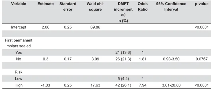
Table 2-'$ $ $/';>$?##$GJ
AIC (Akaike Information Criteria)=228.83 -2LogL=222.83
children’s parents. In order to classify the children’s socioeconomic level, the following variables were collected: home ownership, number of people living in the household, mother’s education, monthly family income, and parents’ occupation.
Data analysis
For univariate and multivariate analyses the DMFT increment >0 was selected as dependent variable. In the univariate analysis (Chi-square or Fisher tests) variables related to treatment group, dental plaque, malocclusion, and socioeconomic level were tested with the dependent variable. Those with p<0.15 were selected for the multiple logistic regression. After adjusting the regression model, the values of Odds Ratio, their 95% 4#! The Kruskal-Wallis and Dunn tests, at a 5% level # between the study groups as regards the increase 6 years. The Mann-Whitney test was used to compare differences between the HR-S and HR-NS groups in relation to DMFT at baseline. All statistical tests were performed using the SPSS (version 13.0) and Statistic (version 6.0) programs.
RESULTS
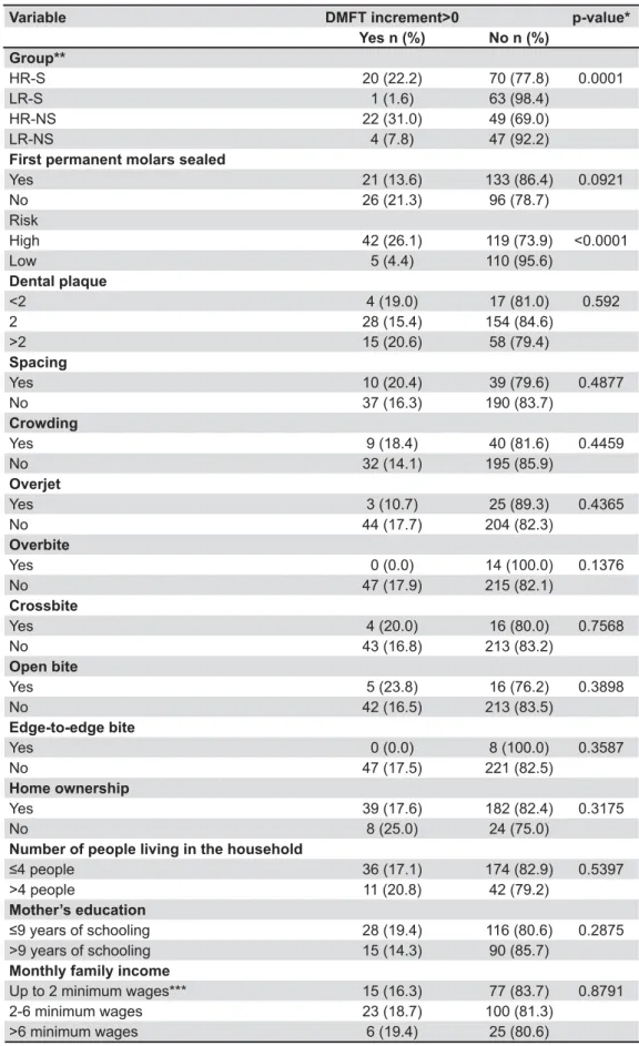
The mean DMFT values at baseline/final examination were 0.40/0.73, 0.00/0.02, 0.46/0.89 and 0.00/0.16 for the HR-S, LR-S, HR-NS and LR-NS groups, respectively. There was no 6 # 3~4K and HR-NS in relation to DMFT at baseline (p>0.05). Approximately 17.0% (n=47) of the children showed a DMFT increment>0 after 2 years (mean DMFT
increment=0.25). Among them, 44.7% (n=21) participated and 55.3% (n=26) did not participate !
Table 1 shows the univariate analysis for association between DMFT increment >0 and independent variables. Only the variables “group”  with DMFT increment>0. Data distribution showed that among those with DMFT increment the majority (31%) belonged to the HR-NS group. However, when testing the participation in the Sealant Program with DMFT increment, no statistical difference was observed (p>0.05).
Table 2 shows the results of the logistic regression analysis. The children who where not submitted to sealant placement were 1.8 more prone to have DMFT increment>0. The high-risk children had 7.94 more chance of developing caries, irrespective of whether or not they had received a sealant in the studied period.
Table 3 shows the increase in the number of 6 8 6 baseline in the different groups. High-risk children not submitted to sealant placement showed the highest increase in the number of decayed teeth. However, they did not differ from high-risk children submitted to sealant placement. Significant differences (p<0.05) were also found between high- and low-risk individuals, irrespective of participation in the sealant program.
Teeth status Group* Mean** Standard Deviation Median Minimum Maximum
Decayed Teeth HR-S 0.09ab 0.32 0 0 2
LR-S 0.02c 0.12 0 0 1
HR-NS 0.14a 0.35 0 0 1
LR-NS 0.04bc 0.2 0 0 1
Filled Teeth HR-S 0.24a 0.32 0 0 2
LR-S 0.00b 0 0 0 0
HR-NS 0.28a 0.7 0 0 3
LR-NS 0.12b 0.2 0 0 1
Decayed +Filled Teeth
HR-S 0.33a 0.69 0 0 3
LR-S 0.02b 0.12 0 0 1
HR-NS 0.42a 0.74 0 0 3
LR-NS 0.16b 0.64 0 0 4
Table 3-K$J ###%#$$$ JQ #$ $
! "#$%$ &" $#$%$ **Means followed by distinct letters are statistically different by the Kruskal-Wallis and Dunn tests (p<0.05)
Group* Tooth Tooth status n (%)
Sound at BL**
Sound at FE**
Decayed at BL
Decayed at FE
Filled at BL
Filled at FE
Sealed at BL
Sealed at FE
HR-S 16 85 (94.4) 24 (26.7) 4 (4.4) 4 (4.4) 0 (0.0) 7 (7.8) 1 (1.1) 55 (61.1)
26 80 (88.9) 15 (16.7) 8 (8.9) 4 (4.4) 0 (0.0) 11 (12.2) 2 (2.2) 60 (66.7)
36 77 (85.6) 11 (12.2) 13 (14.4) 5 (5.6) 0 (0.0) 17 (18.9) 0 (0.0) 57 (63.3)
46 79 (87.8) 11 (12.2) 9 (10.0) 6 (6.7) 1 (1.1) 12 (13.3) 1 (1.1) 61 (67.8)
LR-S 16 64 (100.0) 10 (15.6) 0 (0.0) 1 (1.6) 0 (0.0) 0 (0.0) 0 (0.0) 53 (82.8)
26 64 (100.0) 8 (12.5) 0 (0.0) 0 (0.0) 0 (0.0) 0 (0.0) 0 (0.0) 56 (87.5)
36 64 (100.0) 6 (9.4) 0 (0.0) 0 (0.0) 0 (0.0) 0 (0.0) 0 (0.0) 58 (90.6)
46 64 (100.0) 7 (10.9) 0 (0.0) 0 (0.0) 0 (0.0) 0 (0.0) 0 (0.0) 57 (89.1)
HR-NS 16 59 (91.1) 47 (66.2) 6 (8.4) 7 (9.9) 3 (4.2) 7 (9.9) 3 (4.2) 10 (14.1)
26 62 (87.3) 45 (63.4) 3 (4.2) 9 (12.7) 1 (1.4) 5 (7.0) 5 (7.0) 12 (16.9)
36 58 (81.7) 45 (63.4) 9 (12.7) 6 (8.4) 1 (1.4) 7 (9.9) 3 (4.2) 13 (18.3)
46 56 (87.5) 40 (56.3) 11 (8.3) 9 (12.7) 2 (2.8) 13 (18.3) 5 (7.0) 9 (12.7)
LR-NS 16 46 (90.2) 40 (78.4) 0 (0.0) 1 (2.0) 0 (0.0) 1 (2.0) 5 (9.8) 9 (17.6)
26 46 (90.2) 39 (76.5) 0 (0.0) 1 (2.0) 0 (0.0) 1 (2.0) 5 (9.8) 10 (19.6)
36 43 (84.3) 40 (78.4) 0 (0.0) 0 (0.0) 0 (0.0) 2 (3.9) 8 (15.7) 9 (17.6)
46 45 (88.2) 41 (80.4) 0 (0.0) 0 (0.0) 0 (0.0) 2 (3.9) 6 (11.8) 8 (15.7)
Table 4-"$$X" # #?## #Z#%# #[# # /';>#\ #$ $
]^ #$ _ $$%$ $J#%\$ Q #$ $
! "#$%$ &" $#$%$ `!J;j%\$
DISCUSSION
The results showed that the DMFT increment found in 17% of the children was statistically associated with risk, as shown in Table 1. Moreover,
strong predictor of future caries16-18,29,30
the importance of using the correct risk assessment to identify the most caries-susceptible individuals, thus targeting them for preventive care.
The results also showed a trend towards better results for those submitted to sealant placement (Table 1), which is in line with other studies that demonstrate the effectiveness of sealants in high-risk groups34. On the other hand, the DMFT
increment in 31% of children without sealant placement (HR-NS group) demonstrates that there
and that the population strategy alone (oral health # (Table 1).
As regards low-risk children only 1 child submitted to sealant placement (1.6%) developed caries, while among those without sealant, 4 children (7.8%) developed caries. This difference, ## 6 ! Table 3). Therefore, one must calculate the cost-effectiveness of applying sealants in low-risk groups because there may be other preventive
instructions, capable of achieving the same results. This study also assessed the influence of clinical and socioeconomic variables on caries incidence. The presence of plaque is usually linked to high rates of dental caries17. This study showed
different results (Table 1), probably due to the use KY36 *#6! This was done in order to not disturb the school class routines. Variables related to malocclusions were also not statistically associated with DMFT increment (Table 1).
The socioeconomic level has been strongly associated with caries prevalence27. Our results
differed from published data, possibly because this study was designed to measure caries incidence. However, most children who lived in their own house, with fewer than 4 persons, parents with over 9 years of schooling and families with a monthly income of over 6 minimum wages, showed no increment in DMFT (Table 1), indicating the important role of socioeconomic characteristics in caries development.
The significant increase in the number of 6 86 4 risk individuals (Table 3), with or without sealed permanent molars, demonstrated that these to care targeting all the children. Oulis and Berdouses23 (2009)evaluated the effectiveness of
on caries reduction, in a sample of children with low, moderate and high caries risk. The results demonstrated that the highest percentage of teeth that developed caries was found in the high risk
!" & may demand a great attention from the oral health team, who should bear in mind that high risk and population strategy should be taken together in order to improve the children’s overall health7.
8 molars could be detected in children who had been submitted to the sealant placement (Table 4). The fact that the majority of permanent molars were healthy at baseline demonstrated that this was an excellent time to take preventive measures. * 3~4 K # higher percentage of decayed permanent molars, demonstrating the importance of having a good team of professionals working on preventive care.
It is important to emphasize that this study was developed within an oral health program targeting Portuguese children from Sintra region. The Cacem Health Center was selected because the largest number of children was treated there and it presented organized dental records, while other health centers in the same region had some problems about that. Though this study had been carried out in a bounded area, probably the results can be inferred for whole region because children live in the same socioeconomic conditions. A 6 in controlling the dental treatments that each child received besides the PPSOCA protocol, such as sealants placed by other dentists.
I n c o n c l u s i o n , i t a p p e a r s t h a t s e a l a n t placement was effective in preventing dental caries development. Moreover, the variables “risk” and “sealant placement” were predictors of DMFT increment in the studied children. Therefore, 6 & submitted to sealant placement presented higher risk of developing caries lesions within a 2 year-period. Finally, the past caries experience was an excellent predictor of future caries and can easily be used in oral health programs.
REFERENCES
1- Ahovuo-Saloranta A, Hiiri A, Nordblad A, Mäkelä M, Worthington 3`! 6 permanent teeth of children and adolescents. Cochrane Database Syst Rev. 2008;4:CD001830.
2- Antunes JLF, Narvai PC, Nugent ZJ. Measuring inequalities in the distribution of dental caries. Community Dent Oral Epidemiol. 2004;32:41-8.
4K !% 6 sealants and the effect of fluoridated water consumption. Community Dent Health. 2007;24:4-11.
4- Assaf AV, Castro Meneghim M, Zanin L, Tengan C, Pereira AC. Effect of different diagnostic thresholds of dental caries calibration - a 12-month evaluation. Community Dent Oral Epidemiol. 2006;34:213-9.
6- Badovinac RL, Morgan KE, Lefreve J, Wadhawan S, Mucci L, Schoeff L, et al. Risk assessment criteria applied to a screening *^ 6 program. J Public Health Dent. 2005;65:203-8.
7- Batchelor PA, Sheiham A. The distribution of burden of dental caries in schoolchildren: a critique of the high risk caries prevention strategy for populations. BMC Oral Health. 2006;31;6:3. 8- Batchelor PA, Sheiham A. Grouping of tooth surfaces by susceptibility to caries: a study in 5-16 year-old children. BMC Oral Health. 2004;4:2.
9- Bönecker M, Cleaton-Jones P. Trends in dental caries in Latin American and Caribbean 5-6- and 11-13 year-old children: a systematic review. Community Dent Oral Epidemiol. 2003;31:152-7.
[4!_ K %_ * with a proposal for a new global oral health goal for 12-year-olds. Int Dent J. 2000;50:378-84.
11- Campain AC, Morgan MV, Evans RW, Ugoni A, Adams GG, Conn JA, et al. Sugar-starch combinations in food and the relationship to dental caries in low-risk adolescents. Eur J Oral Sci. 2003;111:316-25.
12- David J, Raadal M, Wang NJ, Strand GV. Caries increment and prediction from 12 to 18 years of age: a follow-up study. Eur Arch Paediatr Dent. 2006;7:31-7.
[4 % % & K ~ % " 6&! of a community sealant program. Pediatr Dent. 2005;27:212-6. 14- Francis R, Mascarenhas AK, Soparkar P, Al-Mutawaa S. ~ # children. Community Dent Health. 2008;25:211-5.
[4 %` ~!"6 *! J Am Dent Assoc. 1964;68:7-13.
16- Kassawara ABC, Tagliaferro EPS, Cortelazzi KL, Ambrosano GMB, Assaf AV, Meneghim MC, et al. Epidemiological assessment of predictors of caries increment in 7-10-year-olds: a 2-year cohort study. J Appl Oral Sci. 2010;18:116-20.
17- Leroy R, Bogaerts K, Lesaffre E, Declerck D. Multivariate survival analysis for the identification of factors associated #6 !YK! 2005;113:145-52.
18- Li Y, Wang W. Predicting caries in permanent teeth from caries in primary teeth: an eight-year cohort study. J Dent Res. 2002;81:561-6.
19- Marthaler TM. Changes in dental caries 1953-2003. Caries Res. 2004;38:173-81.
20- Meneghim MC, Kozlowski FC, Pereira AC, Ambrosano GMB, ! !% Saúde Coletiva. 2007;12:523-9.
21- Meneghim MC, Saliba NA, Pereira AC. The importance of " *! Odontopediatr Odontol Bebe. 1999;2:37-41.
22- Moberg Sköld U, Birkhed D, Borg E, Peterson LG. Approximal caries development in adolescents with low to moderate caries & 464 rising programs. Caries Res. 2005;39:529-35.
23- Oulis CJ, Berdouses ED. Fissure sealant retention and caries with low, moderate and high caries risk. Eur Arch Paediatr Dent. 2009;10:211-7.
24- Pardi V, Pereira AC, Ambrosano GMB, Meneghim MC. Clinical sealant: 24-months results. J Clin Pediatr Dent. 2005;29:133-7. 25- Pereira SM, Tagliaferro EPS, Ambrosano GMB, Cortellazzi KL, Meneghim MC, Pereira AC. Dental caries in 12-year-old schoolchildren and its relationship with socioeconomic and behavioral variables. Oral Health Prev Dent. 2007;5:299-306. 26- Pereira SM, Tagliaferro EPS, Cortellazzi KL, Ambrosano GMB, Mialhe FL, Meneghim MC, et al. An estimate of DMFT using teeth most affected by dental caries in twelve-year-old children. Rev Saude Publica. 2009;43:179-82.
27- Peres KGA, Bastos JRM, Latorre MRDO. Severity of dental caries in children and relationship with social and behavioral aspects. Rev Saude Publica. 2000;34:402-8.
28- Splieth CH, Ekstrand KR, Alkilzy M, Clarkson J, Meyer-Lueckel H, Martignon S, et al. Sealants in dentistry: outcomes of the ORCA Saturday Afternoon Symposium 2007. Caries Res. 2010;44:3-13. 29- Tagliaferro EPS, Ambrosano GMB, Meneghim MC, Pereira AC. Risk indicators and risk predictors of dental caries in schoolchildren. J Appl Oral Sci. 2008;16:408-13.
30- Tagliaferro EPS, Pereira AC, Meneghim MC, Ambrosano GMB. Assessment of dental caries predictors in a seven-year longitudinal study. J Public Health Dent. 2006;66:169-73.
31- Tickle M. The 80:20 phenomenon: help or hindrance to planning caries programmes? Community Dent Health. 2002;19:39-42. 32- Van Wyk PJ, Van Wyk C. Oral health in South Africa. Int Dent J. 2004;54:373-7.
33- Vanobbergen J, Martens L, Lesaffre E, Bogaerts K, Declerck D. Assessing risk indicators for dental caries in the primary dentition. Community Dent Oral Epidemiol. 2001;29:424-34.
4 ! 44& individuals. J Dent Educ. 2001;65:1084-90.