Original contribution
Synovial sarcoma of nerve
Bernd W. Scheithauer MD
a,⁎
, Kimberly K. Amrami MD
c, Andrew L. Folpe MD
a,
Ana I. Silva MD
a, d, Mark A. Edgar MD
e, James M. Woodruff MD
e,
Allan D. Levi MD, PhD
f, Robert J. Spinner MD
ba
Department of Pathology and Laboratory Medicine, Mayo Clinic, Rochester, MN 55905, USA b
Department of Neurosurgery, Mayo Clinic, Rochester, MN 55905, USA c
Department of Radiology, Mayo Clinic, Rochester, MN 55905, USA d
Department of Pathology, Hospital Sao Marcos, Braga, Portugal e
Department of Pathology, Memorial Sloan-Kettering Cancer Center (JMW, Emeritus), New York, NY 10021, USA f
Department of Neurosurgery, University of Miami, Miami, FL 33133, USA
Received 21 June 2010; revised 11 August 2010; accepted 20 August 2010
Keywords:
Nerve; Sarcoma; Synovial sarcoma; Immunohistochemistry
SummaryTumors of peripheral nerve are largely neuroectodermal in nature and derived from 2 elements
of nerve, Schwann or perineurial cells. In contrast, mesenchymal tumors affecting peripheral nerve are rare and are derived mainly from epineurial connective tissue. The spectrum of the latter is broad and includes lipoma, vascular neoplasms, hematopoietic tumors, and even meningioma. Of malignant peripheral nerve neoplasms, the vast majority are primary peripheral nerve sheath tumors. Malignancies of mesenchymal type are much less common. To date, only 12 cases of synovial sarcoma of nerve have been described. Whereas in the past, parallels were drawn between synovial sarcoma and malignant glandular schwannoma, an uncommon form of malignant peripheral nerve sheath tumor, molecular genetics have since clarified the distinction. Herein, we report 10 additional examples of molecularly confirmed synovial sarcoma, all arising within minor or major nerves. Affecting 7 female and 3 male patients, 4 tumors occurred in pediatric patients. Clinically and radiologically, most lesions were initially thought to be benign nerve sheath tumors. On reinterpretation of imaging, they were considered indeterminate in nature with some features suspicious for malignancy. Synovial sarcoma of nerve, albeit rare, seems to behave in a manner similar to its more common, soft tissue counterpart. Those affecting nerve have a variable prognosis. Definitive recommendations regarding surgery and adjuvant therapies await additional reports and long-term follow-up. The literature is reviewed and a meta-analysis is performed with respect to clinicopathologic features versus outcome.
© 2011 Published by Elsevier Inc.
1. Introduction
The vast majority of peripheral nerve tumors are neuroectodermal in nature. Derived mainly from Schwann
cells, most are represented by schwannoma and neurofi-broma. Only in recent years have the 2 variants of perineurioma come to be recognized, a soft tissue form unassociated with nerve [1] and subsequently the intra-neural variety involving multiple fascicles and exhibiting pseudo-onion bulb formation, a highly distinctive histologic feature[2]. Mesenchymal tumors and tumor-like lesions are rare and comprise a wide variety of benign and malignant soft tissue tumors[3,4].
⁎Corresponding author. Department of Pathology and Laboratory
Medicine, Mayo Clinic, Rochester, MN 55905, USA.
E-mail address:[email protected](B. W. Scheithauer).
www.elsevier.com/locate/humpath
0046-8177/$–see front matter © 2011 Published by Elsevier Inc.
doi:10.1016/j.humpath.2010.08.019
Synovial sarcoma is a distinctly rare primary tumor of nerve. Only 12 examples have been described to date
[5-15]. This may not only reflect its true rarity in this anatomical location but may also reflect the natural tendency of pathologists to diagnose as “malignant peripheral nerve sheath tumor” (MPNST) all monomor-phous, spindle cell sarcomas arising in nerve. Alternative diagnoses are infrequently considered. Herein, we report the clinicopathologic features of 10 primary synovial sarcomas of nerve, all with molecular genetic confirma-tion of the synovial sarcoma-specific translocaconfirma-tion t(X;18)
[16].
2. Materials and methods
2.1. Case selection
The consultation archives of 2 of the authors (B. W. S., A. L. F.) were searched for cases coded as “synovial sarcoma”occurring as primary tumors of nerve. All lesions were limited to nerve; no tumors with features of secondary neural involvement were included in this study. The tumor of case 3 was encountered in practice at another institution by one of the authors (M. A. E.). The diagnosis of synovial sarcoma was based largely on its characteristic histology, as well as on clinicopathologic features dissimilar to MPNST, fully half of which occur in association with NF1 and arise in transition from neurofibroma, are far more often high grade (85%), and show far greater histologic diversity [3]. All tumors were found to be positive for either the SS18-SSX1 (4 cases) or SS18-SSX2 (6 cases) gene fusions by reverse transcriptase–polymerase chain reaction (RT-PCR).
2.2. Immunohistochemistry
Formalin-fixed, paraffin-embedded tissue sections were immunostained for vimentin (Dako, Carpinteria, CA; 1:500, V9), epithelial membrane antigen (EMA) (Dako, 1:50, E29), S-100 protein (Dako, 1:1600, polyclonal), CD57 (Becton Dickinson, 1:20; HNK-1), neurofilament protein (Dako, 1:800, 2F11), collagen IV (Dako, 1:25, CIV22), TLE1 (Santa Cruz Biotechnology, Inc, Santa Cruz, CA; dilution 1:100, polyclonal), Fli1 (BD Pharmingen, San Jose, CA; 1:50, BRD, G146-254), and Ki-67 (Dako, 1:30, MIB-1) using heat-induced epitope retrieval and the Dako Envision detection system. Appropriate positive and negative controls were used.
2.3. Molecular genetics
Methods for the characterization of synovial sarcoma including RT-PCR (10 cases) and fluorescence in situ hybridization (FISH) (1 case) have been previously pub-lished elsewhere[17].
3. Results
3.1. Clinicopathologic features
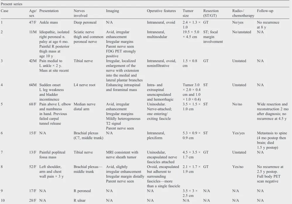
Essential clinical and therapeutic data regarding our 10 cases are summarized inTable 1. The tumors occurred in 7 female and 3 male patients ranging in age from 11 to 68 years (mean, 40 years), inclusive of 4 pediatric cases.
3.2. Imaging
Imaging was available for review in 4 patients (cases 2, 3, 5, 8). All had MRI examinations (3 with gadolinium enhancement) performed at 1.5 T or lower. A positron emission tomography (PET) scan was available in a single patient (case 2). The nerves involved were the distal tibial nerve, peroneal division of the sciatic nerve, median nerve in the distal arm, and the middle trunk of the brachial plexus. All lesions arose from the affected nerve and extended proximally and distally along its length (“tail sign”) (Figs. 1 and 2A). In the case of the tibial nerve lesion (case 3), the mass extended from the common tibial nerve into the medial and lateral plantar branches. All lesions were isointense to muscle on T1-weighted imaging, hyperintense on T2-weighted sequences, and showed avid enhancement in the 3 cases wherein post-contrast images were available (Figs. 1 and 2A). Smaller lesions showed homogeneous enhancement. The large, complex sciatic lesion (case 2) showed a more heterogeneous signal and enhancement in addition to hemorrhage within the largest portion of the tumor. Of the 4 tumors (cases 1, 2, 3, 8), 3 were oval in shape and ranged from slightly less than 1 cm to 3 cm in maximum dimension with irregular margins in each case. The sciatic lesion (case 2) was multinodular, the 4 distinct, ovoid nodules collectively measuring 19 cm in length and 4 cm in maximal axial dimension (Fig. 2A). The PET scan in this case showed avid uptake of the fluorodeoxyglucose (FDG) in all the nodules (Fig. 2B). On reinterpretation of imaging, the 4 tumors, based on the presence of irregular margins, were considered indeterminate in nature and suspicious for malignancy.
3.3. Operative findings
Table 1 Synovial sarcoma of nerve Present series Case Age/ sex Presentation Nerves involved
Imaging Operative features Tumor size Resection (ST/GT) Radio-/ chemotherapy Follow-up
1 47/F Ankle mass Deep peroneal N/A Intraneural, ovoid 2.4 × 1.3 × 1.0
GT No/yes No recurrence
at 8 y 2 11/M Idiopathic, isolated
right peroneal n. palsy at age 6 mo. Painful R posterior thigh mass at age 10 y
Sciatic nerve thigh and common peroneal nerve
Avid, irregular enhancement Irregular margins Parent nerve seen FDG PET strongly positive
Intraneural, multinodular
19.5 × 5.0 × 4.5 cm
ST; focal margin involvement
No/unstated N/A
3 42/M Pain medial to L ankle × 2 y. Mass at site recent
Tibial nerve Irregular, localized enlargement of the nerve with extension into the medial and lateral plantar branches
Intraneural, ovoid, noninfiltrative
1.5 × 0.8 cm
GT Unstated N/A
4 64/M Sudden onset
L leg weakness and bladder incontinence
L4 nerve root Enhancing intraspinal and foraminal mass
Intra- and extraspinal unencapsulated and hemorrhagic
Tumor 3.0 × 2.0 × 0.4 cm and 1.0 × 1.0 × 0.4)
ST Unstated N/A
5 68/F Pain above L elbow and numbness in hand. Previous failed carpal tunnel release Median nerve distal arm Avid, irregular enhancement Irregular margins Mildly heterogeneous T2 signal
Parent nerve seen
Uninodular. Nerve-attached; one entering/ exiting fascicle
3.5 × 1.5 × 1.0 cm
ST No/no Wide resection and
reconstruction 2 mo after diagnosis; no recurrence at 4.5 y
6 15/F N/A Brachial plexus
(C7, middle trunk)
N/A Intraneural,
plexiform
5.3 × 0.9 × 0.9 cm
ST Yes/yes Metastasis to spine (4 mo postop then brain; died 1.5 y postop) 7 13/F Painful popliteal
fossa mass
Tibial nerve MRI consistent with nerve sheath tumor
Uninodular, encapsulated nerve fascicles attached
4.5 × 3.5 × 1.7 cm
GT Unstated N/A
8 52/F Left shoulder, arm and chest wall pain × 3 y
Brachial plexus—
middle trunk
Avid, slightly irregular enhancement Irregular margin distally Parent nerve seen
Ovoid, encapsulated but adherent to surrounding fascicles—more than a single fascicle
2.1 × 1.7 × 1.9 cm
GT Yes/no No recurrence at 2.5 y postop. Full body PET scan negative
9 17/F N/A R peroneal N/A N/A 3.5 × 3 ×
2.5 cm
N/A N/A N/A
10 28/F N/A R ulnar N/A N/A N/A N/A N/A N/A
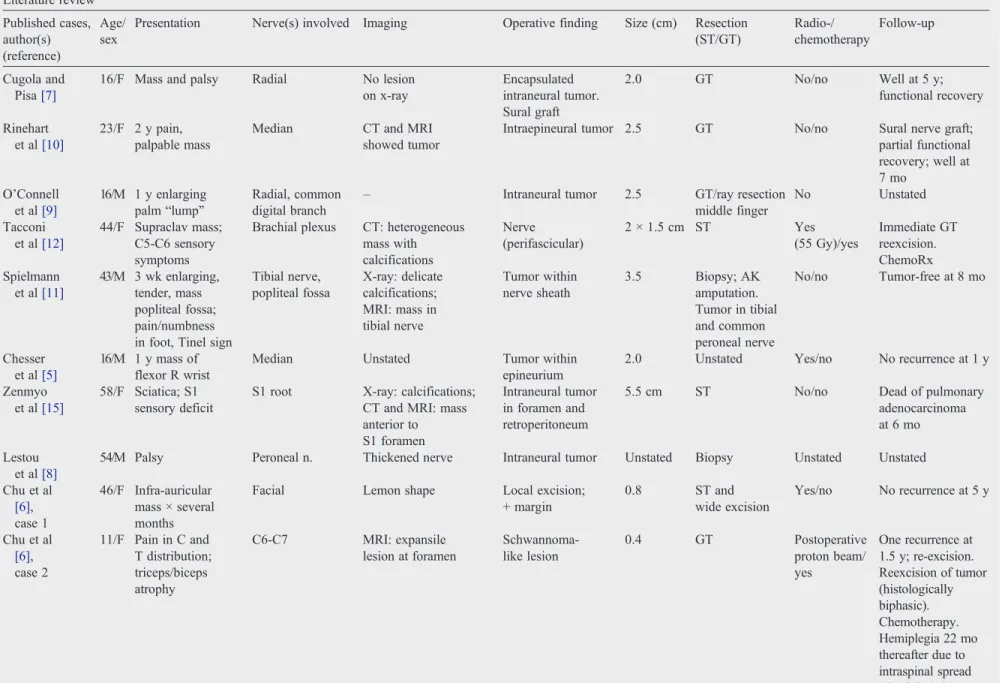
Table 1(continued) Literature review Published cases, author(s) (reference) Age/ sex
Presentation Nerve(s) involved Imaging Operative finding Size (cm) Resection (ST/GT) Radio-/ chemotherapy Follow-up Cugola and Pisa[7]
16/F Mass and palsy Radial No lesion on x-ray
Encapsulated intraneural tumor. Sural graft
2.0 GT No/no Well at 5 y;
functional recovery
Rinehart et al[10]
23/F 2 y pain, palpable mass
Median CT and MRI showed tumor
Intraepineural tumor 2.5 GT No/no Sural nerve graft; partial functional recovery; well at 7 mo
O'Connell et al[9]
16/M 1 y enlarging palm“lump”
Radial, common digital branch
– Intraneural tumor 2.5 GT/ray resection
middle finger
No Unstated
Tacconi et al[12]
44/F Supraclav mass; C5-C6 sensory symptoms
Brachial plexus CT: heterogeneous mass with calcifications
Nerve (perifascicular)
2 × 1.5 cm ST Yes
(55 Gy)/yes
Immediate GT reexcision. ChemoRx Spielmann
et al[11]
43/M 3 wk enlarging, tender, mass popliteal fossa; pain/numbness in foot, Tinel sign
Tibial nerve, popliteal fossa
X-ray: delicate calcifications; MRI: mass in tibial nerve
Tumor within nerve sheath
3.5 Biopsy; AK amputation. Tumor in tibial and common peroneal nerve
No/no Tumor-free at 8 mo
Chesser et al[5]
16/M 1 y mass of flexor R wrist
Median Unstated Tumor within
epineurium
2.0 Unstated Yes/no No recurrence at 1 y
Zenmyo et al[15]
58/F Sciatica; S1 sensory deficit
S1 root X-ray: calcifications; CT and MRI: mass anterior to S1 foramen
Intraneural tumor in foramen and retroperitoneum
5.5 cm ST No/no Dead of pulmonary
adenocarcinoma at 6 mo
Lestou et al[8]
54/M Palsy Peroneal n. Thickened nerve Intraneural tumor Unstated Biopsy Unstated Unstated
Chu et al
[6], case 1
46/F Infra-auricular mass × several months
Facial Lemon shape Local excision; + margin
0.8 ST and
wide excision
Yes/no No recurrence at 5 y
Chu et al
[6], case 2
11/F Pain in C and T distribution; triceps/biceps atrophy
C6-C7 MRI: expansile lesion at foramen
Schwannoma-like lesion
0.4 GT Postoperative
proton beam/ yes
One recurrence at 1.5 y; re-excision. Reexcision of tumor (histologically biphasic). Chemotherapy. Hemiplegia 22 mo thereafter due to intraspinal spread (C4-C6). DOD 6 y after initial diagnosis
(continued on next page)
571
sarcoma
of
3.4. Pathology
Of the 10 tumors, 9 showed classic histologic features of monophasic synovial sarcoma, including a fascicular proliferation of monomorphic, hyperchromatic spindled cells in association with wiry collagen and a branching,
“staghorn”vascular pattern (Fig. 4). In one instance (case 2), a biphasic variant, epithelial differentiation, took the form of small glands lined by cuboidal to slightly columnar cells (Fig. 5). Mitotic activity ranged from 1 to 13/10 high-powered fields (HPF) (mean, 4/10 HPF). No tumor showed calcification. Round cell areas or necrosis, that is, poorly differentiated histology, was not encountered.
3.5. Immunohistochemical findings
The immunohistochemical results are summarized in Table 3 and illustrated inFig. 6. All tumors were diffusely positive for vimentin. Patchy expression of EMA, pan-cytokeratin, and cytokeratin 7 was seen in the spindled cells of 90%, 80%, and 100% of studied cases, respectively. S100 protein and CD57 were positive in a variable number of cells in 50% and 100% of cases. Immunostains for neurofilament protein showed single or grouped and aligned axons within 6 of 8 tumors studied (cases 1, 3, 4, 5, 7, 8), suggesting either intrafascicular origin or extension of the tumor (Fig. 6F). Diffuse nuclear expression of TLE1 protein was present in all cases. No case expressed FLI-1 protein.
3.6. Molecular genetic findings
As noted previously, 6 cases were known to carry the
SS18-SSX2fusion and 4, theSS18-SSX1fusion.
3.7. Literature review
Comparable data regarding the 12 previously published cases are summarized inTables 1 and 2.
4. Discussion
Synovial sarcoma is a relatively common malignant soft tissue neoplasm, comprising approximately 10% of soft tissue sarcomas. Despite its name, synovial sarcoma rarely involves synovium or joints. It is now abundantly clear that this tumor bears no relationship to synovium. Synovial sarcomas most often involve the extremities of adolescents and young adults but may occur in patients of any age and in any soft tissue or even visceral location. Indeed, with the discovery that synovial sarcomas carry a variety of specific translocations, including t(X;18)(p11.23;q11)(SS18-SSX1) (∼65% of cases), t(X;18)(p11.21;q11) (SS18-SSX2) (∼35%
of cases), t(X;18)(p11;q11) (SS18- SSX4) (b1% of cases), and t(X;20)(p11;q13.3) (SS181-SSX1) (b1% of cases), the
Table 1 ( continued ) Present series Case Age/ sex Presentation Nerves involved Imaging Operative features Tumor size Resection (ST/GT) Radio-/ chemotherapy Follow-up Weinreb et al [14] 48/F Intermittent shooting pain L leg, big toe Tibial MRI: heterogeneous mass posterior compartment of L calf Debulking 8.7 ST Unstated Recurrences 3 mo postop. AK
amputation. Recurrence-free at14
mo Uehara et al [13] 3/F Radiating pain R palm. Decreased grasp strength. Atrophy of thenar muscles Median MRI: enhancing spindle-shaped tumor Subcapsular removal 1.5 × 1.2 GT on immediate reexcision Yes/no Local recurrence at 14 mo requiring additional resection Abbreviations: ST, subtotal; GT, gross total; N/A, not available; AK, above knee.
diagnostic application of molecular techniques (RT-PCR, FISH) has made it increasingly apparent that many synovial sarcomas occur in unusual locations[18,19].
Primary synovial sarcomas of nerve are very rare, there being only 22 reported cases, inclusive of those in the present series (Tables 1 and 2)[5-12,14,15]. Extremely rare cases of intraneural metastases from synovial sarcoma have also been reported[20]. As noted in the present study, the morphologic and immunohistochemical features of intraneural synovial sarcomas are essentially identical to those of their more common soft tissue counterparts. However, 9 (90%) of 10 cases in the present series were of monophasic type, as compared with 70% of soft tissue cases, and 6 (60%) of 10 carriedSS18-SSX2fusions, versus the 35% frequency seen in synovial sarcomas at nonneural sites. This suggests that synovial sarcomas of nerve have some unique features. Given the relatively small sample size, however, it is entirely possible that these findings represent a statistical aberration. The radiographic features of primary intraneural synovial sarcoma have not been previously described in depth. Imaging studies were available for 4 of cases in the present series. The lesions had similar imaging characteristics: all had irregular margins and clearly were associated with individual nerves. None had imaging features suggestive of a benign nerve sheath tumor. Indeed, 3 of the 4 cases had MR imaging characteristics consistent with MPNST. One tumor affecting the tibial nerve (case 7) was not as characteristic, presenting with less prominent localized enlargement of the nerve, an appearance indeterminate by imaging criteria; contrast enhancement, which might have improved sensitivity and narrowed the differential diagnosis, was unavailable in this case. The one instance wherein PET imaging was available (case 2) showed avid uptake of the tracer FDG. Although this
is occasionally seen in cellular, benign nerve sheath tumors, the appearance in this case was more consistent with a malignant process. Soft tissue synovial sarcomas are usually heterogeneously isointense on T1, bright on T2-weighted
Fig. 1 A, Case 8. Coronal T2-weighted MR image without fat suppression showing a relatively well-marginated mass in the middle trunk of
the supraclavicular brachial plexus (asterisk). The mass is bright on T2 with mild signal heterogeneity. The normal nerve proximal and distal to the lesion is clearly seen. B, Case 8. Post-contrast T1-weighted image with fat suppression at the same level as A. This image shows the irregular margins of the tumor, especially at its distal aspect (arrows) and avid contrast enhancement.
Fig. 2 A, Case 2. Sagittal T1-weighted image after gadolinium
imaging, and homogeneously enhancing in most cases. Approximately one third of tumors will have associated calcifications, a feature lacking in our 4 cases. On imaging, soft tissue synovial sarcomas are often mistaken for benign nerve sheath tumors given their oval shape and frequent longitudinal orientation relative to surrounding soft tissue. Nonetheless, close inspection shows slight irregularity of their margins and no association with an individual nerve. This is in sharp distinction to the cases reviewed herein, where the parent nerve was always visualized.
The differential diagnosis for primary intraneural synovial sarcoma is mainly with conventional, spindle cell MPNST. The morphologic features of monophasic synovial sarcomas and conventional, spindle cell MPNST are remarkably similar. The only features offering significant discriminatory assistance being tumor occurrence in a patient with documented neurofibromatosis type 1 and/or an origin from a preexisting plexiform neurofibroma. Additional morphol-ogies include the presence of wiry collagen and stromal calcifications in synovial sarcoma and the often greater pleomorphism of MPNST. Even glandular differentiation may be seen in both synovial sarcoma and MPNST, although the glands of biphasic synovial sarcoma tend to be small, lined by cuboidal cells, and filled with eosinophilic debris, in contrast to the enteric-type glands seen in those extremely
rare MPNST with glandular differentiation[21]. Similarly, the immunophenotypes of monophasic synovial sarcoma and MPNST show considerable overlap, with frequent expression of putative nerve sheath markers such as S100 protein and CD57 in synovial sarcoma[22], expression of EMA both in synovial sarcoma and in MPNST with perineurial differen-tiation, rare reported cytokeratin-positive MPNST[23], and very focal or even absent expression of epithelial markers in some monophasic synovial sarcomas. CD34 expression, frequently present in MPNST, particularly low-grade exam-ples[24]but essentially unheard of in synovial sarcoma, may be helpful in this differential diagnosis. TLE1, a WNT pathway-associated transcription factor, has recently been shown to be a highly sensitive marker of synovial sarcomas, typically showing expression in nearly 100% of nuclei in a given tumor[25]. TLE1 is not, however, a perfectly specific marker, as it may be positive in a significant number of benign and malignant peripheral nerve sheath tumors, albeit typically
Fig. 3 Case 2. Gross photo of the specimen showed this unusual
tumor of the peroneal division of the sciatic nerve to be multilobed.
Fig. 4 Case 5. Histologically, most of the lesions were
monophasic, consisting of sheaves of spindle cells associated with collagen bundles (original magnification ×400).
Fig. 5 Case 3. A single tumor of the biphasic subtype, featuring
well-formed glands (original magnification ×600).
in a smaller percentage of tumor nuclei[26]. A large number of other markers, including BCL-2, SYT protein, CD56, p75NTR, nestin, and HMGA2 protein, have been suggested to be of some value in this differential diagnosis but either lack specificity (BCL-2, CD56) or have been studied only in a very small number of cases (nestin, HMGA2).
Given some morphologic and considerable immunophe-notypic overlap between synovial sarcoma and MPNST, demonstration of one of the synovial sarcoma-associated fusion genes has come to be regarded as the“gold standard”
for this differential diagnosis. Although it was initially asserted that MPNST and other types of nerve sheath tumors could contain the (X;18) translocation [27], this has
subsequently been disproved by a number of large, carefully performed studies [28-30]. Positive molecular markers of MPNST, which may also be of value in this differential diagnosis, include NF1 and p16 deletions as well as epidermal growth factor receptor amplification and polysomies for either chromosome 7 or 22 as demonstrated by FISH[31].
In conclusion, we have described the clinical, radiographic, pathologic, and molecular genetic findings in 10 cases of primary intraneural synovial sarcoma, the largest series to date. In general, primary intraneural synovial sarcomas are similar to their more common soft tissue counterparts, although there may be a relative intraneural predominance of monophasic tumors and tumors containingSS18-SSX2fusions. Based on
Table 2 Synovial sarcoma of nerve: histology, immunohistochemistry, genetics
Present cases
Case Histologic type
Mitoses/ 10 HPF
EMA AE1-AE3 S-100 protein
CD57 TLE-1 Fli1 NFP showing nerve association
PCR
1 Monophasic 13 + 1+ + 1+ 4+ – Axons SYT-SSX2
2 Monophasic 10 – 1+ – 1+ 2+ – – SYT-SSX1
3 Biphasic 3 2 2+ – 2+ 4+ – Axons SYT-SSX1
4 Monophasic 2 1 2+ 1+ 2+ 3+ – Axons SYT-SSX1
5 Monophasic 4 2 – 1+ 1+ 1+ – – SYT-SSX2
6 Monophasic 3 1+ 1+ – 3+ 4+ – Axons SYT-SSX2
7 Monophasic 2 2+ 1+ 1+ 1+ 4+ – Axons SYT-SSX2
8 Monophasic 3 2+ 1+ ND 3+ 1+ – Axons SYT-SSX2
9 Monophasic 1 1+ 2+/no 1+ ND ND ND Axons SYT-SSX2
10 Monophasic 2 scant 1+/NA 1+ ND 2+ ND – SYT-SSX1
Literature review
Published cases, author(s) (reference)
Histologic type
EMA KERATIN S100
protein
NFP showing nerve association
Miscellaneous Genetics/PCR
Cugola and Pisa[7] Biphasic NA NA NA NA – SYT-SSX1
Rinehart[10] Monophasic Focal+ Focal+ Focal+ Axons Desmin—focal SYT-SSX2 O'Connell[9] Biphasic +b5% 15%-20%+ Neg Axons SMA, CD34- x;18
Tacconi et al[12] Monophasic + + Focal+ NA – NA
Spielmann et al[11] Biphasic Focal Neg Neg Axons CD99+; MSA, Desmin−
SYT-SSX2
Chesser et al[5] Biphasic NA + NA NA SYT-SSX1
Zenmyo et al[15] Monophasic + + NA NA bcl-2+ t(x;18) (p11;q11)
SYT-SSX - ?later Lestou et al[8] Monophasic Minor+ Minor+ Minor+ NA Vimentin and
CD99+
Cryptic t(x;18), ins (6;18) and SYT-SSX2 gene fusion Chu et al[6], case 1 Biphasic + Tumor and
perineurium
+ Neg Axons – t(x;18)
(SYT-SSX) Chu et al[6], case 2 Monophasic Neg Neg Neg
Nerve fibers+
Axons _ t(x;18)
(SYT-SSX)
Weinreb et al[14] Biphasic + + Neg Neg Desmin, actin,
and CD34−; BCL-2+
SYT-SSX1
Uehara et al[13] Monophasic + + + + SYT-SSX1
Abbreviations: NFP, neurofilament protein; NA, not assessed; ND, not done.
relatively limited follow-up, we see no reason to believe that the behavior of intraneural synovial sarcomas is any different from that of synovial sarcomas at other locations, particularly in extremities. Definitive recommendations regarding possible roles for adjuvant radiation and chemotherapy await the study of additional cases.
The present series, even combined with our literature review, has limitations with regard to assessing recurrence/ survival data. Nonetheless, several clinicopathologic factors appear to affect outcome. Comparing our data with that of Lewis et al[32], a multivariate analysis of prognostic factors in 112 patients with primary localized synovial sarcomas of
Fig. 6 The immunophenotype of the tumors included keratin (A; case 3), epithelial membrane antigen (B; case 3), S-100 protein reactivity
(C; case 5), CD57 (D; case 6), and TLE1 (E; case 6). Axons immunoreactive for neurofilament protein were identified within some tumors and indicated endoneurial involvement (F; case 7) (A-F, original magnification ×400).
the extremities, we believe that small tumor size may affect the prognosis. Of the combined 22 patients in our report and review study, 16 (73%) had small (b5 cm) tumors, a meaningful prognostic factor according to Lewis et al[32]. An early presentation with neurologic symptoms may be the basis of their small size. Indeed, 15 (75%) of 20 patients experienced sensorimotor loss and/or pain, a not surprising presentation for a nerve-based tumor. Thus, synovial sarcomas of nerve may be associated with a more favorable prognosis than nonneural examples. Relative to MPNST of the extremities, particularly lower extremity examples, which have a poor prognosis [33], synovial sarcomas at this site appear to have a more favorable prognosis.
References
[1] Lazarus SS, Trombetta LD. Ultrastructural identification of a benign perineurial cell tumor. Cancer 1978;41:1823-9.
[2] Emory TS, Scheithauer BW, Hirose T, Wood M, Onofrio BM, Jenkins RB. Intraneural perineurioma. A clonal neoplasm associated with abnormalities of chromosome 22. Am J Clin Pathol 1995;103: 696-704.
[3] Scheithauer BW, Woodruff JM, Erlandson RA. Benign and malignant non-neurogenic tumors (chapter 10). In: Rosai J, Sobin LH, editors.
Atlas of tumor pathology—tumors of peripheral nervous system.
Washington, D.C.: Third Series, Fascicle 24. Armed Forces Institute of Pathology; 1999. p. 283-302.
[4] Scheithauer BW, Woodruff JM, Erlandson RA. Primary malignant tumors of peripheral nerve (chapter 11). In: Rosai J, Sobin LH, editors.
Atlas of tumor pathology—tumors of peripheral nervous system.
Washington, D.C.: Third Series, Fascicle 24. Armed Forces Institute of Pathology; 1999. p. 303-72.
[5] Chesser TJ, Geraghty JM, Clarke AM. Intraneural synovial sarcoma of the median nerve. J Hand Surg [Br] 1999;24:373-5.
[6] Chu PG, Benhattar J, Weiss LM, Meagher-Villemure K. Intraneural synovial sarcoma: two cases. Mod Pathol 2004;17:258-63.
[7] Cugola L, Pisa R. Synovial sarcoma: with radial nerve involvement. J Hand Surg [Br] 1985;10:243-4.
[8] Lestou VS, O'Connell JX, Robichaud M, et al. Cryptic t(X;18), ins (6;18), and SYT-SSX2 gene fusion in a case of intraneural monophasic synovial sarcoma. Cancer Genet Cytogenet 2002;138:153-6. [9] O'Connell JX, Browne WL, Gropper PT, Berean KW. Intraneural
biphasic synovial sarcoma: an alternative “glandular” tumor of
peripheral nerve. Mod Pathol 1996;9:738-41.
[10] Rinehart GC, Mustoe TA, Weeks PM. Management of synovial sarcoma of the median nerve at the elbow. Plast Reconstr Surg 1989;83:528-32.
[11] Spielmann A, Janzen DL, O'Connell JX, Munk PL. Intraneural synovial sarcoma. Skeletal Radiol 1997;26:677-81.
[12] Tacconi L, Thom M, Thomas DG. Primary monophasic synovial sarcoma of the brachial plexus: report of a case and review of the literature. Clin Neurol Neurosurg 1996;98:249-52.
[13] Uehara H, Yamasaki K, Fukushima T, et al. Intraneural synovial sarcoma originating from the median nerve. Neurol Med Chir 2008;48: 77-82.
[14] Weinreb I, Perez-Ordonez B, Guha A, Kiehl TR. Mucinous, gland predominant synovial sarcoma of a large peripheral nerve: a rare case closely mimicking metastatic mucinous carcinoma. J Clin Pathol 2008;61:672-6.
[15] Zenmyo M, Komiya S, Hamada T, et al. Intraneural monophasic synovial sarcoma: a case report. Spine 2001;26:310-3.
[16] Sandberg AA, Bridge JA. Updates on the cytogenetics and molecular genetics of bone and soft tissue tumors. Synovial sarcoma. Cancer Genet Cytogenet 2002;133:1-23.
[17] Weinbreck N, Vignaud JM, Begueret H, et al. SYT-SSX fusion is absent in sarcomatoid mesothelioma allowing its distinction from synovial sarcoma of the pleura. Mod Pathol 2007;20: 617-21.
[18] Billings SD, Meisner LF, Cummings OW, Tejada E. Synovial sarcoma of the upper digestive tract: a report of two cases with demonstration of the X;18 translocation by fluorescence in situ hybridization. Mod Pathol 2000;13:68-76.
[19] Pan CC, Chang YH. Primary synovial sarcoma of the prostate. Histopathology 2006;48:321-3.
[20] Matsumine A, Kusuzaki K, Hirata H, Fukutome K, Maeda M, Uchida A. Intraneural metastasis of a synovial sarcoma to a peripheral nerve. J Bone Joint Surg Br 2005;87:1553-5.
[21] Woodruff JM, Christensen WN. Glandular peripheral nerve sheath tumors. Cancer 1993;72:3618-28.
[22] Guillou L, Wadden C, Kraus MD, Dei Tos AP, Fletcher CDM. S-100
protein reactivity in synovial sarcomas—a potentially frequent
diagnostic pitfall: immunohistochemical analysis of 100 cases. Appl Immunohistochem 1996;4:167-75.
[23] Smith TA, Machen SK, Fisher C, Goldblum JR. Usefulness of cytokeratin subsets for distinguishing monophasic synovial sarcoma from malignant peripheral nerve sheath tumor. Am J Clin Pathol 1999;112:641-8.
[24] Zhou H, Coffin CM, Perkins SL, Tripp SR, Liew M, Viskochil DH. Malignant peripheral nerve sheath tumor: a comparison of grade, immunophenotype, and cell cycle/growth activation marker
expres-sion in sporadic and neurofibromatosis 1–related lesions. Am J Surg
Pathol 2003;27:1337-45.
[25] Jagdis A, Rubin BP, Tubbs RR, Pacheco M, Nielsen TO. Prospective evaluation of TLE1 as a diagnostic immunohistochemical marker in synovial sarcoma. Am J Surg Pathol 2009;33:1743-51.
[26] Kosemehmetoglu K, Vrana JA, Folpe AL. TLE1 expression is not specific for synovial sarcoma: a whole section study of 163 soft tissue and bone neoplasms. Mod Pathol 2009;22:872-8.
[27] O'Sullivan MJ, Kyriakos M, Zhu X, et al. Malignant peripheral nerve sheath tumors with t(X;18). A pathologic and molecular genetic study. Mod Pathol 2000;13:1253-63.
[28] Coindre JM, Hostein I, Benhattar J, Lussan C, Rivel J, Guillou L. Malignant peripheral nerve sheath tumors are t(X;18)-negative sarcomas. Molecular analysis of 25 cases occurring in neurofibroma-tosis type 1 patients, using two different RT-PCR-based methods of detection. Mod Pathol 2002;15:589-92.
[29] Ladanyi M, Woodruff JM, Scheithauer BW, et al. Re: O'Sullivan MJ, Kyriakos M, Zhu X, Wick MR, Swanson PE, Dehner LP, Humphrey PA, Pfeifer JD: malignant peripheral nerve sheath tumors with t(X;18). A pathologic and molecular genetic study. Mod pathol 2000;13:1336-46. Mod Pathol 2001;14:733-7.
[30] Tamborini E, Agus V, Perrone F, et al. Lack of SYT-SSX fusion transcripts in malignant peripheral nerve sheath tumors on RT-PCR analysis of 34 archival cases. Lab Invest 2002;82:609-18.
[31] Perry A, Kunz SN, Fuller CE, et al. Differential NF1, p16, and EGFR patterns by interphase cytogenetics (FISH) in malignant peripheral nerve sheath tumor (MPNST) and morphologically similar spindle cell neoplasms. J Neuropathol Exp Neurol 2002;61: 702-9.
[32] Lewis JJ, Antonescu CR, Leung DH, et al. Synovial sarcoma: a multivariate analysis of prognostic factors in 112 patients with primary localized tumors of the extremity. J Clin Oncol 2000;18: 2087-94.