*Correspondence: Song-Tao Ma. Department of Pharmacy, Chengdu Medical College, 610083 - Chengdu - Sichuan, China. E-mail: [email protected]
A
vol. 50, n. 4, oct./dec., 2014 http://dx.doi.org/10.1590/S1984-82502014000400012
Protective effect of mulberry flavonoids on sciatic nerve in
alloxan-induced diabetic rats
Song-Tao Ma
*, Dong-lian Liu, Jing-jing Deng, Yan-juan Peng
Department of Pharmacy, Chengdu Medical College, Chengdu, China
Mulberry leaves (Morus alba L.) are a traditional Chinese medicine for blood serum glucose reduction. This study evaluated the protective effects of mulberry lavonoids on sciatic nerve in alloxan-induced diabetic rats. In this study, 80 Sprague-Dawley rats were divided into ive groups: A (control), B (diabetic treated with saline), C-D (diabetic treated with 0.3, 0.1 g/kg mulberry lavonoids once a day for 8 weeks) and E (diabetic treated with 0.3 mg/kg methycobal). The diabetic condition was induced by intraperitoneal injection of 200 mg/kg alloxan dissolved in saline. At the end of the experimental period, blood, and tissue samples were obtained for biochemical and histopathological investigation. Treatment with 0.3 g/kg mulberry flavonoids significantly inhibited the elevated serum glucose (P< 0.01). The increased myelin sheath area (P< 0.01), myelinated iber cross-sectional area and extramedullary iber number (P< 0.05) were also reduced in alloxan-induced rats treated with 0.3 g/kg mulberry lavonoids. 0.3 g/kg mulberry flavonoids also markedly decreased onion-bulb type myelin destruction and degenerative changes of mitochondria and Schwann cells. These indings demonstrate that mulberry lavonoids may improve the recovery of a severe peripheral nerve injury in alloxan-induced diabetic rats and is likely to be useful as a potential treatment on peripheral neuropathy (PN) in diabetic rats.
Uniterms: Morus alba L./pharmacognosy. Mulberry lavonoids/protective effects/experimental study. Diabetic neuropathy/experimental study. Medicinal plants. Diabetics/treatment/experimental study.
Folhas de amoreira (Morus alba L.) é um medicamento tradicional chinês para a redução da glicose no soro sanguíneo. Avaliaram-se, neste trabalho, os efeitos protetores dos lavonóides de amora no nervo ciático em ratos diabéticos aloxano-induzidos. Dividiram-se 80 ratos Sprague-Dawley em cinco grupos: A (controle), B (diabétidos tratados com solução salina), C-D (diabéticos tratados com 0,3, 0,1 g/kg) e E (diabéticos tratados com 0,3 mg de metilcobal).A diabetes foi induzida por injeção intraperitoneal de 200 mg/kg de aloxana dissolvida em solução salina. No inal do período experimental, obtiveram-se amostras de sangue e de tecido para investigação bioquímica e histopatológica. O tratamento com 0,3 g/kg de lavonóides da amoreira inibiu, signiicativamente, a elevação de glicose no soro (p <0,01). O aumento da área da bainha de mielina (p <0,01), da área de ibra da seção transversal e do número de ibras mielinizadas extramedulares (p <0,05) foi também reduzido em ratos aloxânicos, tratados com 0,3 g/kg lavonóides de amora. Flavonóides da amoreira na dose de 0,3 g/kg também diminuiram, acentuadamente, a destruição da mielina do tipo bulbo de cebola e as alterações degenerativas das células mitocôndrias e das células de Schwann. Estes resultados demonstram que os lavonóides da amoreira podem melhorar a recuperação de uma lesão nervosa periférica grave em ratos com diabetes, induzida por aloxana, e parece ser útil como tratamento potencial para a neuropatia periférica (PN) em ratos diabéticos.
INTRODUCTION
Diabetes mellitus (DM) is a major degenerative ailment in the world today, affecting at least 100 million
people in China (Hu, Zhang, 2011)and resulting in severe
metabolic imbalances and pathological changes in many tissues. Diabetic peripheral neuropathy (DPN) is one of the most common secondary complications of diabetes,
which affects approximately 70-90% of the world’s population suffering from diabetes (Cow, 1997). The histopathology of the condition is characterized by axonal
degeneration, demyelination, and atrophy in association
with failed axonal regeneration, remyelination, and impaired synaptogenesis (Omran, 2012). Glucose control may prevent, stabilize, and even reverse neuropathy
and other chronic diabetic complications
(Ismail-Beigi et al., 2010). Unfortunately, insulin therapy does not prevent the progression of chronic lesions
in the nerves of the diabetic patient (Dandona et al.,
1985). Therefore, there is great interest in investigating
other medicines to protect the peripheral nerves from damage.
In traditional Chinese medicine, several medicinal
plants or their extracts are widely used to cure
“Xiao-ke” (diabetes) such as Mulberry leaves (Morus alba L.).
Mulberry tree is widely grown throughout China. The
extract from Mulberry leaves (Morus alba L.), especially flavonoids, has been demonstrated to have potential
effect on oxidative stress (Naowaboot et al., 2009a),
inflammation(Kim et al., 2012) and cardiovascular
protection (Naowaboot et al., 2009b). Importantly, there
has been scientific evidence showing that mulberry flavonoids has antihyperglycemic effects on patients
with type 2 diabetes or diabetic animal model (Andallu,
Varadacharyulu, 2002; Katsube et al., 2010) and protective effects on ocular functions of pups of diabetic rats (El-Sayyad et al., 2011). However, to date there has not been a
study to assess the role of mulberry lavonoids on diabetic
peripheral neuropathy.
Since Mulberry leaf is a potent natural blood
glucose lowing activity agent, it was hypothesized that
it has a neuroprotective role on sciatic nerve in diabetic
rats. Hence, this investigation was conducted to examine whether mulberry lavonoids may counteract the effects of alloxan on peripheral neuropathy, speciically sciatic
nerve damage. To the best of our knowledge, this research is the first report on the effects of mulberry
lavonoids on sciatic nerve in alloxan-induced diabetic
rats, which may contribute to the development of new phytotherapies in counteracting peripheral neuropathy of diabetes.
MATERIAL AND METHODS
Drug and Animals
The dried Mulberry leaves were collected in Chengdu, the provincial capital of Sichuan province,
in China. Mulberry lavonoids (purity>98% by HPLC, No. 100601) was extracted from dried Mulberry leaf by
Chengdu Yiquan Science and Technology Development Co., Ltd. (Chengdu, China). Methycobal pill (No.
101271A) was manufactured by Eisai Co., Ltd (Japan).
7-8 weeks Sprague-Dawley rats (male, n=40; female, n=40, respectively) weighing 180-220 g were
purchased from the Experimental Animal Center of
Chengdu University of Traditional Chinese Medicine
(Chengdu, China). Animals were allowed free access to
standard diet and sterile water in a restricted access room with temperature-controlled (20±1 °C) and
humidity-controlled (60±10%) rooms under 12 h light/dark. All the experimental procedures were performed in accordance with the guidelines of the Experimental Research Institute
of Chengdu University of Traditional Chinese Medicine.
Forty rats were included in this study after conirmation
of success of diabetic models and randomly divided into
5 groups: A (control), B (diabetic treated with saline),
C (diabetic treated with 0.3 g/kg mulberry flavonoids, equivalent to 15 times of an adult human dosage), D (diabetic treated with 0.1 g/kg mulberry flavonoids, equivalent to 5 times of an adult human dosage) and E (diabetic treated with 0.3 mg/kg methycobal, equivalent to 10 times of an adult human dosage); each group contain ten animals.
Induction of diabetes
Diabetes rat model was induced in B-E group by intraperitoneal injection of alloxan dissolved in saline at
the dose of 100 mg/kg body weight, twice a day. Control rats were injected with the same volume of saline as the
diabetic animals that received alloxan. The serum glucose
levels of each of the rats were checked everyday from
the 3rd day with glucose oxidase method. After 5 days,
animals with serum glucose levels of 16.7 mmol/L and above were considered to be diabetic and were used for the study. Serum glucose levels in control animals remained normal for the duration of the study.
The rats in B-E groups were given corresponding
drug once a day orally for 8 weeks starting 5 days after
alloxan injection, respectively. Control rats were given
the same volume of saline once a day orally for 8 weeks.
TABLE I - Changes of rats’ fasting blood glucose (FBG) and body Weight (BW) before/after the 8 weeks treatment (–x ±s, n=10)
Group Dosage
(mg/kg)
FBG (mmol/L) BW (g)
before after before after
Control (A) — 5.54±1.10 6.01±1.13 182.64±12.18 391.3±11.2
Diabetic model (B) — 22.51±2.36** 19.86±3.89** 195.87±5.46 286.5±34.5**
Mulberry lavonoids (C) 300 21.67±2.32** 16.01±2.41** 194.45±4.41 358.7±36.2*#▲
Mulberry lavonoids (D) 100 22.30±3.10** 18.12±2.65** 190.12±9.85 319.4±41.3**
Methycobal (E) 0.3 21.82±3.69** 18.35±3.10** 194.68±7.76 322.7±35.2**
*P<0.05, **P<0.01 vs group A; P<0.05, P<0.01 vs group B; ▲P<0.05 vs group D; # P<0.05, ## P<0.01 vs group E
groups were recorded. Blood samples were collected by orbital venous and the initial and inal serum glucose levels of the various groups were measured by glucose oxidase
method. Segments of the right sciatic nerve were obtained
from each rat after anaesthetized with 4% pentobarbital
(25 mg/kg, i.p) and used for light and electron microscopy.
Histopathological examination
Sciatic nerve was fixed in 10% (v/v)
neutral-buffered formalin for 24 h at 4 °C. Tissues were embedded
in paraffin, sectioned at 4 μm thickenss, and stained
with chromotrope 2R-brilliant green solution. In the
sciatic nerve, the myelin sheath area, myelinated fiber
cross-sectional area and extramedullary iber area were quantiied using MIAS2000 software.
Electron microscopy
For electron microscopy, sciatic nerves specimens
were fixed with 3% glutaraldehyde in 0.1 M sodium
phosphate buffer (pH 7.2) for 3 h at 4 °C, washed in the
same buffer for 1h at 4 °C and post-ixed with 1% osmium
tetraoxide in sodium phosphate buffer for 1h at 4 °C. The tissues were then dehydrated in graded series of acetone and embedded in Epon812. Ultrathin sections (60 nm) were stained with both lead citrate and uranyl acetate for the transmission electron microscope (H-600IV,
Japan) evaluation. Seven different images from each specimen were taken to calculate myelinated ibre area
and density using an image analysis system (Image-Pro
Plus 6.1, Media Cybernetics, Silver Spring, MD, USA).
The investigator was blind to group identity during the morphometry process.
Statistical analysis
Values are presented as means ± standard deviation. Statistical significance was determined by one-way
ANOVA and Student’s t-test using SPSS13.0 for Windows. P<0.05 was considered to indicate statistical signiicance.
RESULTS
Effect of mulberry flavonoids on blood glucose level and body weight in diabetic rats
Blood glucose level and body weight were shown in
Table I. The baseline weights of the rats at the beginning of the study were similar in all groups. The diabetic
animals exhibited obviously decreased body weight and
consistently hyperglycaemia relative to non-diabetic controls (P<0.01) (Table I). Treatment with 0.3g/kg mulberry flavonoids caused a visible decrease in the
elevated serum glucose (P<0.05) in alloxan induced diabetic rats and a signiicantly difference in weight gain
compared to the model group (P<0.01) (Table I).
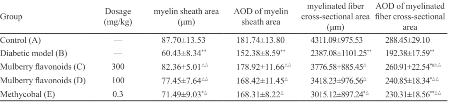
Effects of mulberry flavonoids on morphological changes of myelinated fiber of sciatic nerve
The myelin sheath area and myelinated iber
cross-sectional area were decreased in diabetic rats (group
B) compared with control rats (group A) and these changes were alleviated by mulberry lavonoids (group
C, D) and methycobal (group E). Likewise, the average optical density of myelin sheath area and myelinated
iber cross-sectional area were decreased in diabetic rats (group B) compared with control rats (group A) and these changes were also alleviated by mulberry lavonoids and
methycobal administration (P <0.05 or 0.01; Table II).
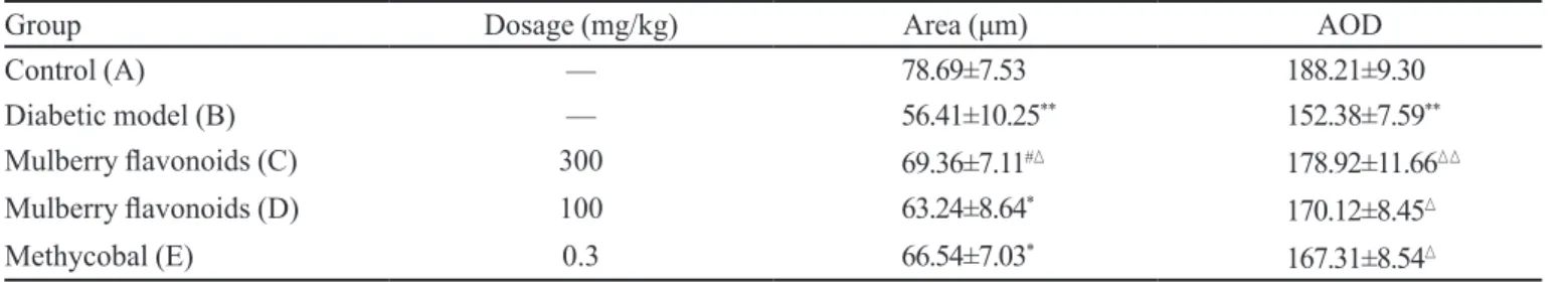
Effects of mulberry flavonoids on extramedullary fiber of sciatic nerve
In control rats, histology of sciatic nerves was
(Figure 1B), whereas the numbers of extramedullary iber
of sciatic nerves were markedly increased in group C-E
(Figures 1C, 1D and 1E), especially mulberry lavonoids
treated groups (Figure 1C). The same results can be
obtained by quantified using MIAS2000 software. In diabetic rats, the extramedullary iber area and AOD were signiicantly reduced compared with control rats. Treated with mulberry lavonoids and methycobal can alleviate
these changes (P < 0.05 or 0.01; Table III).
Effects of mulberry flavonoids on changes of ultrastructural of sciatic nerve
Ultrastructural evaluation of sciatic nerves confirmed the light microscopy findings. The sciatic nerves in control group showed normal structure
(Figure 2A). Sciatic nerve electron micrograph of diabetic rats shows shrunken axons, myelin destructions
with onion-bulb form protrusion on the myelin sheath
TABLE II - Changes of the myelin sheath area and myelinated iber cross-sectional area of rat’s sciatic nerve (–x ±s, n=10)
Group Dosage
(mg/kg)
myelin sheath area
(μm) AOD of myelin sheath area
myelinated iber
cross-sectional area
(μm)
AOD of myelinated iber cross-sectional
area
Control (A) — 87.70±13.53 181.74±13.80 4311.09±975.53 288.45±29.10
Diabetic model (B) — 60.43±8.34** 152.38±8.59** 2387.08±1101.25** 192.38±17.59**
Mulberry lavonoids (C) 300 82.36±5.01 178.92±11.66 3776.58±885.45 260.91±22.54*#
Mulberry lavonoids (D) 100 77.45±7.64 168.42±11.45 3418.23±976.56 240.85±18.34*
Methycobal (E) 0.3 71.49±9.03* 168.31±8.22 3015.12±897.24* 230.31±18.56**
*P<0.05,**P<0.01 vs group A;P<0.05, P<0.01 vs group B; # P<0.05, ## P<0.01 vs group E
FIGURE 1 - Histology of the sciatic nerve of each experimental rat by staining with chromotrope 2R-brilliant green (×200) (A)
TABLE III - Change of extramedullary iber of rat’s sciatic nerve (–x ±s, n=10)
Group Dosage (mg/kg) Area (μm) AOD
Control (A) — 78.69±7.53 188.21±9.30
Diabetic model (B) — 56.41±10.25** 152.38±7.59**
Mulberry lavonoids (C) 300 69.36±7.11# 178.92±11.66
Mulberry lavonoids (D) 100 63.24±8.64* 170.12±8.45
Methycobal (E) 0.3 66.54±7.03*
167.31±8.54
*P<0.05, **P<0.01 vs group A;P<0.05, P<0.01 vs group B; # P<0.05, ## P<0.01 vs group E
and the axonal myelins were vacuolization and lamellar
separation. Degenerative changes were also observed in
mitochondria and Schwann cells (Figure 2B). Histologic
evaluation of the tissues in animals pretreated with 0.3 g/kg mulberry flavonoids showed ultrastructural
features of myelin and axons remarkable improved and
myelin breakdown decreased markedly (Figure 2C).
Likewise, vacuolization and lamellar separation of the
FIGURE 2 - Representative transmission electron micrographs showing the ultrastructure of sciatic nerves (×7000) (A) Control
rats: intact myelinated axon. (B) Diabetic neuropathy rats: serious demyelination. (C) Diabetic neuropathy rats treated with 0.3 g/kg mulberry lavonoids: tiny segmental demyelination and local axon separation from sheaths is seen. (D) Diabetic neuropathy rats treated with 0.1 g/kg mulberry lavonoids: mild axon separation from the sheaths is seen. (E) Diabetic neuropathy rats treated with methycobal: mild axon separation from the sheaths is seen.
axonal myelin was less obvious. The ine structure of
Schwann cells was seemingly normal.
DISCUSSION
The present study is the first to demonstrate that
led to improvement of myelinated iber and extramedullary iber. Ultrastructural evaluation of sciatic nerves conirmed that mulberry lavonoids restored the axonal degeneration
and demyelination.
Alloxan is one of the usual substances used to induce diabetes mellitus. It caused pancreatic β cells suficient damage thus resulted to a notable increase in
blood glucose levels (Verma et al., 2010). In our study,
we irst evaluated the antidiabetic activity of mulberry lavonoids in alloxan-induced diabetic rats by testing fasting blood glucose level by glucose oxidase method. Our indings, in accordance with earlier studies, indicate that treatment with 0.3 g/kg mulberry lavonoids (equal
to 15-fold the clinic dosage) significantly inhibit the
alloxan-induced hyperglycemia of rats. On the other
hand, our results also indicated that 0.3 g/kg mulberry
lavonoids caused a signiicantly difference in weight
gain compared to the model group.
As the most common secondary complications of diabetes, DPN is characterized by progressive axons loss in humans (Calcutt, Backonja, 2007). Axon
demyelination and loss were also presented in the sciatic nerve of diabetic mice by electron microscopy
examination (Kang et al., 2011). The proper function
and maintenance of axons depended on myelin formation
(Lappe-Siefke et al., 2003). Consequently, myelin
loss results in pathological conditions, such as DPN.
Moreover, the axon degeneration is relation with
pathogenesis of negative signs of DPN(Thomas,
1999). As reported before (Veiga et al., 2006), the most abundant nerve abnormality observed in our study was
that both myelinated iber and extramedullary iber of the
sciatic nerve were markedly decreased in diabetic rats, as opposed to the control group.
Flavonoids extracted from Mulberry leaves (Morus
alba L.) were described for anti-diabetes effects in rats
(Singab et al., 2005) and demonstrated marked intestinal
maltase inhibitory activity (Adisakwattana et al., 2012) in vitro. In this study, we further evaluated the role of
mulberry lavonoids on diabetic peripheral neuropathy
in rat, and our morphological study of sciatic nerve revealed that administration of mulberry flavonoids, especially at the dose of 0.3 g/kg, notably increased the
area of myelinated iber and number of extramedullary iber to close to the control group. Schwann cells, which
are susceptible to hyperglycemia, play a crucial role in maintenance of peripheral nerve myelin. The sciatic nerve electron micrograph of diabetic rats in the present result shows degenerative changes of Schwann cells, onion-bulb type myelin destructions and lamellar separation.
With 0.3 g/kg mulberry lavonoids treatment, the axonal
and Schwann cells degeneration were ameliorated, too. In conclusion, we show that 0.3 g/kg mulberry
flavonoids exert a substantial protective effect against alloxan-induced diabetic neuropathy in sciatic nerve of rats. These indings provide a therapeutic potential for
future treatment of diabetic peripheral neuropathy.
ACKNOWLEDGEMENTS
This study was supported by Scientific Research
Fund of Sichuan Provincial Traditional Chinese Medicine (No. 2008-05).
REFERENCES
ADISAKWATTANA, S.; RUENGSAMRAN, T.; KAMPA, P.; SOMPONG, W. In vitro inhibitory effects of plant-based foods and their combinations on intestinal α-glucosidase
and pancreatic α-amylase. BMC Complement. Altern. Med.,
v.12, p.110, 2012.
ANDALLU, B.; VARADACHARYULU, N.C.H. Control of
hyperglycemia and retardation of cataract by mulberry
(Morus indica L.) leaves in streptozotocin diabetic rats.
Indian J. Exp. Biol., v.40, p.791-795, 2002.
CALCUTT, N.A.; BACKONJA, M.M. Pathogenesis of pain
in peripheral diabetic neuropathy. Curr. Diab. Rep., v.7,
p.429-434, 2007.
COW, A.P. The roles of oxidative stress and antioxidant
treatment in experimental diabetic neuropathy. Diabetes,
v.46, suppl.2, p.38-40, 1997.
DANDONA, P.; BOLGER, J.P.; BOAG, F.; FONESCA, V.; ABRAMS, J.D. Rapid development and progression of
proliferative retinopathy after strict diabetic control. Br. Med. J. (Clin. Res. Ed.), v.290, p.895-896, 1985.
EL-SAYYAD, H.I.; EL-SHERBINY, M.A.; SOBH, M.A.; ABOU-EL-NAGA, A.M.; IBRAHIM, M.A.; MOUSA, S.A. Protective effects of Morus alba leaves extract on ocular
functions of pups from diabetic and hypercholesterolemic mother rats. Int. J. Biol. Sci., v.7, p.715-728, 2011.
HU, J.; ZHANG, X.E. Impact of epidemic rates of diabetes on
ISMAIL-BEIGI, F.; CRAVEN, T.; BANERJI, M.A.; BASILE, J.; CALLES, J.; COHEN, R.M.; CUDDIHY, R.; CUSHMAN, W.C.; GENUTH, S.; GRIMM, R.H.J.R.; HAMILTON, B.P.; HOOGWERF, B.; KARL, D.; KATZ, L.; KRIKORIAN, A.; O’CONNOR, P.; POP-BUSUI, R.; SCHUBART, U.; SIMMONS, D.; TAYLOR, H.; THOMAS, A.; WEISS, D.; HRAMIAK, I.; ACCORD TRIAL GROUP. Effect of
intensive treatment of hyperglycaemia on microvascular
outcomes in type 2 diabetes: an analysis of the ACCORD
randomised trial. Lancet, v.376, p.419-430, 2010.
KANG, T.H.; MOON, E.; HOMG, B.N.; CHOI, S.Z.; SON, M.; PARK, J.H.; KIM, S.Y. Diosgenin from Dioscorea
nipponica ameliorates diabetic neuropathy by inducing
nerve growth factor. Biol. Pharm. Bull., v.34, p.1493-1498,
2011.
KATSUBE, T.; YAMASAKI, M.; SHIWAKU, K.; ISHIJIMA, T.; MATSUMOTO, I.; ABE, K.; YAMASAKI, Y. Effect
of lavonol glycoside in mulberry (Morus alba L.) leaf on
glucose metabolism and oxidative stress in liver in
diet-induced obese mice. J. Sci. Food Agric., v.90, p.2386-2392,
2010.
KIM, H.J.; YOON, K.H.; KANG, M.J.; YIM, H.W.; LEE, K.S.; VUKSAN, V.; SUNG, M.K. A six-month supplementation
of mulberry, korean red ginseng, and banaba decreases
biomarkers of systemic low-grade inlammation in subjects
with impaired glucose tolerance and type 2 diabetes. Evid. Based Complement. Alternat. Med., v.2012, p.735191, 2012.
LAPPE-SIEFKE, C.; GOEBBELS, S.; GRAVEL, M.; NICKSCH, E.; LEE, J.; BRAUN, P.E.; GRIFFITHS, I.R.; NAVE, K.A. Disruption of Cnp1 uncouples oligodendroglial
functions in axonal support and myelination. Nat. Genet.,
v.33, p.366-374, 2003.
N A O WA B O O T , J . ; P A N N A N G P E T C H , P. ; KUKONGVIRIYAPAN, V.; KONGYINGYOES, B.; KUKONGVIRIYAPAN, U. Antihyperglycemic, antioxidant and antiglycation activities of mulberry leaf
extract in streptozotocin-induced chronic diabetic rats. Plant
Foods Hum. Nutr., v.64, p.116-121, 2009a.
N A O WA B O O T , J . ; P A N N A N G P E T C H , P. ; KUKONGVIRIYAPAN, V.; KUKONGVIRIYAPAN, U.; NAKMAREONG, S.; ITHARAT, A. Mulberry leaf extract restores arterial pressure in streptozotocin-induced chronic
diabetic rats. Nutr. Res., v.29, p.602-608, 2009b.
OMRAN, O.M. Histopathological study of evening primrose
oil effects on experimental diabetic neuropathy. Ultrastruct.
Pathol., v.36, p.222-227, 2012.
SINGAB, A.N.; EL-BESHBISHY, H.A.; YONEKAWA, M.; NOMURA, T.; FUKAI, T. Hypoglycemic effect of Egyptian Morus alba root bark extract: Effect in diabetes and lipid
peroxidation of streptozocin-induced diabetic rats. J.
Ethnopharmacol., v.100, p.333-338, 2005.
THOMAS, P.K. Diabetic neuropathy: mechanisms and future
treatment options. J. Neurol. Neurosurg. Psychiatry, v.67,
p.277-279, 1999.
VEIGA, S.; LEONELLI, E.; BEELKE, M.; GARCIA-SEGURA, L.M.; MELCANGI, R.C. Neuroactive steroids
prevent peripheral myelin alterations induced by diabetes. Neurosci. Lett., v.402, p.150-153, 2006.
VERMA, L.; SINGOUR, P.K.; CHAURASIYA, P.K.; RAJAK, H.; PAWAR, R.S.; PATIL, U.K. Effect of ethanolic extract
of Cassia occidentalis Linn. for the management of
alloxaninduced diabetic rats. Pharmacognosy Res., v.2,
p.132-137, 2010.
Received for publication on 12th May 2013