*Correspondence: N. G. Delgado. Department of Pharmacology, Center of Marine Bioproducts (CEBIMAR), AMA, CITMA. Loma y 37, Al-turas del Vedado, Plaza de la Revolución, 10400 -Havana, Cuba. E-mail: [email protected]; [email protected]
A
vol. 49, n. 1, jan./mar., 2013
Anti-inflammatory and antinociceptive activities of methanolic
extract from red seaweed
Dichotomaria obtusata
Neivys García Delgado
1,*, Ana Iris Frías Vázquez
2, Hiran Cabrera Sánchez
3, Roberto Menéndez
Soto del Valle
1, Yusvel Sierra Gómez
4, Ana María Suárez Alfonso
51Department of Pharmacology, Center of Marine Bioproducts (CEBIMAR), Havana, Cuba, 2Department of Human and Animal
Biology, Faculty of Biology, University of Havana, Cuba, 3Pharmacology Central Laboratory, Faculty of Medical Sciences
Dr. Salvador Allende, Havana, Cuba, 4Center for Protein Studies, Faculty of Biology, University of Havana, Cuba, 5Center of
Marine Researches, University of Havana, Cuba
The aim of the present work was to investigate the anti-inlammatory and antinociceptive effects of
methanolic extract from D. obtusata using classic models in mice (croton oil-induced ear edema and acetic acid-induced writhing) and a phospholipase A2 activity test. Qualitative analysis of the chemical composition of seaweed was also determined by extraction with solvents of increasing polarity and precipitation and color tests. Results of qualitative chemical study showed the presence of lactonic and
phenolic compounds, reduced carbohydrates, other sugars, lavonoids, fatty compounds, triterpenes and steroids. The extract inhibited mouse ear edema in a dose-dependent manner with an eficacy higher than 90% and a mean effective dose of 4.87μg/ear, while intraperitoneal administration presented a moderate
activity. The extract did not inhibit phospholipase A2 activity. In the writhing test, the intraperitoneal administration of the extract showed a strong antinociceptive activity (80.2%), while the oral route showed
a lower eficacy. In conclusion, this study demonstrated the anti-inlammatory and antinociceptive effects
of methanol extract of D. obtusata in experimental models, suggesting its therapeutic potential in the
treatment of peripheral painful and/or inlammatory pathologies.
Uniterms: Dichotomaria obtusata/pharmacognosy. Dichotomaria obtusata/anti-inlammatory activity. Dichotomaria obtusata/antinociceptive activity. Mouse ear edema test. Writhing test.
O objetivo do presente trabalho foi investigar os efeitos antiinlamatórios e antinociceptivos de um
extrato metanólico de D. obtusata, utilizando modelos clássicos em ratos (teste do edema de orelha induzido por óleo de cróton e teste de contorções induzidas por ácido acético) e um teste de atividade de fosfolipase A2. A análise qualitativa da composição química das algas foi também determinada através
de extração com solventes de polaridade crescente e testes de precipitação e cor. Os resultados do estudo de química qualitativa mostraram a presença de compostos lactônicos e fenólicos, hidratos de carbono
reduzidos e outros açúcares, lavonoides, compostos graxos, triterpenos e esteroides. O extrato inibiu o edema de orelha dos ratos de um modo dependente da dose com eicácia superior a 90% e dose média efetiva de 4.87μg/orelha, enquanto a administração intraperitoneal apresentou atividade moderada. O
extrato não inibiu a atividade da fosfolipase A2. No teste de contorção, a administração intraperitoneal
do extrato mostrou forte atividade antinociceptiva (80,2%), enquanto a administração oral mostrou
menor eicácia. Em conclusão, este estudo demonstrou os efeitos antiinlamatórios e antinociceptivos
do extrato metanólico de D. obtusata em modelos experimentais, sugerindo seu potencial terapêutico
no tratamento de patologias dolorosas periféricas e/ou inlamatórias.
INTRODUCTION
Marine environment may contain over 80% of world’s species of plants and animals (Kumar, Xi-rong, 2004). Particularly, seaweeds have been of great interest for humans as marine food sources since old times (Ni-sizawa et al., 1987). Nowadays, these organisms represent a promising source of useful products awaiting discovery for the prevention or treatment of several pathologies
(Faulkner, 2002). Ecological pressures, including ight for
space and predation, have contributed to the evolution of secondary metabolites with diverse pharmacological prop-erties. In fact, the discovery of metabolites with biological
activities from seaweeds has increased signiicantly in the
past three decades (Smit, 2004).
On the other hand, inlammation is a defensive re -action of organisms to antigenic stimulation or physical injuries. This response involves the activation of complex metabolic pathways and the intervention of chemical and cellular mediators (Pruzanskim, Vadas, 1991; Abbas et al.,
2008). Nevertheless, an exacerbated response can conduce to the development of multiple pathologies, including rheumatoid arthritis, acute gout, asthma,
neurodegenera-tive diseases and cancer (Kazłowska et al., 2010).
Further-more, pain is a classic sign of this inlammatory response
(Flórez, Reig, 1994).
Pharmaceutical anti-inlammatory drugs are gener
-ally used to treat inlammation and pain. In general, these drugs reduce inlammatory response by suppressing the
production of pro-inflammatory mediators, which are
involved in the pathogenesis of inlammatory diseases
(Guslandi, 1998). Nevertheless, reports on the increased risk of digestive, cardiovascular and renal diseases with
long-term use of several non-steroidal anti-inlammatory
drugs and the serious systemic side effects of glucocor-ticoids, have raised concerns about using the drugs (Jug-dutt, 2007). To overcome this limitation, a considerable amount of researches have promoted the discovery and development of new bioactive natural products with
anti-inlammatory and antinociceptive properties which do not
show any side effects.
An increasing number of studies have demonstrated that certain extracts and compounds from seaweeds have
potential anti-inlammatory uses. For example, Ganovski
et al. in 1979 reported the anti-inlammatory effect of an
aqueous extract from Cystosina barbata, Ulva lactuca and
Zostera nona. Also, Payá et al. in 1993 studied the
anti-inlammatory potential of methanol and dichloromethane
extracts of seven species of seaweeds. A group of inhibitors of phospholipase A2 were isolated from different microalgae (Mayer et al., 1993). Recently, other metabolites with
anti-inlammatory activity have been obtained from seaweeds,
including a glycosterol (Awad, 2000), a phlorotannin (Sugi-ura et al., 2006), polyphenols (Jung et al., 2009; El Gamal, 2010) and polysaccharides (Ananthi et al., 2010). Moreover, various seaweeds have antinociceptive properties (Anca et al., 1993; Guzmán et al., 2001; Viana et al., 2002).
Dichotomaria obtusata (J. Ellis, Solander) Lamarck, a red seaweed found in tropical and subtropical shores, is one of the most common species of the phylum Rhodophyta (Suárez, 2005). This species has been used in a small
number of biological studies. We have recently reported the intraperitoneal anti-inlammatory and antinociceptive
properties of aqueous extract of D. obtusata in two classic animal models (Frías et al., 2011). However, anti-inlam
-matory eficacy of the aqueous extract was limited and the
mechanisms of action have not been examined yet. In order
to obtain a greater eficacy, a new extract of this seaweed
was prepared. Therefore, the aim of the current study was to
evaluate the anti-inlammatory and antinociceptive potential
of methanolic extract of D. obtusata employing different routes of administration. Before that, we have carried out a preliminary acute toxicological study of our extract with the purpose of knowing the safety of the working material. Also, one of the possible mechanisms of action of bioactive components of the extract was investigated in an in vitro
assay. We present novel anti-inlammatory and antinocicep -tive activities of red seaweed D. obtusata.
MATERIALS AND METHODS
Drugs and reagents
Tested drugs (indomethacin, dexamethasone, ace-tylsalicylic acid) and reagent (croton oil) were purchased from Sigma Chemicals.
Material
The red alga Dichotomaria obtusata was collected at Jaimanitas Beach, Havana, in November 2008, and
was identiied by Dr. Ana María Suarez, researcher of the
Center of Marine Researches, University of Havana. A voucher specimen was deposited at the Herbarium from Center of Marine Researches under the acquisition number r-189. Specimens were washed with common water, dried at room temperature and kept at 4 °C until the obtaining of the methanolic extract.
Compositional analysis of D. obtusata
of D. obtusata was conducted according to Chabra’s meth-od (Chabra et al., 1984), which is based on extraction with solvents of increasing polarity (dichloromethane, ethanol and water) and tests of precipita tion and color solutions.
Preparation of the methanolic extract
Specimens of D. obtusata (50 g) were dissolved in 500 mL of methanol and soaked at room temperature during 8 hours. The methanol extract was concentrated in a rotary evaporator (BUCHI, Model B48C) and stored at 4 °C until its use.
Animals
All animal experiments were conducted according to the Guide for the Care and Use of Laboratory Animals of the Center for Laboratory Animals Production (CEN-PALAB), Cuba. For one week before the experiments, male Cenpalab mice: OF-1 weighing 23-25 g were main-tained in ventilated plastic cages in a soundproofed room
at 22 °C, with an artiicial 12:12 h light:dark cycle. Food
and sterile water were supplied ad libitum. Animals were randomized into treatment groups and deprived of food and water during the experiment.
Acute Toxicity Study
The acute toxicity study of the methanolic extract
of D. obtusata was carried out according to the
acute-toxic-class method as alternative to the LD50 test (Schlede
et al., 1992). Graded doses (25, 200 and 2000 mg/kg) of
methanolic extract were administered intraperitoneally to various groups, each group containing six mice. On the
irst hour after administration, the animals were evaluated
every 10 minutes for any changes in respiratory frequency, writhing, piloerection, spontaneous motor activity, etc. Animals were observed up to 24 hours for any mortality.
Mouse ear edema test
All procedures of mouse ear edema bioassay were carried out following the established method of Tubaro et al. (1986). Edema was induced in right ear of each mouse by the topical application, to both the inner and outer sur-faces, of 10 µL of a 0.47% croton oil solution in acetone
to induce inlammation, and the same volume of acetone
was applied to the left ear. Methanolic extract dissolved in ethanol was administered topically (0.5 x 10⁻³, 1 x 10⁻³,
1.5 x 10⁻³, 7.5 x 10⁻³, 15 x 10-3, 30 x 10-3, 0.5, 1 and 2 mg/ear) and intraperitoneally (i.p.) (12.5, 25, 50 and 100 mg/kg)
simultaneously and 30 min before croton oil applica-tion, respectively. As positive controls for comparative purposes, other groups of animals were treated with
in-domethacin (0.5 mg/ear and 10 mg/kg) dissolved in 5%
NaHCO3 and dexamethasone (0.1 mg/ear and 0.5 mg/kg),
following the same conditions of extract administration. Five hours after croton oil application, the animals were
sacriiced by cervical dislocation and the ears were cut off.
In all experiments, a disk was cut from the middle part of each ear using a punch 7 mm in diameter. The edema size was determined in relation to the weight of untreated left ear and percentage of inhibition was calculated using the following expression:
% Inhibition = (ΔPc - ΔPt)/100 x ΔPc
where: ΔPc → mean weight variation in the control group; ΔPt → mean weight variation in the treated group.
Phospholipase A2 activity test
The ability of methanolic extract to inhibit phospho-lipase A2 was evaluated according to the indirect radial
hemolysis in agar plate method proposed by Gutiérrez
et al., in 1988. A Petri plate was prepared with agar con-taining egg yolks (substrate of phospholipids), calcium chloride (enzymatic cofactor), sodium azide (preserva-tive) and human erythrocytes for visualizing the effect.
Methanolic extract (2.5, 5 and 10 mg/mL) was dissolved
in PBS (0.12 M NaCl, 0.04 M NaHPO42-, pH 7.2) and
ap-plied by triplicate in the holes (6-mm diameter) made in the plate. The phospholipase A2 puriied from sea anemone
Condylactis gigantea was used as control. This enzyme
was donated by the Center for Protein Studies, University of Havana. The plate was maintained at 37 ºC by 12 hours. The enzymatic activity was determined by the presence of halos as an indicative of hemolysis.
Acetic acid-induced writhing test
The acetic acid-induced writhing test was conducted as originally described by Koster et al in 1959. Mice were randomly distributed in control and test groups of ten animals each. In this test, an intraperitoneal injection of
acetic acid (0.8%, 10 ml/kg b.w.) was given 30 and 60 min after the i.p. (12.5, 25, 50 and 100 mg/kg) and oral (100, 200, 400 and 800 mg/kg) administrations of test extract,
respectively. Also, the reference drug acetylsalicylic acid
(68 mg/kg) was administrated by oral route 1 hour before
writhing movements in the mice was counted for 20 min. The antinociceptive activity of the methanolic extract was expressed as percentage of pain reduction in treated mice with respect to control, according to the relation:
% Reduction = (ΔCc - ΔCt)/100 x ΔCc
where: ΔCc → mean of number of writhes of control group (vehicle-injected animals); ΔCt → mean of number of
writhes in the treated group.
Statistical analysis
Data are expressed as mean ± SEM. One way analy-sis of variance (ANOVA) was applied for the analyanaly-sis of results using software packages Statistica (version 7.0) and Past (version 1.99). P-values less than 0.05 and 0.001
were considered signiicantly different for Tukey-Kramer
and Bonferroni multiple comparison tests, respectively.
RESULTS
Compositional analysis of D. obtusata
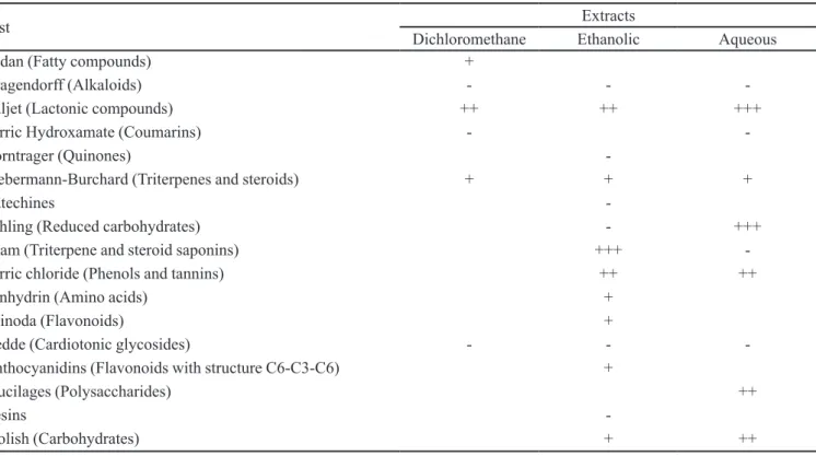
The results of the qualitative chemical study of D. obtusata (Table I) showed the notable presence of lactonic
and phenolic compounds, reduced carbohydrates and other
sugars. However, the presence of lavonoids, fatty com -pounds, triterpenes and steroids was low.
Acute Toxicity Study
Acute toxicity results of the intraperitoneal adminis-tration of methanolic extract are presented in Table II. As can be appreciated, no mortality of animals was observed after 24 hours of administration of extract at 25, 200 and
2000 mg/kg. The most signiicant signs of toxicity were
swing and writhing, which were presented at all doses. Swing showed a percentage of appearance superior at 65%. Also, the periods of latency of appearance of these
signs were dose-dependent at doses of 25 and 200 mg/kg. However, this parameter decreased at 2000 mg/kg. In
addition, the stoop of back train was observed at doses of
200 mg/kg (33.3%) and 2000 mg/kg (66.7%). Finally, a
sedative effect (33.3% of appearance and 75 sec of latency) and the increase of respiratory frequency (100% of ap-pearance and 56 sec of latency) were observed in animals administered with the maximum dose of extract.
Croton oil-induced ear edema test
Table III shows the anti-inflammatory effect of
TABLE I - Qualitative phytochemical analysis of Dichotomaria obtusata
Test Extracts
Dichloromethane Ethanolic Aqueous
Sudan (Fatty compounds) +
Dragendorff (Alkaloids) - -
-Baljet (Lactonic compounds) ++ ++ +++
Ferric Hydroxamate (Coumarins) -
-Borntrager (Quinones)
-Liebermann-Burchard (Triterpenes and steroids) + + +
Catechines
-Fehling (Reduced carbohydrates) - +++
Foam (Triterpene and steroid saponins) +++
-Ferric chloride (Phenols and tannins) ++ ++
Ninhydrin (Amino acids) +
Shinoda (Flavonoids) +
Kedde (Cardiotonic glycosides) - -
-Anthocyanidins (Flavonoids with structure C6-C3-C6) +
Mucilages (Polysaccharides) ++
Resins
-Molish (Carbohydrates) + ++
TABLE II - Percentage of mortality and signs of toxicity observed in OF-1 mice after i.p. administration of methanolic extract from
D. obtusata (n=6 animals/dose)
Doses (mg/kg) Mortality (%) Signs of Toxicity Appearance of signs (%) Latency (seconds; mean ± SEM)
25 0 Swing 66.7 58,8 ± 12,6
Writhing 83.3 73,2 ± 6,1
200 0 Swing 100 21,8 ± 2,0
Writhing 100 39,2 ± 4,9
Stoop of back train 33.3 45,5 ± 0,5
2000 0 Swing 66.7 27,5 ± 8,0
Writhing 50 51,7 ± 4,4
Stoop of back train 66.7 31,5 ± 5,3
Increase of respiratory frequency
100 55,8 ± 17,3
Sedative effect 33.3 75,0 ± 45,0
TABLE III - Effect of topical administration of methanolic extract from D. obtusata on croton oil-induced ear edema in mice(n=6
animals/doses)
Doses (mg/ear) Edema (mg)
(mean ± SEM)
Edema reduction (%)
Control 18.25 ± 0.23
0,5×10⁻³ 18.37 ± 1.03* 0
1×10⁻³ 17.62 ± 0.85** 3.45
1,5×10⁻³ 15.52 ± 0.72** 14.96
7,5×10⁻³ 9.32 ± 0.52** 48.93
0.015 8.05 ± 0.97** 55.89
0.03 5.02 ± 0.60** 72.49
0.5 2.83 ± 0.65** 84.49
1 1.38 ± 0.43** 92.44
2 3.2 ± 0.18** 82.47
Dexamethasone (0.1) 2.17 ± 0.26* 88.11
Indomethacin (0.5) 7.12 ± 0.60* 60.99
* p < 0.0001 compared with control; **p < 0.0001 compared with indomethacin and dexamethasone (Bonferroni’s test)
topical administration of methanolic extract (0.5 x 10⁻³,
1 x 10⁻³, 1.5 x 10⁻³, 7.5 x 10⁻³, 15 x 10-3, 30 x 10-3, 0.5, 1 and 2 mg/ear) on croton oil-induced ear edema in mice. The ear edema in control group, ive hours after croton
oil application, was 18.25 ± 0.23 mg. All doses of extract
decreased signiicantly (p<0.0001) the action of the irritat -ing agent except the dose of 0.5 x 10⁻³ mg/ear.
As can be seen, ive of the evaluated doses exerted
a significant inhibition of edema (> 50%). The extract
at 1 mg/ear showed the greatest inhibition of edema
(92.44%), which was similar to reference drug
dexa-methasone (88.11% at 0.1 mg/ear) and far superior to indomethacin (60.99% at 0.5 mg/ear).
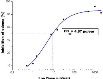
Methanolic extract showed a dose-dependent
anti-inlammatory effect. Figure 1 illustrates the dose-response
relation of topical application of extract. The behavior of curve indicates that the apparent maximum dose is
1 mg/ear. Also, the Mean Effective Dose (dose inducing
a 50% edema inhibition, ED50) was calculated, which was 4.87 µg/ear.
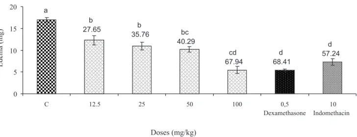
In the same way, the anti-inlammatory activity of
extract was evaluated using the systemic administration (i.p.). The results obtained at different doses of extract
FIGURE 1 - Dose-response relation of topical application of
(12.5, 25, 50 and 100 mg/kg) are shown in Figure 2. All groups of treatment were able to decrease signiicantly
(p < 0.05) the croton oil-induced ear edema compared to control group (which received 0.9% saline solution). Simi-lar to topical application, the i.p. administration of extract inhibited this edema in a dose-dependent manner. The
maximum inhibition was 67.94% at dose of 100 mg/kg,
which was not different from response of reference drugs indomethacin (57.24%) and dexamethasone (68.41%).
Phospholipase A2 activity test
One of the probable mechanisms of action of
anti-inlammatory compounds is mediated by the inhibition of
phospholipase activity. For this reason, methanolic extract of D. obtusata was tested for a possible inhibitory activity of phospholipase A2, using the indirect radial hemolysis
in agar plate method. The effect of extract (2.5, 5 and
10 mg/mL) in this assay is shown in Figure 3. The pres -ence of halos of hemolysis around the holes of the plate, where extract and control phospholipase A2 were applied,
indicates the extract did not inhibit the enzymatic activity. Phospholipase A2 hydrolyzes the phospholipids of egg
yolk, which are liberated into medium as fatty acids and glycerophospholipids. After that, the pH of culture me-dium decreases and this promotes the lysis of erythrocytes and the appearance of halos of hemolysis.
Acetic acid-induced writhing test
Antinociceptive activity of methanolic extract was examined using writhing test. Intraperitoneal
administra-tion of the extract at the doses of 12.5, 25, 50 or 100 mg/kg
body weight decreased significantly (p < 0.0001) the action of acetic acid used to induce writhes in mice, in comparison to the control animals that received only 0.9% saline solution.
The effects of i.p.-administered extract were dose-dependent. There results can be seen in Figure 4.
Treat-ment with 12.5 mg/kg reduced the number of writhes
in 47.72%, which was further decreased in 80.2% with
100 mg/kg of the extract. The reference analgesic drug, acetylsalicylic acid (68 mg/kg) caused inhibition of
writhes in 49.49%.
The results of the acetic acid-induced writhing responses in mice one hour after oral administration of
FIGURE 2 - Effect of intraperitoneal administration of methanolic extract from D. obtusata on croton oil-induced ear edema in mice(n=6 animals/dose).
FIGURE 3 - Effect of methanolic extract from D. obtusata (10,
FIGURE 4 - Effect of intraperitoneal administration of methanol extract from D. obtusata on acetic acid-induced writhing in mice
(n=10 animals/dose). ASA: ace tylsalicylic acid (reference drug).
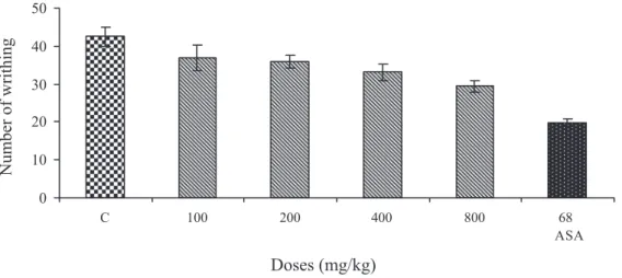
FIGURE 5 - Effect of oral administration of methanol extract from D. obtusata on acetic acid-induced writhing in mice (n=10
animals/dose). ASA: ace tylsalicylic acid (reference drug).
the extract are presented in Figure 5. In this case, only
the maximum dose of the extract (800 mg/kg) had a clear antinociceptive effect, with a signiicant reduction
of the number of writhes (p<0.05) compared to control animals. The percentage of inhibition of writhes at this dose was 30.87%, which was lower than the reference drug (53.25%).
DISCUSSION
The purpose of this paper was to evaluate
anti-inlammatory and antinociceptive effects of methanolic
extract from the red seaweed Dichotomaria obtusata using classic models in mice and an in vitro test.
Red seaweeds are considered the most important source of many biologically active me tabolites in
com-parison to other algal classes (El Gamal, 2010). As a irst
step toward identifying the major chemical groups present
in D. obtusata, we have developed a qualitative chemical study of its composition. Results showed the presence of lactonic and phenolic compounds, flavonoids, fatty compounds, triterpenes, steroids, reduced carbohydrates and other sugars. In agreement with our results, Frías et al. in 2011 demonstrated the presence of carbohydrates, proteins, triterpenes and steroids in the aqueous extract from D. obtusata. Some of these compounds, such as phenols, terpenes, polysaccharides and steroids, have been
reported to possess anti-inlammatory and antinociceptive
effects (Silva, Scheuer, 1980; Chong, Parish, 1985; Awad, 2000; Lucas et al., 2003; Rodríguez et al., 2004; Jung et al., 2009).
alternative acute toxicity test. The animals intraperitone-ally administered showed some signs of toxicity (swing and writhing) at all tested doses. Other signs appeared at the highest dose (increased respiratory frequency and sed-ative effect). However, the animals in all groups survived, signifying the absence of lethality under our experimental conditions. This extract was much less toxic than aque-ous extract from the same seaweed (unpublished data), suggesting that the majority of toxic metabolites from D. obtusata are of polar nature.
In a irst set of pharmacological experiments, we
investigated the effects of the extract on croton oil-induced ear edema using i.p. and topic single administrations. The extract dose dependently reduced edema formation and showed potency similar to that of reference drugs by topi-cal route. In fact, its maximum effect was superior to 90% and its ID50 value was 4.87 µg/ear. Taking into account that
the extract is a complex mixture of several components,
these values indicate its elevated pharmacological eficacy
and potency.
Topical application of croton oil produced a
longer-lasting edema associated with marked inlux of neutrophils and predominant formation of LTB4 along with signiicant
changes in levels of TXB2. Also, earlier studies have re-vealed that histamine, serotonin and prostaglandins could play a role in the development of edema induced by this irritant agent (Gabor; Razga, 1990; Blazso, Gabor, 1994). The advantages of this model include its good predictive
value for screening topical anti-inlammatory activity and
its sensitivity to both steroidal and non-steroidal drugs. Nevertheless, this sensitivity is dependent on the time course of the response (Tubaro et al., 1986).
Although the extract intraperitoneally administered was able to reduce the croton oil-induced ear edema at all
evaluated doses, the eficacy was lower than that of topical
route. Here, bioavailability factor might also be involved, because after topical application a large concentration of the extract could be available to the target tissues whereas this could be limited with the systemic administration. This is in agreement with previous studies of our group on the aqueous extract of this seaweed, which demonstrated a
maximum anti-inlammatory activity greater than 65% in
TPA-induced ear edema in mice (Frías et al., 2011). The results obtained in this acute inflammation
model and the topical eficacy of the extract compared to
dexamethasone suggest an interaction with metabolites of the arachidonic acid pathway, mediated mainly by phospholipase A2 (PLA2) and cyclooxygenase (COX) enzymes (Chen et al., 1994). Thus, the anti-inlammatory
mechanism of action of the active components of the extract would involve PLA2. For this reason we tested
the ability of the extract to inhibit PLA2 activity by the indirect radial hemolysis in agar plate method. However, this study showed absence of inhibitory effect on secretor PLA2 activity of C. gigantea. These results partially agree with previous studies on the inhibitory activity of PLA2 of various extracts from seaweeds, revealing inhibitory activity only in the extract of Dictyota dentata (Llanio et al., 1998). In contrast, Mayer, in 1993, isolated 12 com-pounds from seaweeds with powerful inhibitory activity of PLA2 from bee venom. Our results indicate that this study must be extended to other secretor PLA2, including PLA2 from mammals.
We also investigated the antinociceptive activity of
the extract in the acetic acid-induced writhing test in mice. The abdominal constriction response induced by acetic acid has been mainly used as screening tool for the
assess-ment of analgesic or anti-inlammatory properties of new agents as well as a typical model for visceral inlammatory
pain (Tjølsen, Hole, 1997). The local irritation provoked by a test agent in the intraperitoneal cavity triggers a diversity of mediators, such as bradykinin, substance P and prostaglandins, particularly PGI2, as well as some cytokines such as IL-1β, TNF-α and IL-8 (Correa et al., 1996; Ikeda et al., 2001). These mediators activate chemo-sensitive nociceptors that contribute to the development
of inlammatory pain.
The methanolic extract inhibited signiicantly the
number of writhes suggesting that its antinociceptive ef-fect could be related to inhibition of mediators released in response to acetic acid. Nevertheless, the intraperito-neal route was more effective than oral at inhibiting this
response at all evaluated doses (12.5-100 mg/kg), while only the maximum oral dose (800 mg/kg) reduced the
abdominal constrictions. This result may be due to the lim-ited bioavailability of the active constituents of the extract
provoked by a poor absorption or the irst-pass effect in
liver. According to the test employed, this antinociceptive
effect is probably of peripheral origin. We have previously
demonstrated the antinociceptive activity of aqueous ex-tract of this seaweed in the same test Frías et al. (2011), although in that study we obtained an effect more potent using oral administration. Inhibition of number of writhes in extracts and components obtained from seaweeds were also demonstrated by Guzmán et al. (2001), Viana et al., (2002) and Frías et al. (2011).
In conclusion, it can be suggested that the methano-lic extract from D. obtusata has powerful topical
anti-in-lammatory and antinociceptive activities. These observed
and to determine the possible mechanism of actions of these compounds and their potential for therapeutic use
in the treatment of inlammatory and pain pathologies.
REFERENCES
ABBAS, A.K.; LITCHMAN, A.H.; PILLAI, S. Inmunología
básica y clínica. Philadelphia: Saunders Elsevier, 2008. 215 p.
ANANTHI, S.; BALAJI, H.R.; GOPALAN, A.; GAVATHRI,
V.; RAMAKRISHNAN, G.; VASANTHI, H.R. In vitro
antioxidant and in vivo anti-inlammatory potential of crude
polysaccharide from Turbinaria ornate (Marine Brown
Alga). Food Chem. Toxicol., v.48, p.187-192, 2010.
ANCA, J.M.; LAMELA, M.; CALLEJA, J.M. Activity on the central nervous system of Himanthalia elongata. Planta Med., v.59, n.3, p.218-220, 1993.
AWAD, N.E. Biologically active steroid from the green alga
Ulva lactuca. Phytother. Res., v.14, n.8, p.641-643, 2000.
BLAZSO, G.; GABOR, M. Anti-oedematous action of some Hi-receptor antagonists. Agents Actions, v.42, p.13, 1994.
CHEN, I.L.; GERWICK, W.H.; SCHATZMAN, R.; LANEY,
M. Isorawsonol and related IMO dehydrogenase inhibitors from the tropical alga Avrainvillea rawsoni. J. Nat. Prod., v.57, p.947-952, 1994.
CHONG, A.S.F.; PARISH, C.R. Non-immune lymphocyte macrophage interaction II. Evidence that the interaction
involves sulfated polysaccharide recognition. Cell
Immunol., v.92, n.2, p.277-289, 1985.
CORREA, C.R.; KYKE, D.J.; CHAKRAVERTY, S.; CALIXTO,
J.B. Antinociceptive profile of the pseudopeptide B2 bradykinin and receptor antagonist NPC 18688 in mice. Br. J. Pharmacol., v.117, n.3, p.552-558, 1996.
EL GAMAL, A.A. Biological importance of marine algae.
Saudi. Pharm. J., v.18, n.1, p.1-25, 2010.
FAULKNER, D.J. Marine natural products. Nat. Prod. Rep.,
v.19, p.1-48, 2002.
FLOREZ, J.; REIG, E. Terapéutica farmacológica del dolor.
Pamplona: Ediciones Universidad de Pamplona, 1994. 331 p.
FRIAS, A.I.; DUTOK, C.M.; GARCIA, N.; SUAREZ, A.M.;
SANTOS, Y.; CABRERA, H. Anti-inflammatory and
analgesic activities of red seaweed Dichotomaria obtusata.
Braz. J. Pharm. Sci., v.47, n.1, p.111-118, 2011.
GABOR, M.; RAZGA, Z. Effects of non-steroidal antiphlogistics on mouse ear oedema induced with dithranol. Acta Physiol.
Hung., v.75, p.287, 1990.
GANOVSKI, K.H.; SHIPOCHLIEV, T.; BRATOVA, K.
Anti-inlammatory action of extracts from marine algae collected
in the area of Burgas seacoast. Vet. Med. Nauki, v.16, n.7, p.54-61, 1979.
GUSLANDI, M. Nitric oxide and inlammatory bowel diseases.
Eur. J. Clin. Invest., v.28, p.904-907, 1998.
GUTIERREZ, J.M.; ÁVILA, C.; ROJAS, E.; CARDAS, L. An alternative in vitro method for testing the potency of polyvalent antivenom produced in Costa Rica. Toxicon., v.26, n.3, 1988.
GUZMÁN, S.; GATO, A.; CALLEJA, J.M. Antiinlammatory,
analgesic and free radical scavenging activities of the marine
microalgae Chlorella stigmatophora and Phaeodactylum
tricornutum. Phytother. Res., v.15, n.3, p.224-230, 2001.
IKEDA, Y.; UENO, A.; NARABA, H.; OH-ISHI, S. Involvement
of vanilloid receptor VR1 and prostanoids in the acid-induced writhing responses of mice. Life Sci., v.69, n.24, p.2911-2919, 2001.
JUGDUTT, B.I. Cyclooxygenase inhibition and adverse remodeling during healing after myocardial infarction.
Circulation, v.115, p.288-291, 2007.
JUNG, W.; CHOI, I.; OH, S.; PARK, S.W.; SEO, S.K.; LEE, S.W.; LEE, D.S.; HEO, S.J.; JEON, Y.J.; JE, J.Y.; AHN,
C.B.; KIM, J.S.; OH, K.S.; KIM, J.M.; MOON, C.; CHOI,
I.W. Anti-asthmatic effect of marine red alga (Laurencia undulata) polyphenolic extracts in a murine model of asthma. Food Chem. Toxicol., v.47, n.2, p.293-297, 2009.
KAZLOWSKA, K.; HSU, T.; HOU, CH.-CH.; YANG, J.-CH.;
TSAI, G.-J. Anti-inflammatory properties of phenolic compounds and crude extract from Porphyra dentata. J. Ethnopharmacol., v.128, p.123-130, 2010.
KUMAR, R.; ZI-RONG, X. Biomedical Compounds from Marine organisms. Mar. Drugs, n.2, p.123-146, 2004.
LLANIO, M.; FERNANDEZ, M.D.; CONCEPCION, A.R.; MUSTELIER, E.; CABRERA, B. Pesquisaje de
propiedades antiinlamatorias y analgésicas en extractos
de origen marino de Cuba. Rev. Cubana Plant. Med., v.3, n.2, p.69-71, 1998.
LUCAS, R.; GIANNINI, C.; D’AURIA, M.V.; PAYÁ,
M. Modulatory effect of bolinaquinone, a marine sesquiterpenoid, on acute and chronic inflammatory processes. J. Pharmacol. Exp. Ther., v.304, n.3, p.1172-1180, 2003.
MAYER, A.M.S.; PAUL, V.J.; FENICAL, W.; NORRIS, J.N.;
DE CARVALHO, M. S.; JACOBS, R.S. Phospholipase A2
inhibitors from marine algae. Hidrobiología, v.260/261,
n.???, p.521-529, 1993.
NISIZAWA, K.; NODA, H.; KIKUCHI, R.; WATAMABA, T.
The main seaweed foods in Japan. Hydrobiol. J., v.151/152,
n.1, p.5-29, 1987.
PAYÁ, M.; FERRÁNDIZ, M.L.; SANZ, M.J.; BUSTOS, G.;
BLASCO, R.; RIOS, J.L.; ALCARAZ, M.J. Study of the antioedema activity of some seaweed and sponge extracts from the mediterranean coast in mice. Phytother. Res., v.7, p.159-162, 1993.
PRUZANSKI, W.; VADAS, P. Phospholipase A2-a mediator between proximal and distal effectors of inflammation.
Immunol. Today, v.12, n.5, p.143-146, 1991.
RODRÍGUEZ, I.I.; SHI, Y.P.; GARCIA, O.J.; RODRIGUEZ, A.D.; MAYER, A.M.; SANCHEZ, J.A.;
ORTEGA-BARRIA, E.; GONZALEZ, J. New pseudopterosin and secopseudopterosin diterpene glycosides from two Colombian isolates of Pseudopterogorgia elisabethae and their diverse biological activities. J. Nat. Prod., v.67, n.10, p.1672-1680, 2004.
SCHLEDE, E.; MISCHKE, V.; ROLL, R.; KAYSER, D. A
national validation study of the acute-toxic-class method as alternative to the LD50 test. Arch. Toxicol., v.66, p.455-70, 1992.
SILVA, E.D.; SCHEUER, P.J. Manoalide, an antibiotic
sesterterpenoid from the marine sponge Luffariella
variabilis (Poleajaeff). Tetrahedron Lett., v.21, n.17, p.1611-1614, 1980.
SMIT, A.J. Medicinal and pharmaceutical uses of seaweed natural products: a review. J. Appl. Phycol., v.16, p.245-262, 2004.
SUÁREZ, A.M. Lista de Macroalgas Marinas. Rev. Invest. Mar., v.26, n.2, p.93-148, 2005.
SUGIURA, Y.; MATSUDA, K.; YAMADA, Y.; NISHIKAWA, M.; SHIOYA, K.; KATSUZAKI, H.; IMAI, K.;
AMANO, H. Isolation of a newanti-allergic phlorotannin, phlorofucofuroeckol-B, from an edible brown alga Eisenia arborea. Biosci. Biotech. Biochem., v.70, p.2807-2811, 2006.
TJOLSEN, A.; HOLE, K. Animal models of analgesia. In: The
pharmacology of pain. DICKENSON, A.; BESSON, J. (Eds.). Berlín: Verlag, 1997. v.130, p.1-20.
TUBARO, A.; DRI, P.; MELATO, M.; MULAS, G.; BIACHINI, P.; DEL NEGRO, P.; DELLA-LOGGIA, R. In the croton oil
ear edema test the effects of non-steroidal anti-inlammatory
drugs (NSAIDs) are dependent on the dose irritant. Agents Actions, v.19, p.371-373, 1986.
VIANA, G.S.B.; FREITAS, A.L.P.; LIMA, M.M.L.; VIEIRA, L.A.P.; ANDRADE, M.C.H.; BENEVIDES, N.M.B. Antinociceptive activity of sulfated carbohydrates from the red algae Bryothamnion seaforthii (Turner) Kutz. and
B. triquetrum (SG Gmel)M. Howe. Braz. J. Med. Biol. Res., v.35, n.6, p.713-722, 2002.