Serviço de Neurocirurgia do Hospital da Força Aérea do Galeão, Rio de Janeiro RJ, Brasil (HFAG):1Assistente do Serviço de
Neuro-c i ru rgia do HFAG;2Graduação em Medicina, Universidade Federal do Rio de Janeiro (UFRJ); 3Chefe do Serviço de Neuro c i ru rgia do
HFAG, Professor Adjunto da Disciplina de Neurocirurgia da Universidade Federal Fluminense (UFF).
Received 13 July 2004, received in final form 17 December 2005. Accepted 9 March 2005.
Dr. José Alberto Landeiro M.D - Rua Conde de Bomfim 211 / 310 - 20520-050 Rio de Janeiro RJ - Brasil. E- mail: [email protected]
THE HISTORY OF SPINAL SURGERY FOR DISC DISEASE
An illustrated timeline
Igor de Castro
1, Daniel Paes dos Santos
2,
Daniel de Holanda Christoph
2, José Alberto Landeiro
3ABSTRACT - This article presents the evolution in medical history which leads to the surgical treatment for ru p t u red discs. Only at the last century the precise diagnosis of a ru p t u red lumbar disc could be made after t remendous eff o rts of the many medical pioneers in the study of the spine. The experience gained with the lumbar spine was rapidly transferred to the cervical spine. We describe the evolution of the clinical and surgical aspects about ru p t u red discs in the lumbar and cervical spine. An illustrative timeline of the major events re g a rding the surgical treatment for ruptured disks is outlined in a straight forw a rd manner. Our understandings of the relation between symptoms and signs and of that between anatomy and patho-physiology have led to more successful surgical treatment for this disease. Nowadays lumbar and cervical discectomies are the most frequent operations carried out by neurosurgeons. Our current care of patients with this kind of spinal disorders is based on the work of our ancient medical heroes.
KEY WORDS: history, disc disease, spine.
A história da ciru rgia da coluna vertebral aplicada à doença discal: uma linha do tempo ilustrada
RESUMO - Esse artigo apresenta a evolução da história médica que nos conduziu ao tratamento cirúrgico da doença discal. Somente no século passado o diagnóstico preciso de ruptura de disco lombar pode ser feito, após os esforços de vários pioneiros no estudo da coluna vertebral. A experiencia obtida no estudo da coluna lombar foi rapidamente transferida para coluna cervical. Uma revisão ilustrada dos principais eventos relacionados ao tratamento cirúrgico do disco roto na coluna lombar e cervical é apresentada de f o rma objetiva. Nosso conhecimento sobre a relação entre os sinais e sintomas, da anatomia e fisiopatolo-gia levaram ao tratamento cirúrgico mais eficaz das lesões discais. O tratamento atual dessas anorm a l i-dades da coluna vertebral é fruto do trabalho de verdadeiros médicos e heróis.
PALAVRAS-CHAVE: história, doença do disco, coluna vertebral.
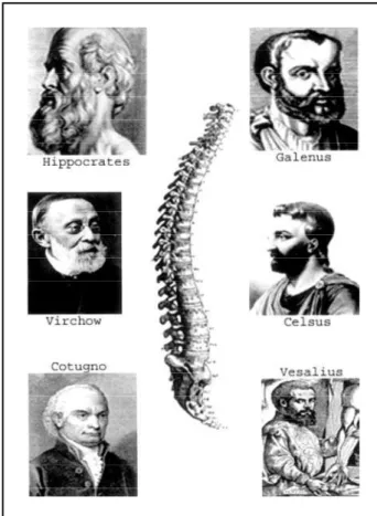
The search for cures to health problems such as spinal disorders most likely dates to the begin-ning of human history. A review of early Egyptian (1550 BC), Greek, Roman, and Arabic texts on med-icine reveal an ongoing interest in treating spinal d i s o rd e r s1. Hippocrates (circa 460-370 BC) was pro b-able the first to mentioned sciatica and low-back pain. He also was the first to correlate that the in-j u ry to the vertebra is related to limb paralysis and made a remarkable observation for that time: that paralysis is always on the same side as the lesion of the cord. For those reasons Hippocrates is con-s i d e red for con-some authorcon-s acon-s the “Father of the con- spi-ne surgery”2(Fig 1).
In the first century AD, Aulus Aurelius Corn e l i u s Celsus (25 BC-AD 50) noted death quickly followed when the spinal injury involved the cervical area. More than this he was the first to recognize that the effects of spinal injury were mediated thro u g h injury of the spinal cord3(Fig 1).
sec-702 Arq Neuropsiquiatr 2005;63(3-A)
ond thoracic segment affected the diaphragm and the muscles supplied by the intercostal nerves pro-g ressively less. He anatomically correctly conclud-ed that the arm remaisn intact when there is a le-sion at the second thoracic vertebra. For the rea-sons mentioned above some authors claim that Galen was the pioneer of spine research4(Fig 1).
In the 4t hC e n t u ry AD, Caelius Aurelianus made the first clinical description of sciatica. He did also the association with heavy lifting, described the radiation of pain to buttock and leg, and the mus-cle wasting in advanced cases. Medical writers of the We s t e rnRoman Empire, used to consider him the greatest Greco-Roman physician after Galen. Caelius probably practiced and taught in Rome and is now thought to rank second only to the physician Celsus as a Latin medical writer. His most famous work is theDe morbis acutis et chro n i c i s5. A n d reas Vesalius (1514-1564) was the first to des-cribe the interv e rtebral disc.“De humani Corporis
Fabrica”(1543) had a plate depictions of the spinal column and the interv e rtebral disc spaces5 , 6( F i g 1). After Domenico Cotugno (1736-1822) mentio-ned sciatica as a clinical entity, related the pain in the leg to disease of the sciatic nerve, and pub-lished in his monograph,De ischiade nervosa com -m e n t a r i u s ,sciatica was known as Cotugno’s dis-ease for many years1,6(Fig 1).
Although early physicians such as Hippocrates and Galen attempted to correlate the level of in-j u ry with the neurological deficit of the trauma victim, localization was not of primary concern un-til almost the 18t hc e n t u ry, when paralysis of the lower extremeties was correlated with spinal cord dysfunction. Giovanni Morgagni (1682-1771), the father of modern pathological anatomy, coment-ed on the paralysis of the lower extremeties pro-duced by intraspinal growths placing pre s s u reon the cord. The cases of Cowper and Saltzmann to which Morgagni refers were probably examples of Pott’s disease and not actually tumors1,6.
The lumbar disc
A.G. Smith was the first to perform a laminec-tomy in 18295,6in the United States probably the first description of a traumatic ru p t u re of an intervertebral disc was made by Rudolf Vi rc h o w ( 1 8 2 1 -1902) in 1857. Vi rchow published a discussion of disc pathology that included ruptured disc which became know as “Virchow’s Tumor”5(Fig 1).
Ernest Lasègue (1816-1883) in 1864 comment-ed on the physical signs of patients with sciatic neurits. In fact he recognized the close association between sciatica and low back pain. He also des-cribed a maneuver that nowadays bear his name5 (Fig 2).
Kocher made the earliest re p o rt of an actual posterior displacement of interv e rtebral disc mate-rial in 1806. Finding at post mortem of disc dis-placement at L1-L2 in a case of a man who had fallen 100 feet. Kocher considered the possibility that the protusion of an interv e rtebral disc might compress the spinal cord6.
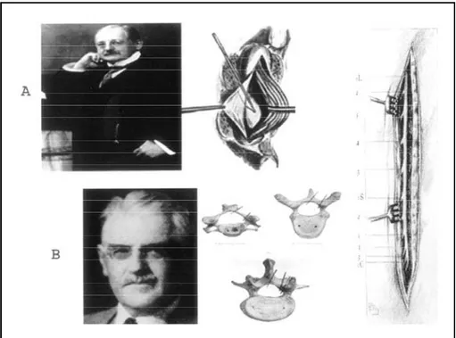
Fedor Krause in 1909 probably made the first successful removal of a ru p t u red disc. He published with Oppenheim a description of a removal what can be regarded with certainty as a ruptured disc. He made a low lumbar midline incision and re f l e c t-ed the paravertebral muscles from the laminae, which then were removed in one piece. The lesion which was resected transdurally , was thought to be an “enchondro m a ”7(Fig 3). In the same year
atica in 1911. He discussed a patient with re c u r-rent sciatica who had been operated on by Harv e y Cushing, but no lesion had been found. He belie-ved that the pain was from re c u rrent dislocation of the disc into the vertebral canal, and he explai-ned the negative exploration by assuming that the A l f red Taylor perf o rmed the first unilateral
lami-n e c t o m y. The ulami-nilateral lamilami-nectomy ilami-n the lum-bar and sacral, was first performed on a cadaver4 (Fig 3).
Joel E. Goldhwaite (1866-1961) was the first to describe a relationship between the disc and
sci-Fig 2. Ernest Lasègue (1816-1883). In this picture we appreciate the maneuver to pro v o k e the so-called Lasègue sign.
704 Arq Neuropsiquiatr 2005;63(3-A)
disc had slipped back into place. According to him, “such a condition could produce the symptoms of sciatica low back pain”5. This theory was far in advance of his time, however, and failed to aro u s e much interest.
In the same year, Middleton and Teacher descri-bed a case of paraplegia of sudden onset in a pa-tient lifting a heavy weight due to re t ropulsion of the disc between T12 and L1, confirmed at autop-s y. The patient died 16 dayautop-s later due to “bedautop-sore autop-s and septic cystitis”. The autopsy confirmed the spinal cord compression from a disc herniation. In this re p o rt they stated:“The following case is of i n t e rest, because it seems to throw light upon cer -tain cases of spinal myelitis and haemorrhage into the spinal cord arising out of strains and racks of the back in men engaged upon heavy work. So far as we are aware, the lesion is one which has not hithero been observed, as we have been unable, after considerable search in literature and enquiry among pathologists and surgeons, to find any record of an exactly similar case”8.
Charles Harrison Frazier (1870-1936) in 1913 dis-cussed problems and pro c e d u res in the surg e ry of the spinal column. He addressed the surgical tech-niques employed by him and demonstrated the patient position for midthoracic operation4 , 5. Tw o years late, while operating on a patient with sci-atica, Elsberg found “a ru p t u redligament of sub-flavum” compressing the fourth lumbar nerve ro o t . The patient’s pain disappeared after resection9.
S i c a rd in 1916 postulated that “sciatica” was commonly due of the roots of the sciatic nerve in their intraspinal course. He used the term “neu-rodochitis” to describe this condition5. In 1922, Al-fred Adson reported a laminectomy and removal of a protrusion of the fourth intervertebral disc4. Putti in 1927 suggested that inflammations of the sciatic nerve were due to an irritation of the nerve roots in the spinal foramina. This irritation was secondary to arthritis of the posterior inter-vertebral articulations. Sciatic pain could be satis-f a c t o ry correlated with the associated low back d i s o rd e r s1 0. Between 1927 and 1931 Schmorl, a ger-man pathologist, made an intensive investigation of the anatomy and pathology of the interverte-bral disc. The findings were based on radiological examination or on post-mortem dissection on the spinal columns. He established the modern basis for understanding the interv e rtebral disc, by pro-viding very clear discussions of herniations as well as degenerations11.
Dandy re p o rted two cases on which he oper-ated for low back and leg pain in 1929. He found c a rtilaginous fragments lying loose in the spinal canal (extruded, sequestrated disc material). He made several important points about the “lumbar disc syndrome”: a) relationship to trauma; b) pre-disposition of the lumbar region for such tions; c) propensity for the posterolateral hernia-tion due to deficiency of the posterior longitudi-nal ligament; d) the disc was affected by a pro c e s s like osteochondritis dessicans with fragments act-ing as a sequestrum12.
The idea that the disc herniation was neopla-sic, however, still was prevalent. Even Paul C. Bucy (1904-1992) in 1930 re g a rded a disc problem as “a typical cartilaginous neoplasm”1 3. Although in his publication Dandy mentioned that this piece of cartilage was simulating a tumor of the spinal ca-nal the full concept of what was the real disc mate-rial did not came up until 1933. Nonetheless, evi-dence rapidly accumulated in favor of a traumat-ic origin for pro t ruded disc material and its role in neurological disturbances.
Mixter and Barr presented their surgical find-ings at the Annual Meeting of the New England S u rgical Society in 1933. Their historical commu-nication was later re p o rted in the New England Journal of Medicine, August 1934. They made the following assertions: 1) ru p t u re of the interv e rt e-bral disc is a common cause of symptoms; 2) the lesion had previously been confused with cartila-ginous neoplasms; 3) disc ru p t u re is far more com-mon than cartilaginous neoplasms; 4) surgical de-compression is the preferred treatment14.
After this publication, lumbar discectomy be-came one of the most frequent operations carr i e d out by neuro s u rgeons. Altough they explore the ru p t u red disc intradurally. The surgical pathophys-iology and pathway were well defined. Initially the pro c e d u res were perf o rmed without magni-fication but subsequently the technique employ-ing the surgical microscope was described.
s-cectomy has became a standard operation for the treatment of herniated lumbar disc.
The microscope stimulated re s e a rch for pro g e s-sively less invasive techniques to treat lumbar disc disease. Two pro c e d u res were introduced: 1) the p e rcutaneous discectomy, and 2) the intradiscal in-jection of enzymes that theoretically promotes the biochemical degradation of the nucleous pulpous. Hijikata in 1977 was the first to design the instru-mentation for percutaneous removal of lumbar disc herniation18. In 1985 Maroon and Onik desig-ned an automated percutaneous discectomy sys-tem that uses a re c i p rocating section-cutter for re-moving disc material, re p o rting a success rate in 1987 of 79% in patients with symptomatic lumbar herniated disc19.
In 1969 Lyman Smith found that the intrathe-cal injection of enzimes in rabbits discretely re m o-ved the nucleous pulposus but left the annulus intact. He consequently injected the first patient in 196920.
The Cervical Disc - Posterior Approach
With the lumbar disc syndrome well under-stood, the concept was rapidly extended to the rest of the spinal canal. In 1905, Walton and Paul p e rf o rmed a posterior exploration for neoplasm of the cervical spinal cord with negative findings. The patient died few days later. At autopsy the spinal cord was deeply indented anteriorly by an extradural mass that arose from the sixth interv e r-tebral space, presumably the abnormal disc21.
The removal of cervical disc by the posterior approach probably was first described by Charles E l s b e rg in 1925 in his bookTumors of the Spinal Cord.He performed a cervical laminectomy from the fifth to the seventh segment and removed a “chondroma ” in a 57-year-old man who had a 10-week history of progressive quadriparesis22.
In 1928, Stookey reporte d a group of cerv i c a l extradural chordomas, which he removed via hemi-l a m i n e c t o m y. By anahemi-lysing his cases on the basis of symptoms and operative findings, Stookey defi-ned three classes of symptoms according o the site of pro t rusion: 1) those with unilateral ventral pre s-s u re on the cord, 2) thos-se with bilateral ventral c o m-pression, and 3) those with nerve - root pressure23.
R. Eustace Semmes (1885-1982) and Francis Mur-phy (1906-1994) wrote a classic paper correlating neck and arm pain wit cervical root compro m i s e at the interv e rtebral foramen2 4in 1943. This work was verified in 1953 by Spurling and Segerberg , who championed the posterior keyhole appro a c h
for removal of the lateral disc2 5. They stressed con-s e rvative treatment, with which theyhave had 70% success rate; only 30% of their patients re q u i re d surgery25.
The Cervical Disc - Anterior Approach
C e rvical discs were routinely removed by the posterior approach. Little interest was expre s s e d in the anterior cervical approach until 1955 when Robinson and Smith re p o rted anterior disc re m o v a l and subsequent interbody fusion using autograft b o n e2 6. This was followed by Cloward ’s re p o rt , which introduced ingenious instruments to insert a circular graft for anterior cervical interbody fu-sion after the discs had been removed27.
In 1960, Hirsch described an anterior cerv i c a l discectomy that was not followed by fusion. His technique consisted of incising the anterior annu-lus and removing the disc, leaving the posterior annulus and the ligament intact2 8. Subsequent se-ries emphasized a more radical decompression of the neural structures without fusion and, eventu-ally, the use of the microscope29.
In summary in the lumbar spine, according with Sonntag, the evolution of the management of disc disease had a lot of controversies1. Once a patient is diagnosed as having ru p t u red disc with associ-ated sciatica the amount of non surgical tre a t m e n t and (if its fails) which of the above surgical tech-niques should be employed are arguable. In the c e rvical spine the controversies are not less, but rather more extensive. The kind of approach, the use or not of bone and plates make the literature re s e a rch endless. The understanding of the evo-lution brings us upon only a small amount of k n o w-ledge. We should be grateful and give more atten-tion to the past because there sometimes one can find the key of future ’s door. The daring, coura-geous, and brilliant eff o rts of our early medical pioneers built the guideline in the way we should treat our patients.
REFERENCES
1. Sonntag VKH. History of degenerative and traumatic disease of the spine. In Samuel H. Greenblatt (Ed); T. Forcht Dagi and Mel H. Epstein, contributing editors. A history of neuro s u rgery: in its scientific and p rofessional contexts. Park Ridge: American Association of Neuro l o g i c a l Surgeons, 1997.
2. Marketos SG, Skiadas P. Hippocrates: the father of the spine surg e r y. Spine 1999; 24:1381-1387.
3. Knoeller SM, Seifried C. Historical perspective: history of spinal sur-gery. Spine 2000;25:2838-2843.
4. P a t w a rdhan RV, Hadley MN. History of surgery for ru p t u red disk. Neurosurg Clin N Am 2001;12:173-179.
6. Kocher T. Die Verletzungen der Wirbelsäule zugleich als Beiträg zur Physiologie des menschlichen Rückenmarks. Mitt Grenzgeb Med Chir 1896;1:415-480.
7. Oppenheim H, Krause F. Uber Einklemmung bzw. Strangulation der cauda equine. Dtsch Med Wochenschr 1909;35:697-700.
8. Midleton GS, Teacher JH. Injury of the spinal cord due to ru p t u re of an intervertebral disc during muscular effort. Glasgow Med J 1911 ; 7 6 : 1 - 6 . 9. E l s b e rg CA. Extradural ventral chondromas (ecchondroses), their favorite sites, spinal cord and root symptoms they produce, and their surgical treatment. Bull Neurol Inst NY 1931;1:350-388.
10. Putti V. New conceptions in the pathogenesis of sciatic pain. Lancet 1927;2:53.
11. Schmorl G. Über die pathologische Anatomie der Wi r b e l b a n d s c h e i b e n . Beitr Klin Chir 1931;151:360-368.
12. Dandy WE. Loose cartilage from intervertebral disk simulating tumor of the spinal cord. Arch Surg 1929;19:660-672.
13. Bucy PC. Chondroma of intervertebral disc. JAMA 1 9 3 9 ; 9 4 : 1 5 5 2 - 1 5 5 4 . 14. Mixter WJ, Barr JS. Rupture of the intervertebral disk with
involve-ment of the spinal canal. N Engl J Med 1934;211:210-215.
15. Ya s a rgil MG. Micro s u rgical operation of herniated lumbar disc. A d v Neurosurg 1977;4:81.
16. Caspar W. A new surgical procedure for lumbar disc herniation caus-ing less tissue damage through a micro s u rgical approach. Adv Neuro-surg 1977;4:74-80.
17. Williams RW. Microlumbar discectomy: a conservative surg i c a l approach to the virgin herniated lumbar disc. Spine 1978;3:175-182. 18. Hijikata S. Percutaneous nucleotomy. A new concept technique and 12
years experience. Clin Orthop 1989;238:9-23.
19. M a roon JC, Onik G. Percutaneous automated discectomy: a new me-thod for lumbar disc removal. Technical note. J Neuro s u rg 1987;66: 143-146.
20. Smith L. Chemonucleolysis. Clin Orthop 1969;67:72-80.
21. Walton GL, Paul WE. Contribution to the study of spinal surgery: one successful and one unsuccessful operation for the removal of tumor. Bost Med Surg J 1905;153:114-117.
22. Elsberg CA. Tumors of the spinal cord and the symptoms of irritation and compression of the spinal cord and nerve roots: pathology, sympto-m a t o l o g y, diagnosis and treatsympto-ment. New York: Paul B Hoeber, 1925: 195-198.
23. Stookey B. Compression of the spinal cord due to ventral extradural cervical chondromas: diagnosis and surgical treatment. A rch Neuro l Psychiatry 1928;20:275-291.
24. Semmes RE, Murphey F. The syndrome of unilateral ru p t u re of the sixth cervical intervertebral disk with compression of the seventh cer-vical nerve root: report of four cases with symptoms simulating coro-nary disease. JAMA 1943;121:1209-1214.
25. Spurling RG, Segerberg LH. Lateral intervertebral disc lesions in the lower cervical region. JAMA 1953;151:354-359.
26. Robinson RA, Smith GW. A n t e rolateral cervical disc removal and inter-body fusion for cervical disc syndrome. Bull Johns Hopkins Hosp 1955;96:223.
27. C l o w a rdRB. The anterior approach for removal of ru p t u red cervical discs. J Neurosurg 1958;15:602-617.
28. Hirsch C. Cervical disc ru p t u re: diagnosis and therapy. Acta Orthop Scand 1960;30:172-186.
29. Hankinson HL, Wilson CB. Use of the operating microscope in anteri-or cervical discectomy without fusion. J Neurosurg 1975;43:452-456.