Arq Neuropsiquiatr 2001;59(2-B):358-361
BEHAVIOUR OF OLIGODENDROCYTES
AND SCHWANN CELLS IN AN EXPERIMENTAL
MODEL OF TOXIC DEMYELINATION OF THE
CENTRAL NERVOUS SYSTEM
Dominguita Lühers Graça
1, Eduardo Fernandes Bondan
2,
Luis Antonio Violin Dias Pereira
3, Cristina Gevehr Fernandes
4,
Paulo César Maiorka
5ABSTRACT - Oligodendrocytes and Schwann cells are engaged in myelin production, maintenance and repairing respectively in the central nervous system (CNS) and the peripheral nervous system (PNS). Whereas oligodendrocytes act only within the CNS, Schwann cells are able to invade the CNS in order to make new myelin sheaths around demyelinated axons. Both cells have some limitations in their activities, i.e. oligodendrocytes are post-mitotic cells and Schwann cells only get into the CNS in the absence of astrocytes. Ethidium bromide (EB) is a gliotoxic chemical that when injected locally within the CNS, induce demyelination. In the EB model of demyelination, glial cells are destroyed early after intoxication and Schwann cells are free to approach the naked central axons. In normal Wistar rats, regeneration of lost myelin sheaths can be achieved as early as thirteen days after intoxication; in Wistar rats immunosuppressed with cyclophosphamide the process is delayed and in rats administered cyclosporine it may be accelerated. Aiming the enlightening of those complex processes, all events concerning the myelinating cells in an experimental model are herein presented and discussed.
KEY WORDS: demyelination, remyelination, oligodendrocytes, Schwann cells, ethidium bromide, immuno-suppression, cyclophosphamide, cyclosporine.
Comportamento de oligodendrócitos e células de Schwann em modelo experimental de desmielinização tóxica do sistema nervoso central
RESUMO - Oligodendrócitos e células de Schwann realizam a produção e manutenção das bainhas de mielina, respectivamente no sistema nervoso central (SNC) e periférico (SNP). As células de Schwann, à diferença dos oligodendrócitos, são capazes de invadir o SNC para remielinizar axônios desmielinizados, sempre que os astrócitos tenham sido destruídos. O brometo de etídio é uma droga gliotóxica usada para induzir desmielinização com o desaparecimento precoce de astrócitos, de modo que as células de Schwann têm liberdade para invadir o SNC. Em ratos Wistar normais, a remielinização é detectada treze dias após desmielinização; em ratos Wistar imunossuprimidos com ciclofosfamida a reparação do tecido é tardia, enquanto que em animais tratados com ciclosporina ela é acelerada. O objetivo do artigo é discutir todas as etapas do processo de destruição e reparação da mielina em um modelo experimental de desmielinização em ratos.
PALAVRAS-CHAVE: desmielinização, remielinização, oligodendrócito, células de Schwamm, brometo de etídio, imunossupressão, ciclosporina, ciclofosfamida.
1Departamento de Patologia, Universidade Federal de Santa Maria (UFSM), Santa Maria RS, 2Universidade Bandeirante (UNIBAN), São Paulo, SP; 3Departamento de Histologia e Embriologia, Universidade Estadual de Campinas (UNICAMP), Campinas, SP; 4Departamento de Patologia Animal, Universidade Federal de Pelotas (UFPel), Pelotas, RS; 5Departamento de Patologia Veterinária, Universidade São Paulo (USP), São Paulo SP, Brasil.
Received 6 October 2000, received in final form 24 January 2001. Accepted 1 February 2001.
Dra. Dominguita L. Graça - Departamento de Patologia UFSM - 97105-900 Santa Maria RS - Brasil. FAX 55 220 8284. E-mail dlgraç[email protected]
Oligodendrocytes and Schwann cells perform the unique task of producing and maintaining myelin sheaths around selected axons respectively in the central nervous system (CNS) and in the peripheral
nervous system (PNS)1,2. Although both cells have
been exhaustively investigated either in normalityor in disease1,3, many questions remain unanswered
oligodendro-Arq Neuropsiquiatr 2001;59(2-B) 359
cytes within demyelinating lesions3-5 and the origin
of those Schwann cells that invade the CNS to repair the lost myelin sheaths6-8. To get a deeper insight
into those issues, the behaviour of both cells in vari-ants of the Etmidium Bromide (EB) model of demyeli-nation was studied6,9-13. EB is an intercalating gliotoxic
dye extensively used to induce demyelination in the CNS6,7,10,13. The area of demyelination is larger than in
other models (i.e. lysolecithin14) and glial cells, namely
astrocytes, are destroyed early in the process. Central axons are chiefly remyelinated by Schwann cells, re-flecting the degree of astrocytic damage 14 and
dis-ruption of the glial limiting membrane15,16.
METHOD
Animals and ethidium bromide injection - Immunossu-pressed9,10 and normal7 Wistar rats of different ages12,17 were injected with either single or multiple local doses of 0.5 to 10 µl of 0,1 % ethidium bromide in saline within the brainstem9,10,12,13 and the spinal cord6,7. (Table 1).
Immunosuppressive treatment - The administratiom of the immunosuppressive drugs began at the same time of the EB injection. Animals treated with cyclophosphamide (CY) had an intraperitoneal injection of two weekly doses (respectively 50 and 30 mg/kg every three days), along the length of the experiment. Animals immunosuppressed with cyclosporine (CsA) had a daily injection of 10 mg/kg in the first week and thereafter the injections were given three times a week, every 48 hours, along the length of the experiment.
Perfusions and sampling of the tissues - The rats were killed by intraaortic perfusion of 4 % glutaraldehyde from 24 h to up to 150 days after injection in the spinal cord and from 7 to 30 days in the brainstem. Transverse slices 1 mm thick of the brain and the spinal cord from the area of the lesions were sampled for light and electron micros-copy studies.
Processing of the samples - The samples were washed in phosphate buffer and post-fixed in Millonig´s Osmium tetroxide for 2 hours. Routine dehydration and resin em-bedding procedures were performed 7. Semi-thin sections
were stained with Toluidine blue (Methilene blue) and thin sections selected from the former were stained with Ura-nyl acetate and Lead citrate and examined under an elec-tron microscope.
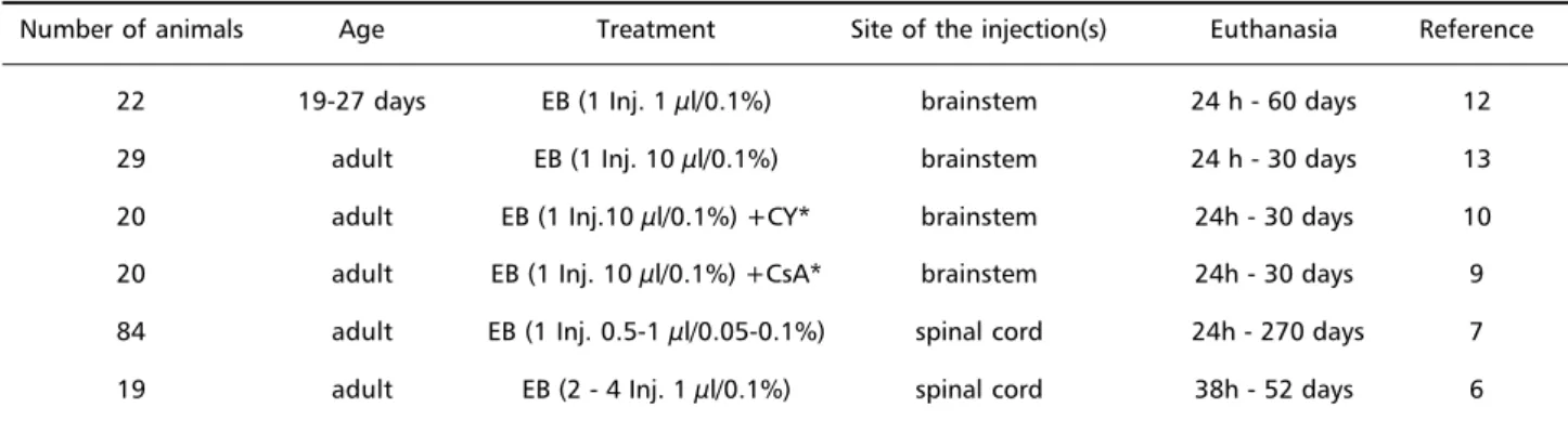
Results - Table 1 summarizes the results which are il-lustrated in Figure 1.
DISCUSSION
Demyelinated lesions were induced because of the early disappearance of glial cells7,10,13,17. The loss
of astrocytes determined a breach in the glial limit-ing membrane (GLM) that allowed the free entry of Schwann cells within the CNS territory2,7,8, feature
reported elsewhere15,16 .The bulk of the invading
Schwann cells is larger than in most toxic demyeli-nating models where the target of the chemical is the myelin sheath itself14. In the spinal cord the
num-ber of invading Schwann cells was much more mar-ked than in the brainstem8,18. The origin of the
in-vading Schwann cells was not definitely sorted out. The proposed origins include the pial and cranial nerves, the dorsal roots and the sympathetic nerves that surround the blood vessels7,18.
In the spinal cord the GLM was restored after the recovery of astrocytes by day 13 after injection in the spinal cord7 and by day 15 in the brainstem10.
The new myelin sheaths were easily detected because of their reduced thickness related to axonal diam-eter7,10,19. The sheaths produced by Schwann cells
also showed a basement membrane and collagen fibres around the cell boundaries7,9,18. Excluding
mi-nor differences in the morphology and timing of remyelination between the brainstem and the spi-nal cord, the whole process was very much alike. The burden of myelin repair was carried out by Schwann cells in areas away from the normal tis-sue7-10 – chiefly in subpial and perivascular areas.
Oligodendrocytes produced the new sheaths close to the normal tissue where astrocytic processes
Table 1. Data from the rats and the experimental procedures.
Number of animals Age Treatment Site of the injection(s) Euthanasia Reference
22 19-27 days EB (1 Inj. 1 µl/0.1%) brainstem 24 h - 60 days 12
29 adult EB (1 Inj. 10 µl/0.1%) brainstem 24 h - 30 days 13
20 adult EB (1 Inj.10 µl/0.1%) +CY* brainstem 24h - 30 days 10 20 adult EB (1 Inj. 10 µl/0.1%) +CsA* brainstem 24h - 30 days 9 84 adult EB (1 Inj. 0.5-1 µl/0.05-0.1%) spinal cord 24h - 270 days 7 19 adult EB (2 - 4 Inj. 1 µl/0.1%) spinal cord 38h - 52 days 6
360 Arq Neuropsiquiatr 2001;59(2-B)
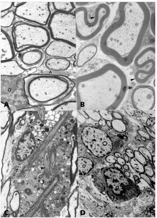
were conspicuous (Fig 1A). Oligodendrocytes showed different degrees of activation: in weanling rats they produced huge intracytoplasmic scrolls12
(Fig 1C); in normal adult rats it was detected the degree of slight activation reported in most experi-mental models when they repair lost myelin
shea-Fig 1. Ethidium bromide (EB) model of demyelination. (A) Oligodendrocytes (O) remyelinate axons in an area where many astrocytic (A) processes are detected. 150 days after one local injection in the spinal cord. 4500 X.
(B) Schwann cells remyelinated axons showing redundant myelin loops (arrowheads). 30 days after one local injection in the spinal cord. 4275 X. (C) Intracytoplasmic scrolls (arrowheads) in an intoxicated oligodendrocyte of a weanling rat 48 h after intracisternal injection of EB. 5580 X. (D) Reactive oligodendrocytes (O) 21 days after intracisternal injection in a rat treated with cyclosporine. 4875 X.
A
B
Arq Neuropsiquiatr 2001;59(2-B) 361
ths10; in cyclosporine immunosuppressed adult rats
oligodendrocytes were more conspicuous and al-though exhibited a round appearance, they showed a marked increase in the number of rough endo-plasmic cisternae9 (Fig 1D). When cyclophosphamide
was administered to the rats the whole process of removal of dead cells and disrupted myelin sheaths was delayed, suggesting an interference with mac-rophagic scavenging activities10.
Glial transplantation studies have shown that minor cell migration is detected when cell cultures enriched for oligodendrocytes precursors are placed within areas of normal and X-irradiated adult CNS20,
a situation that mimics what is observed in the EB-induced lesions. Thus, intended transplants must be placed within the lesion area in spontaneous demy-elinating diseases. In the EB-induced lesions a thin rim of oligodendrocyte remyelinated axons lined the normal tissue. Although it is known for more than a decade that totipotent neural stem cells occur in the CNS of adult mammals21 full detection of those
cells has been elusive. Therefore the origin of the oligodendrocytes that remyelinated the naked axons in our model remains to be defined.
Schwann cells kept a similar behaviour despite the anatomical location and age of the rats, producing myelin for CNS axons in a fast and efficient manner, frequently forming redundant myelin loops (Fig 1B). Other than promoting axonal regeneration in the CNS Schwann cells are extremely effective in repairing areas of central demyelination and restoring conduc-tion. They do so by their remarkably ability to produ-ce trophic factors and produ-cell adhesion molecules22.
In most lesions, either in the brainstem or in the spinal cord lymphocytes have been depicted. They were interpreted as part of the general inflamma-tory influx induced by the chemical11.Those
experi-ments using multiple injections in the spinal cord23
confirmed the suspicion that there is no immune interference in the EB lesions. The presence of lym-phocytes in the early stages of demyelination ad-dresses these cells as a component of the whole in-flammatory response that have a brief interaction with macrophages who might be the actual effec-tor cells within the CNS24.
The EB model of experimental demyelination has been useful to demonstrate that Schwann cells are able to repair CNS axonal sheaths of myelin in any situation when astrocytes and the GLM are destroyed even when they are induced to produce their own collagen fibres7,8. Likewise it was possible to assess
oligodendrocytes activation according to the age of
the rats and under selective immunosuppression and also their dependence on astrocytes to produce the new myelin sheaths.
Acknowledgments - The authors are indebted to the Electron Microscopy Laboratories of the Veterinary Pathol-ogy Unit, UFSM, Santa Maria, RS, HistolPathol-ogy and EmbriolPathol-ogy Department, UNICAMP, Campinas, SP and Department of Veterinary Pathology, FMVZ/USP, São Paulo, SP, Brazil
REFERENCES
1. Bignami A, Dahl, D. Glial cells in the central nervous system and their reaction to injury. Austin: Landes, 1994.
2. Graça DL. Mielinização, desmielinização e remielinização no sistema nervoso central. Arq. Neuropsiquiatr 1988;46:292-297
3. Norton WT. Do oligodendrocytes divide? Neurochem Res 1996;21:495-503 4. Rosano C, Felipe-Cuervo E, Wood PM. Regenerative potential of adult
O1+ oligodendrocytes. Glia 1999;27:189-202
5. Tokumoto YM, Durand B, Raff MC. An analysis of the early events when oligodendrocyte precursor cells are triggered to differentiate by thyroid hormone, retinoic acid, or PDGF withdrawal. Dev Biol 1999;213:327-329
6. Gevehr FC, Graça DL, Pereira LAVD. Desmielinização e remielinização após múltiplas injeções intramedulares de brometo de etídio em ratos Wistar. Arq Neuropsiquiatr 1997;55:452-459
7. Graça DL, Blakemore WF. Delayed remyelination in rat spinal cord following ethidium bromide injection. Neuropath Appl Neurobiol 1986;12:593-605
8. Graça DL. Desmielinização tóxica do sistema nervoso central: II. Aspectos biológicos das células de Schwann observados durante o processo de reparação do tecido. Arq Neuropsiquiatr 1989;47:298-273 9. Bondan EF, Lallo MA, Graça DL. Efeitos do brometo de etídio no tronco encefálico de ratos Wistar imunossuprimidos com ciclosporina. Cadernos de Estudos e Pesquisas, UNIP 1998;IV.
10. Bondan EF, Lallo MA, Sinhorini IL, Pereira LAV, Graça DL. The effect of cyclophosphamide on brainstem remyelination following local ethidium bromide injection in Wistar rats. J Submicr Cytol Pathol 2000;32:431-438
11. Graça DL. The presence of lymphocytes in a toxically induced demy-elinating process of the central nervous system. Micr Electr Biol Cell 1988;12:17-22.
12. Graça DL, Gevehr FC, Pereira LAVD. Morphological changes of myelinating oligodendrocytes in the ethidium bromide model of de-myelination. Rev Esp Patol 1997;30:297-301
13. Pereira LAV, Dertkigil MSJ, Graça DL, Cruz-Höfling MA. Dynamics of remyelination in adult rat brain after exposure to ethidium bromide. J Submicr Cytol Pathol 1998;30:341-348
14. Woodruff RH, Franklin RJ. Demyelination and remyelination of the caudal cerebellar peduncle of adult rats following stereotaxic injections of lysolecithin, ethidium bromide and complement/anti-galactocere-broside: a comparative study. Glia 1999;25:216-228
15. Franklin RJ, Blakemore WF. Requirements for Schwann cells migra-tion within CNS environments: a viewpoint. Int J Dev Neurosci 1993;11:641-649
16. Franklin RJ, Blakemore WF. Reconstruction of the glia limitans by sub-arachnoid transplantation of astrocyte-enriched cultures. Microsc Res Tech 1995;32:295-301
17. Graça DL. Desmielinização tóxica do sistema nervoso central: I. Efeitos de uma droga intercalante gliotóxica na medula espinhal de ratos Wistar. Arq Neuropsiquiatr 1989; 47:263-267
18. Pereira LAV, Cruz-Höfling MA, Dertkigil MSJ, Graça DL. Biology of the repair of central nervous system demyelinated lesions. Arq Neuropsiquiatr 1996;54:331-334
19. Pereira LAV, Cruz-Höfling MA, Dertkigil MSJ, Graça DL. Biology of the repair of central nervous system demyelinated lesions. Arq Neuropsiquiatr 1996;54:331-334.
20. Franklin RJ, Bayley SA, Blakemore WF. Transplanted CG4 cells (an oligodendrocyte progenitor cell line) survive, migrate, and contribute to repair areas of demyelination in X-irradiated and damaged spinal cord but not in normal spinal cord. Exp Neurol 1996;137:263-276 21. Peters A, Palay SL, Webster HF. The fine structure of the nervous
sys-tem. 3Ed. New York: Oxford Univ Press 1991.
22. Bunge RP. The role of the Schwann cell in trophic support and regen-eration. J Neurol 1994;241:S19-S21
23. Fernandes CG, Graça DL, Pereira LAVD. Inflammatory response of the spi-nal cord to multiple episodes of blood-brain barrier disruption and toxic demyelination in Wistar rats. Braz J Med Biol Res 1998;31:933-936 24. Zsuzsa F, Raine CS, Hart MN. Nervous tissue as an immune