NEUROPSYCHOLOGICAL AND PHONOLOGICAL
EVALUATION IN THE APERT’S SYNDROME
Study of two cases
Sylvia Maria Ciasca
1, Ana Paula Araujo
1, Adriana Nobre De Paula Simão
1,
Simone Aparecida Capellini
1, Paula Scalla Chiaratti
1, Edwaldo Eduardo Camargo
2,
Allan De Oliveira Santos
2, Elba Cristina Sá De Camargo
2ABSTRACT - This study evaluated two cases of Apert’s syndrome, through phonological, cognitive, and neuropsychological instruments and correlated the results to complementary exams. In short, this study reveals the necessity of application of neuropsychological, cognitive and phonological evaluation and correlation of the results with complementary testings because significant differences can be present in the Apert’s syndrome.
KEY WORDS: Apert syndrome, neuropsychological evaluation, phonological evaluation.
Avaliação neuropsicológica e fonológica na síndrome de Apert: estudo de dois casos
RESUMO - O objetivo deste estudo é apresentar a avaliação fonológica, cognitiva e neuropsicológica de dois casos com síndrome de Apert e correlacionar os achados destas avaliações com o resultado de exames complementares. Este estudo nos possibilitou verificar a necessidade da realização dessas avaliações em decorrência de diferenças significativas presentes nos casos com síndrome de Apert .
PALAVRAS-CHAVE: síndrome de Apert, avaliação neuropsicológica, avaliação fonológica.
Faculdade de Ciências Médicas da Universidade Estadual de Campinas, Campinas SP, Brasil (FM/UNICAMP): 1Child neurology Subject, Neurology Department FCM/UNICAMP; 2Nuclear Medicine Service FCM/UNICAMP.
Received 2 June 2000, received in final form 9 January 2001. Accepted 15 January 2001.
Dra. Sylvia Maria Sciasca - Praça XV de Novembro 40 / 41 - 13024-180 Campinas SP - Brasil.
Apert’s syndrome was described by Wheaton1,
in 1894. In 1906, Apert2 published a summary on
nine cases, and in 1920 Park and Powers3 wrote an
excellent monography on this disease. In 1960, Blank4
registered a total of 150 published cases. Apert’s syndrome is a genetic pathology of dominant auto-somal inheritance and it has as main characteristics: the acrocephalia due to synostosis of the coronary suture and the syndactilism which most of the time is symmetrical involving the four extremities.
The cranium is shortened in the antero-posterior diameter and prolonged vertically, taking the cha-racteristic acrocephalic aspect. The face is generally flattened and it can be asymmetric. There are front bossas and the occipital is flattened. Most of times there are big fontanels, closing with lateness. The jaws are frequently hipoplasic and a prominent jaw resulting in a moderate prognatism. There is ocular hypertelorism and the orbits are sidelong shallow and oblique, with antimongoloid inclination and protusion of the ocular globes. Frequently there is
strabismus. The nose is short and enlarged. The na-sal bridge is flattened. The pavilion headphones are generally moved away of the head and they seem big. The palate is arched, narrow and it can be cleft. The maxillary arcade can present a configuration in V with superimposed teeth and protuberant gums. The dental eruption is late. It presents bony and/or cutaneous syndactilism, going from the partial coa-lition of the fingers, it is generally observed total coalition of the second, third and fourth fingers. Sometimes the last phalanges of the thumbs come increased in volume and with valga position . The fingers can be short. Cutaneous syndactilism of all the ankles, with or without bony syndactilism. The extremity of the great ankle is sometimes thick and malformed. Moderated or serious acne in the teen-agers, including in the forearms.
Gonçalves and Silva5 discussed how a
pre-vent the mental deficiency. There can be mental de-ficiency, but cases of normal intelligence were also registered; the incidence of the mental deficiency is unknown.
The objectives of the present stydy are: to accom-plish phonological intellectual and neuropsychologi-cal evaluations in two children with Apert’s syndro-me; to compare the discoveries of the phonoaudiolo-gical, intellectual and neuropsychological evaluations among the two children with Apert’s syndrome.
METHOD
Subjects - Two female children took part in this study, the first child (Case 1) is 8 years and 5 months of age, she attended the second year of first grade in 1998, her eco-nomic social level is low; the second child (Case 2 ) is 12 years and 9 months, she attended APAE, her economic social level is low.
Procedures - The following instruments of evaluation were used: - neuropsychological evaluation –Luria ques-tionary –Nebraska6; - the Wechsler Intelligence Scale for
Children – WISC7; - phonological child evaluation8; -
aware-ness phonological test (APT)9; - brain scintillography - SPECT.
RESULTS
The first case obtained the following results in the WISC7: she presented intellectual revenue within
the standart average in relation to the children of the same age group, as it shows the following chart (Table 1).
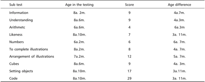
It was obtained the following results in the sec-ond case: the WISC7 presented the intellectual
out-put below the average in relation to the children of the same group as in chart II (Table 2).
The WISC data in both cases were significant showing that just Case 1 demonstrated higher
re-Table 1. Evaluation of the Case 1 – WISC.
Sub test Age in the testing Score Age difference
Information 7a. 6m. 8 11 m.
Understanding 7a. 10m. 8 7 m.
Arithmetc 7a. 6 m. 5 11 m.
Likeness 9a. 10m. 8 1a. 5m.
Numbers 7a. 2m. 7 1a.2m.
To complete illustrations 8a. 2m. 8 3 m.
Arrangement of illustrations 8a. 2m. 20 3 m.
Cubes 10a. 2m. 18 1a. 8m.
Setting objects 8a. 10m. 17 5 m
Code 7a. 2m. 18 1a.2m.
Table 2. Evaluation of the Case 2 – WISC.
Sub test Age in the testing Score Age difference
Information 8a. 2m. 9 4a.7m.
Understanding 8a.6m. 9 4a.3m.
Arithmetc 6a.6m. 4 6a.3m
Likeness 8a.10m. 7 3a. 11m.
Numbers 6a.2m. 6 6a. 7m.
To complete illustrations 8a.2m. 8 4a. 7m.
Arrangement of illustrations 7a.2m. 12 5a. 7m.
Cubes 8a.6m. 9 4a. 3m.
Setting objects 8a.10m. 17 3a.11m.
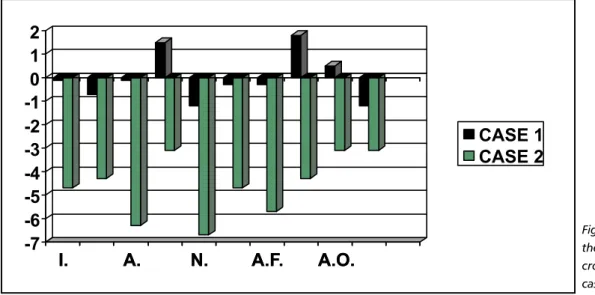
sults in the age group in the tests of likeness, cubes and setting objects, which measure respectively asso-ciative thought, abstraction, as it represents Fig 1.
In the neuropsychological evaluation, Case 1 ob-tained better results in tactile ability, rhythm, speech, expressive language, mathematical reasoning and memory. And in Case 2, it was obtained better re-sults in moving and right ability, writing and read-ing, as it represents Figure 2.
Concerning the phonological evaluation, the child of the Case 1 presented substitution of deaf pho-nemes for sound amethodically such as / k / - /g /; / ƒ / - /v /, besides distortion in the phonemes /s / and / s /. The child of Case 2 presented distortion in the pho-nemes /s / and / s /, systematic omission of the pho-neme /l/ in syllabic clamping and asystematic omis-sion of this phoneme in consonantal group, substi-tution asystematic of /l / for /r / in consonantal group and systematic substitution of /n / for / l /, and lan-guage earliness in the phoneme /t /. Although in both cases the children presented lexicon suited to the age.
In concern to the results of the Awareness Phono-logical Testing (APT)9, the child Case 1 obtained score
of 22 points and the child Case 2, 26 points. In both cases, the children presented difficulties in the ex-ecution of the subtests which involved the phone-mic and sylabic abilities, indicating therefore those phonological difficulties (Fig 3).
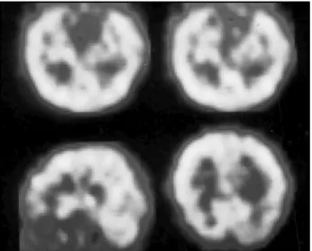
In SPECT the results were as shown in Figures 4 and 5.
DISCUSSION
Gonçalves and Silva5 described normal
neurologi-cal evaluation in the clinineurologi-cal suggestive case reported. In previous studies, Patton et al.10, Léfèvre et al.11, it
was observed among 70 patients, with no sex preva-lence, being 50% of men and 50% of women, re-spectively. After these studies it can be concluded that most of the patients with Apert’s syndrome is mental faulty in different levels. The most common causes observed to determine the mental deficiency are: perinatal factors, hydrocephalus, malformation
Fig 1. Difference between the achieved age and the cronological age in both cases.
I.
A.
N.
A.F.
A.O.
-7
-6
-5
-4
-3
-2
-1
0
1
2
I.
A.
N.
A.F.
A.O.
CASE 1
CASE 2
Fig 2. Comparision of the neuropsychological evaluation in both studied cases.
20 21 22 23 24 25 26 27
Score
Case I
Case II
of the central nervous system, and the increase of the intrabrain pressure.
The data were correlated to exams of neuroimage (SPECT). According to the accomplished evaluations, it was observed that the results of the second eva-luation case confirms the characteristic of mental deficiency presented by the Apert’s syndrome shown in the literature, the patient revealed difficulty in the tests : rhythm, tactile ability, memory, mathematical reasoning, arithmetic, numbers and arrangement of illustrations compatible with the hipoperfusion in the right temporal lobe shown by SPECT. However, the first case differs from studies by presenting nor-mal intellectual revenue, without other deficits and presenting a normal SPECT, being an atypical case to the situation, proving that inside the same symp-tomatology there can be significant differences which justify the neuropsychological diagnosis and the phonological difficulties.
In concern to the phonological evaluation, in both cases it was possible to verify the presence of pho-nological deviation and difficulty in the manipula-tion of the sounds of the speech. On the other hand in the second case it was verified that the phono-logical deviation is compatible with the hypoper-fusion in the temporal area shown in the SPECT; in the first case, the SPECT is normal and so there is no link between the findings of the neuroimage and the phonological findings in this case. But it is neces-sary to perform the neuroimage to stablish the pres-ence or not of the hypoperfusion in the temporal
area, mainly because the phonological deviation can be present in both cases : with the presence of the hypoperfusion of this brain area (Case 2) and in cases with no evidence of the same however with the evi-dence of lateness language development (Case 1).
In concern to the language, few are the studies which were found in the literature. However we can mention the study accomplished by Misquiatti12,
which evaluated 8 individuals’ language with the Apert’s syndrome, 4 with Crouzon’s syndrome and 3 with Pfeiffer’s syndrome. As procedures it was ac-complished genetic evaluations, psychological evalu-ation, audiological evaluation and phonoaudiological evaluation. The findings revealed that 9 individuals presented conductive hearing loss with degrees vary-ing from light to moderate and only 2 with normal hearing; just 4 individuals with light mental defi-ciency; most of the individuals with current syndrome of mutation in the gene FGFR2 presented complaints with relation to the language and hearing, and most were related to the oral delivery; the language alter-ations related to the emission and oral reception, perceptual processes and basic functions were pre-sented in the great majority of individuals, in differ-ent commitmdiffer-ent the delay in the acquisition of the language frequently happened in the appraised in-dividuals. The author concludes from this study the
Fig 4. SPECT of Case 1. Superior half of the temporal axis: perfu-sion of the temporals. Sagittal inferior axis to the left: homoge-neous and normal perfusion of the brain cortex and other struc-tures. Inferior transversal axis to the left: homogeneous and nor-mal perfusion of the brain cortex.
need of specific phonoaudiological studies both in area of the language as of the audiology, in order to determine phonoaudiological manifestations in these syndromes, besides the need to direct these individuals precociously to rehabilitation of the dis-turbances of communication.
In short, this study reveals the necessity of appli-cation of neuropsychological, cognitive and phono-logical evaluation and correlation of the results with complementary testings because significant differ-ences can be present in the Apert’s syndrome.
REFERENCE
1. Wheaton, SM. Two specimens of congenital cranial deformity in infantis asociated with fusion of fingers and toe. Trans Pathol Soc , London, 1894;45:238-241.
2. Apert, E. De l’acrocephalosyndactylie. Bull Soc Méd Paris 1906;23: 1310-1330.
3. Park EA, Powers GF. Acrocephaly and scaphocephaly with symmetri-cally distributed malformations of the extremities. Am J Dis Childd 1920;20:235-315.
4. Blank, CE. Apert’s syndrome (a type of acrocephalosyndactyly) obser-vations on a british series of trirty nine cases. Ann Hum Genet 1960;24:151-164.
5. Gonçalves da Silva JA. Discussão genético – clínica sobre a síndrome de Apert: a propósito de um caso. Arq Neuropsiquiatr 1976;34:293-297 6. Lúria A. Fundamentos de neuropsicologia. Porto Alegre: Artes Médicas,
1991.
7. Wechsler D. Manual for the Wechsler intelligence scale for children. New York: Psychological Corporation, 1974.
8. Yavas M, Hernandorena CLM, Lamprecht RR. Avaliação fonológica da criança. Porto Alegre: Artes Médicas, 1991.
9. Capovilla AGS, Capovilla FC. Prova de consciência fonológica: desenvolvimento de dez habilidades da pré-escola à segunda série. Temas sobre Desenvolvimento 1998;37:14-20.
10. Patton MA, Goodship J, Hayword R, Lansdown R. Intellectual devel-opment in Apert’s syndrome: a long term follow up of 29 patients. J Med Genet 1988;25:164-167.
11. Lefèvre A, Travis F, Arndt EM, Munro IR. A psychiatric profile before and after reconstructive surgery in children with Apert’s syndrome. Br J Plast Surg 1986;39:510-513.