Relatório Final de Estágio
Mestrado Integrado em Medicina Veterinária
CRYOPRESERVATION AND THAWING PROTOCOL OF
MESENCHYMAL STEM CELLS FOR USE IN VETERINARY MEDICINE
CELL THERAPY
Cláudia Cristina Lopes Simões Melo Ferreira e Costa
Orientador
Professora Doutora Ana Colette Pereira de Castro Osório Maurício
Co-Orientador
Mestre Ana Rita Caseiro Santos
i Abstract
The field of regenerative and reparative medicine is an expanding and rapidly growing one. In order to create cell-based therapies it is crucial to standardize techniques and establish protocols.
The purpose of this study was to establish cryopreservation and thawing protocols of animal cells as well as to evaluate the viability loss of mesenchymal stem cells upon thawing, and up to 72 hours later under three different environmental conditions.
Using the trypan blue method, cell viability was determined under three different temperature settings post-thaw: room temperature at 21ºC, incubation at 37ºC and refrigeration at 4ºC for up to 72 hours. The preservation of the cells in plastic and glass vials was also compared as well as the thaw method using human umbilical cord plasma and canine serum.
It was possible to determine that there was no difference in cell viability results between the use of fetal bovine serum and human umbilical cord blood plasma at either 90 or 95%, as medium supplements during cryopreservation, which also enables the use of only 5 to 10% dimethyl sulfoxide.
On the other hand, canine serum does not serve as an appropriate protein source during thawing since it causes cellular agglomeration resulting in lower viabilities.
Results also show that the material of the vials used to preserve cells post thaw does not influence viability significantly, whereas the temperature at which they are conserved does. Refrigeration at 4ºC appears to be the preferable temperature for maintenance of these cells post-thaw.
Nonetheless, there is still room for protocol improvement since many questions remain unanswered and much work is needed to be done in the laboratory and the clinic to understand how to fully apply these cell-based therapies to treat disease.
ii Acknowledgements
To everyone that somehow contributed to this thesis directly or indirectly, I thank you.
A realização deste mestrado nunca teria sido uma possibilidade, nem sequer uma consideração minha se não tivesse tido a sorte de ter a Professora Doutora Ana Colette Maurício como professora no meu percurso inicial pelo ICBAS. Obrigada por me compreender e me dar sempre uma oportunidade. Foi a minha escolha óbvia para orientadora, nem pestanejei, não só pela sua permanente disponibilidade e paciência para nós alunos, mas também pela admiração que tenho por si e pelo trabalho que tem desenvolvido ao longo dos anos.
Gostaria de agradecer também à Mestre Rita Caseiro, minha co-orientadora, pela sua preciosa disponibilidade, orientação, apoio, sentido de humor e simpatia.
À Doutora Sílvia Pedrosa, pela sua eterna paciência, explicações pormenorizadas, ajuda e boa disposição.
Aos colegas envolvidos também neste projecto pela ajuda no laboratório e que através da sua ligação a este grupo de investigação contribuíram de alguma forma para este projecto, a todos o meu muito obrigado.
Agradeço o apoio e as oportunidades proporcionadas neste percurso e em especial ao bem-disposto ambiente de trabalho que permitiu a continuação do meu enriquecimento profissional. Ao Fernando, o meu mundo, meu porto de abrigo sem cuja sabedoria e serenidade isto nunca teria sido possível. Obrigada por acreditares sempre em mim mesmo quando nem eu acreditava.
À minha filha Cristina cujos sorrisos e risos constantes iluminam a minha vida e a tornam tão mais simples, mesmo arrancando o cabo do portátil num momento fulcral de elaboração da tese!
iii Abbreviations
AIDS - acquired immune deficiency syndrome ASC - adult stem cell
cSM - canine synovia cDP - canine dental pulp
DMSO – dimethyl sulfoxide (Sigma-Aldrich) ºC – degree Celsius
ESC - embryonic stem cell FBS – fetal bovine serum
HIV – human immunodeficiency virus iPSC - induced pluripotent stem cells µl - microliter
ml - milliliter
MSC - mesenchymal stem cell % - percentage
PBS - phosphate buffered saline rpm – revolutions per minute rSM - rat synovia
SEM - standard error of the mean
iv TABLE OF CONTENTS Abstract ... i Acknowledgements ... ii Abbreviations ... iii I. Introduction ... 1
II. Literature Review ... 2
1. Regeneration in nature ... 2
2. Stem cells ... 2
3. Mesenchymal stem cells ... 3
Sources of Mesenchymal stem cells ... 4
4. Cell therapy and the uses and applications of Mesenchymal stem cells ... 4
5. Cryopreservation of mammalian cells ... 6
III. Research Aim ... 7
Cell viability assay ... 9
Statistical analysis ... 10
1. Cryopreservation of MSCs of different sources... 10
a) Materials and Methods ... 11
b) Results and discussion ... 12
2. Thaw method ... 13
a) Materials and Methods ... 13
b) Results and discussion ... 14
3. Vials and post-thaw preservation of the cSM samples ... 16
a) Materials and Methods ... 16
b) Results and discussion ... 16
4. Post-thaw preservation of the rSM and cDP samples ... 20
a) Materials and Methods ... 21
b) Results and discussion ... 21
VI. Conclusion ... 23
1 I.INTRODUCTION
As in human medicine, the search for ways to increase longevity in animal species and maintaining their health has gained increasing interest in veterinary medicine studies and advances. Mechanisms of natural and biological repair (both in physical damage as well as in illness) of living organisms using cells and tissues are of most interest.
During the 19th century, it was postulated that certain tissues, comprised of short-lived cells, had the ability to self-renew for the lifetime of an organism. Research advanced and culminated in the discovery of stem cells. This capacity to differentiate into specialized cells makes stem cells valuable in therapeutic applications designed to regenerate and repair cellular or tissue damage. This discovery opened a whole new realm of possibilities in the field of biological repair offering new potential for treating diseases as a result of those unique regenerative abilities (Ramalho-Santos & Willenbring, 2007; Bianco et al., 2008). These were the early days of regenerative medicine.
Since then and to this day, there has been a rapid growth in this field, accelerating in the last 30 years and expanding from research to clinical trials (Dominici et al., 2006). Regenerative medicine as a cell-based therapy refers to therapeutic approaches in which stem cells are induced to differentiate under controlled conditions to be later injected into or transferred to a patient to repair damaged or destroyed cells or tissues (Lane et al., 2014; Mao & Mooney, 2015). Here lies the potential to not only treat, but cure disease.
Specific and consistent protocols for the preparation and application of such therapies in preclinical research, clinical trials and final treatments are essential (Heathman et al., 2015). Protocols for mesenchymal stem cell cryopreservation and thawing processes remain suboptimal and inconsistent (Ikebe & Suzuki, 2014). Following this concept, the final aim of this study was to determine cellular number and viability loss of mesenchymal stem cells after undergoing a cryopreservation and thawing process and possibly perfect these protocols. The number of cells lost during these processes could then be taken into account when determining the number of viable cells needed to be cryopreserved, according to the final number of cells required for a given treatment. Post-thaw loss over time was also determined.
Although this field is advancing at a hasty pace, there are still a few obstacles that must be overcome for the application of certain cell-based therapies, such as ethical and legal questions due to the use of embryonic stem cells, particularly human, and the conduction of stem research in general (Hyun, 2010).
2 II.LITERATURE REVIEW
1. Regeneration in nature
Regeneration and repair processes are naturally observed in humans and animals. These processes can also be found in plants and simpler animals, such as planaria and crayfish as well as in embryos (Metcalfe & Ferguson, 2007; Gurtner et al., 2008; Wagner et al., 2011; Rouhana & Tasaki, 2016).
Planarians are flat, soft-bodied worms that have evolved a remarkable stem cell regenerative system, giving them the ability to rapidly self-renew the entire animal within a matter of weeks (Wagner et al., 2011; Rink, 2013). Crayfish, on the other hand, have the ability to shed or sever an appendage to escape predators and then regenerate it (Juanes & Smith, 1995; Cooper, 1998; Maginnis, 2006). There is a cost that comes with this process as discussed by Maginnis 2006.
In nature, there appears to be a reverse relationship between complexity and regenerative ability; the more complex the organism, the more decreased its capacity for regeneration is. Tissue and organ regeneration in postnatal mammals is more limited than that found in more basic animals, and different tissues have different renewal abilities. The gut, blood, epidermis and liver have the highest regenerative ability, followed by bone and muscle whereas heart, ligaments and the brain have a low capacity (Schroeder, 2008). Reservoirs of specific stem cells can be found in these tissues (Meirelles et al., 2006). Only some of these tissues, for example blood, can be restored to their original state after injury, but most tissues can only be repaired but not completely restored (Metcalfe & Ferguson, 2007; Schroeder, 2008).
2. Stem cells
Stem cells are the basis for nature’s reparation system. They are found in several tissues and are capable of self-renewal and differentiation, both in vitro and in vivo, into different cell lineages. This means that when a stem cell divides, it may produce a new stem cell (self-renewal) or a tissue or organ specific cell (differentiation). Even after long periods of inactivity, these unspecialized stem cells are capable of renewing themselves or be induced, under certain physiologic or experimental conditions, to become tissue or organ specific cells with specific functions.This ability to self-renew is essential in the organism for both the expansion of several tissue cells as well as cellular turnover and repair (Wei et al., 2013; Melton, 2014). Whereas, in vitro, most somatic cells display a finite number of replications prior to senescence or replicative arrest (Hayflick & Moorhead, 1961; Hayflick, 1965; Cristofalo et al., 2004; Melton,
3
2014), stems cells exhibit a seemingly unlimited proliferative capacity. In vivo, stem cells exhibit sufficient proliferative capacity to last throughout the lifetime of the organism (Melton, 2014). In some tissues of postnatal mammals, such as bone marrow, muscle, and brain, discrete populations of adult stem cells exist. They self-renew and differentiate into a number of specialized cells formed by these tissues to generate replacements for cells that are lost through normal wear and tear, injury, or disease (Meirelles et al., 2006; El-Hashash, 2016). In some organs, such as the gut and bone marrow, stem cells regularly replicate to repair and replace worn out or damaged tissues whereas in other organs, namely the pancreas and the heart, stem cells only divide under specific conditions (Bhattacharyya & Khanduja, 2010).
Until recently, research was done mainly with two kinds of stem cells from animals and humans: pluripotent embryonic stem cells (ESCs) and multipotent non-embryonic or adult stem cells (ASCs), as the cells used in this study. ESCs have certain ethical limitations, as well as the risk of teratoma formation whereas ASCs have a more limited differentiation potential (Salem & Thiemermann, 2010). Researchers have now developed genetically reprogrammed multipotent adult cells that function like embryonic stem cells; these are called induced pluripotent stem cells (iPSCs), from mouse and human adult stem cells. These are capable of generating cells characteristic of all three germ layers; the ectoderm, mesoderm and endoderm but as with ESCs, they also pose the risk of teratoma formation (Hou et al., 2013; Wei et al., 2013; Ross & Akimov, 2014).
3. Mesenchymal stem cells
Mesenchymal stem cells (MSCs) are a highly investigated population of stem cells, due to their unique biological properties, that were first isolated from bone marrow and characterized by Friedenstein and his colleagues (Afanasyev et al., 2009). They are multipotent stem cells, found in both embryonic and postnatal mammals, with differentiation potential towards mesoderm-derived lineages (i.e., adipogenic, chondrogenic, myogenic, and osteogenic) (Caplan & Dennis, 2006; Dominici et al., 2006).
It has been found that not only do they have the ability and potential to differentiate into adypocytes, chondrocytes, myocytes and osteoblasts but also neurons, hepatocytes and pancreatic islet cells (Squillaro et al., 2016). Recent studies suggest that the differentiation capabilities of MSCs into diverse cell types vary between those of different origins.
MSCs from different species and/or sources share the same primary characteristics and have similar morphology, yet clear differences in functional features have been verified in MSCs
4
isolated from synovial fluid versus bone marrow, as well as differences between species, such as ovine and equine were demonstrated (De Schauwer et al., 2014; Hillmann et al., 2016). Although the golden standard as a source of MSCs for research continues to be the bone marrow, since bone marrow derived MSCs are the most investigated and hence the best characterized of this cell type, other sources of MSCs are now being used (Fortier & Travis, 2011; Mafi et al., 2011; Giai Via et al., 2012), such as dental pulp and synovia as was the case in this study.
Sources of Mesenchymal stem cells
MSCs can be found and isolated from several tissues in postnatal mammals, such as bone marrow (where they were originally identified), adipose tissue, peripheral blood, skeletal muscle, olfactory mucosa, synovium, dental pulp and hair follicles, as well as from fetal (i.e., bone marrow, liver, blood), and extraembryonic tissues (i.e., placenta, amnion) (DiMarino et al., 2013; Wei et al., 2013; Taran et al., 2014). Once isolated, they may be proliferated, expanded, differentiated and preserved in vitro.
Since different methods of isolation and expansion of MSCs are used by researchers, it became necessary to characterize the cells and create standards to define human MSCs for both laboratory-based scientific investigations and for pre-clinical studies. To be classified as MSCs they must obey certain minimal criteria such as: they must be plastic-adherent when maintained in standard culture conditions using tissue culture flasks, ≥ 95% of the MSC population must express certain surface antigens and lack expression of others and finally, the cells must be able to differentiate to osteoblasts, adipocytes and chondroblasts under standard in vitro differentiating conditions (Dominici et al., 2006). Although Dominici’s paper served to characterize human MSCs, this also became the standard for characterizing mammalian MSCs and served as a basis for extrapolation to other species.
4. Cell therapy and the uses and applications of Mesenchymal stem cells
When Friedenstein et al. first described and isolated MSCs from bone marrow, showing their multipotent capacities to form bone, cartilage, and adipose tissue, they gave us the ingredients to explore the possibility of using these cells for regenerative medicine (Afanasyev et al., 2009). MSCs are advantageous over other stem cells types in regenerative medicine due to a variety of reasons making them, at present time, the most commonly used adult stem cells, and our cells of choice in this study. As previously mentioned, they avoid the ethical issues surrounding ESCs and are easily obtainable in sufficient quantities with minimal invasive methods since they
5
are present in several different tissues of postnatal mammals. Due to this, they are a well-characterized population of adult stem cells and researchers now have advanced knowledge on how to grow them in culture and protocols for their isolation, expansion, and differentiation have been established. Isolation of MSCs and their in vitro maintenance has had increasing application in both experimental and clinical setups, owing to their easy isolation, high expansion properties in addition to their reproducible results. The idea that the infusion of MSCs might benefit organ and tissue repair given their ability to differentiate into cells of the targeted tissue and replace damaged resident cells has been sustained by several studies (Squillaro et al., 2016).
So far, advancements in MSCs research show how these cells may have several clinical applications in both immunological disorders, such as prevention of graft-versus-host disease, and non-immunological disorders, and cell replacement therapies for mesenchymal tissues, for instance, bone and cartilage (Wei et al., 2013; Ikebe & Suzuki, 2014; Lane et al., 2014; Squillaro et al., 2016). It has also been demonstrated that local infusion is the preferred method of administration to take advantage of their differentiation potential since following systemic injection most MSCs are trapped in capillary beds of various tissues, especially the lungs (Squillaro et al., 2016).
In recent years, some medical and biological issues such as rejection of donor stem cells by the patient’s immune system have arisen (de Almeida et al., 2013; Tang et al., 2013) but appear to be close to being solved by new cell therapies which will also help in solving transplant organ rejection. Mesenchymal stem cells also seem to be the core solution for this problem due to their immunomodulatory response reducing both the risks of rejection and complications of cellular, tissue and organ transplantation (Salem & Thiemermann, 2010; Taran et al., 2014). The ultimate goal for applying stem cell research is the cure of fatal or debilitating diseases in both humans and other animals. The search for the application in the eradication of malignant stem cells (cancer cells) and the cure for HIV/AIDS in humans is ongoing. The possibility of nervous system regeneration opens the door for cell therapies of both neurodegenerative conditions as well as spinal cord injuries, and the cure of other motor neuron and demyelinating diseases (Taran et al., 2014).
To date there are 340 MSC-based clinical trials either complete or ongoing (obtained from
https://clinicaltrials.gov/) worldwide.
In clinical veterinary medicine, so far, stem cells are most commonly used in the treatment of musculoskeletal injuries of horses and dogs. Small and large animal species also serve as valuable models for preclinical evaluation of stem cell applications in human beings and in veterinary patients in areas such as spinal cord injury and myocardial infarction. There are also
6
new technologies of assisted reproduction undergoing development to apply the properties of spermatogonial stem cells to preserve endangered animal species (Fortier & Travis, 2011).
5. Cryopreservation of mammalian cells
The first mammalian cells to be cryopreserved successfully were spermatozoa in 1949 by Polge and his team (Polge et al., 1949). Since then, many methods and protocols have been developed for various types of cells, tissues and organs.
The preservation of cells is an extremely important aspect of cell culture and the only effective means of preservation of animal cells is by freezing. Cryopreservation is a method whereby cells are frozen, maintaining their viability, until they are defrosted or thawed. Increased understanding of the causes of cryoinjury has continually led to improve cryopreservation methods and much progress in the field.
The main reasons and advantages for the cryopreservation of cells are: - to avoid loss by microbial contamination
- to reduce risk of cross contamination with other cell lines
- to minimize genetic change in continuous cell lines (prone to genetic drift and morphological changes)
- to enable storage of stocks of cells preventing the need to have all cell lines in culture at all times (which is invaluable when dealing with cells of a limited life span)
- to carry out studies at a consistent and known passage number
- to avoid aging and transformation into finite cell lines (fated for senescence)
By careful preparation and strict procedures, once at ultralow temperatures, cells are biologically inert and can be maintained viable in this way for many years (typically ten years or more).
The basic principle for success when cryopreserving and thawing cells is a slow freeze and a quick thaw. As a general guide, cells should be cooled at a rate of -1ºC to -3ºC per minute and thawed quickly by incubation in a water bath for 1-2 minutes (Liu et al., 2010; Hunt, 2011; Martinello et al., 2011). This principle was respected in this study and this procedure followed.
7 III.RESEARCH AIM
The use of MSCs has spread worldwide and its importance has gained recognition in the past 30 years. Therefore, it has become essential to establish well documented protocols for each step of the cell-based therapies, including their preservation.
In general terms, it isn’t possible to follow every step required in cell-based treatments, from the harvest and isolation of MSC through their expansion to their final administration, without the need to cryopreserve, making this a vital step (Liu et al., 2010).
Although there are reasonably well established and reproducible protocols for the cryopreservation of human stem cells in general (Hunt, 2011), there is a lack of confirmed cryopreservation protocols for stem cells of other mammals and of different sources (Mitchell et al., 2015). Nonetheless, even with established protocols, there is always room for improvement especially in light of new technologies, that are constantly becoming more readily available, and of new research results. Additionally, a very useful and significant data is that of the total loss of cell number and viability that occurs post cryopreservation and thaw processes.
Therefore, the main aim of this study was to improve MSCs cryopreservation and thawing protocols using MSCs of three different sources; canine synovia (cSM), canine dental pulp (cDP) and rat synovia (rSM).
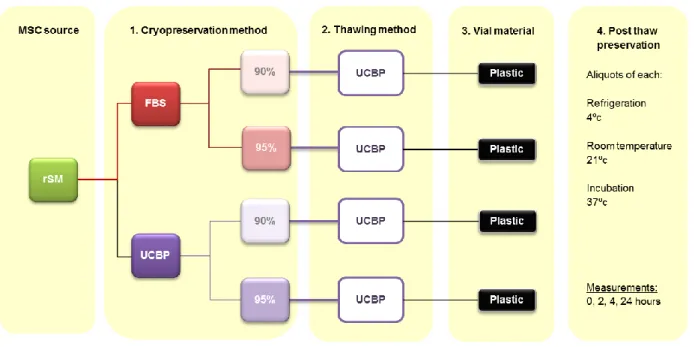
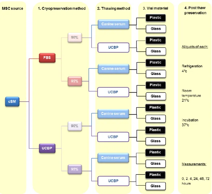
In order to achieve the aim of this study, four main objectives were proposed as described below and shown in figures 1a), 1b) and 1c):
- Improve cryopreservation protocols of MSCs by using two different media supplements, fetal bovine serum and human umbilical cord blood plasma (UCBP) at different percentages 90 and 95%;
- Determine the best thaw method for cryopreserved MSCs by thawing the samples using two different protein sources, canine serum and UCBP;
- Define the optimum preservation temperature of thawed MSCs over time;
- Establish the best vial material for conservation and transportation of thawed MSCs comparing results using plastic and glass vials.
To determine cell viability the trypan blue exclusion test of cell viability was performed on each sample.
In light of constant arising of new technologies and preclinical research results it is always possible to perfect existing cellular preservation techniques and that is what was attempted here.
8
Figure 1a) – Schematic representation of the experiments performed on the rSM MSCs sample.
9
Figure 1c) – Schematic representation of the experiments performed on the cSM MSCs sample.
Cell viability assay
To evaluate cell viability, the trypan blue exclusion test of cell viability was performed. A 1:1 dilution of the suspension was prepared by mixing 10µl of the cell suspension with 10µl of a 0.4% trypan blue solution and then filling the Countess™ II FL reusable glass slide. The viability of the total number of cells (a percentage of the viable unstained cells in suspension related to the total number of cells counted) was obtained by reading the slide in a Countess® II FL Automated Cell Counter (Thermo Fisher Scientific).
For each reading of the samples, four different fields of the slide were read and the mean of the percentage of viable cells was calculated.
10 Statistical analysis
All statistical tests were performed with GraphPad Prism version 6.00 for Mac OS X (GraphPad Software, La Jolla California USA).
In each sample reading, data were taken in quadruplicates. Data were presented as mean ± standard error of the mean (SEM). The SEM is an indication of the reliability of the mean. The smaller the SEM, the more accurate the sample mean as a reflection of the actual population mean.
Probabilities of significant differences were performed using ANOVA with a significance level of 0.05 and with p<0.05 to be considered statistically significant.
1. Cryopreservation of MSCs of different sources
One of the objectives of this study was to compare the cryopreservation of MSCs of three different sources, canine synovial (cSM) MSCs, canine dental pulp (cDP) MSCs and rat synovial (rSM) MSCs, using two different protocols, with either fetal bovine serum (FBS) or human umbilical cord blood plasma (UCBP), at a final concentration of 90 or 95% (figures 1a)-1c)). Synovial MSCs are an attractive cell source due to their high expansion and chondrogenic potentials when compared to MSCs of other sources (Nakagawa et al., 2015). They require minimally invasive harvesting techniques and can be used in autologous stem cell therapies (Hatakeyama et al., 2017).
Dental pulp MSCs, also represent an interesting adult stem cell source since they are easily recovered in large amount in a non-invasive manner from teeth extracted during routine dental procedures (Collart-Dutilleul et al., 2015; Potdar & Jethmalani, 2015) and have been isolated from several different species including humans (Iohara et al., 2006; Cheng et al., 2008; Alge et al., 2010; Mrozik et al., 2010; Nakatsuka et al., 2010; Potdar & Jethmalani, 2015). Another reason that makes them an interesting source involves their multipotent capabilities and the high plasticity they exhibit (Ledesma-Martínez et al., 2016).
The most frequent method for cryopreserving mammalian cells minimizing cryoinjury is by storing them in liquid nitrogen in complete medium comprising of basal medium supplemented with 10-20% FBS or solely in FBS, and in the presence of a cyoprotective agent (Martinello et al., 2011; Naaldijk et al., 2012). Our intention was to measure the effects of the use of two different media supplements, FBS and UCBP, at different supplementation concentrations (90 or 95%) and indirectly the use of low concentration of DMSO (10 or 5% respectively), on cellular number and viability .
11
The medium serves as a protein source for the cells and for the stabilization of the cell membrane during the crystallization process throughout the freezing or thawing process. FBS also acts as a protein shield for the toxic action of dimethyl sulfoxide (DMSO) in the early or late stages of the freezing and thawing processes, respectively (Liu et al., 2010).
FBS is still used as a standard media supplement for animal cell expansion and cryopreservation since it is a low-cost and easily to obtainable supplement that provides an optimal culture medium. It contains an abundance of proteins, growth factors, enzymes and other chemical components that make it ideal for exceptional growth enhancement (Ikebe & Suzuki, 2014).
Serum is the liquid portion of the blood after coagulation has occurred, thus fibrin and fibrin associated proteins are not present in serum. UCBP is an alternative to FBS as a media supplement but being plasma, and contrary to FBS, it still contains anti-coagulants and thus retains the proteins involved in the coagulation cascade.
Our cryopreservative of choice was DMSO at a final concentration of 5 or 10% accordingly. Cryoprotective agents reduce the freezing point of the medium and also allow a slower cooling rate, greatly reducing the risk of ice crystal formation, which can damage cells by rupture and cause cell death. The detergent action of the DMSO increases cell membrane permeability, increasing its resistance rupture from ice crystals formation (Liu et al., 2010). This study also served to indirectly evaluate the use of low DMSO concentrations.
a) Materials and Methods
Prior to cryopreservation, cellular concentration was determined by trypan blue staining using a Countess® II FL Automated Cell Counter (Thermo Fisher Scientific). The MSCs were resuspended in a solution containing either FBS at 90 or 95% or UCBP at 90 or 95% with a final concentration of DMSO of 5 or 10% accordingly. The cells were then aliquoted into cryogenic storage vials at a concentrations of 0.25 x 10⁶ cells/ml (rSM and cDP) and 1 x 10⁶ cells/ml (cSM) to be frozen slowly at 1°C/min. The vials, containing 1ml each, were placed in a pre-cooled Mr. Frosty® Cryo 1°C Freezing Container (Nalgene®), appropriately filled with 100% isopropyl alcohol and subsequently placed in a -80°C freezer. They were left undisturbed for a minimum of 4 hours and finally the cryovials were transferred to permanent, long-term liquid nitrogen storage, at -195.8ºC, until needed.
The samples remained cryopreserved in liquid nitrogen storage for a minimum of 9 days and a maximum of 30 days before being thawed and cell viability was assessed by trypan blue staining method immediately after thawing and removal of freezing solutions.
12
Good laboratory practices were followed at all times, from harvesting to cryopreserving the cells, to avoid contamination of the samples as well as cross-contamination of cell lines avoiding the need for the use of antibacterial and anti-mycotic agents.
b) Results and discussion
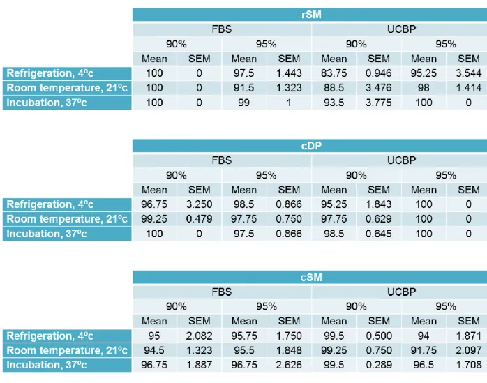
To interpret the effects of the two different media supplements, FBS or UCBP, at the different concentrations of 90 and 95% on post-thaw viability, the viability results immediately post-thaw with canine serum into plastic vials at 0 hours of all three sample sources, must be compared. The viability results of rat synovia MSCs, canine dental pulp MSCs and canine synovia MSCs are shown in table 1 as well as represented in figures 2, 8 and 9.
Table 1 – Post-thaw viability mean and SEM of rSM, cDP and cSM samples. Results are given as a percentage (%).
From these results, there appears to be better thawing results with the rSM samples that were cryopreserved with FBS at 90% and those with UCBP at 95%, when compared to those cryopreserved with FBS 95% and UCBP 90% yet they were not statistically significant.
13
rSM was the first batch used to perfect our technique so their results cannot be taken too much into account, since a proper thawing and counting technique had not been previously established.
In the cDP sample, on the other hand, cryopreservation with UCBP at 95% appears to yield the highest viability at 100±0% upon thawing but again these results were not statistically significant.
The cSM sample does not have statistically significant differences and that comes through in the results represented in table 1 since they are quite evenly distributed.
Therefore, there appears to be no effect on the viability of MSCs of these three sources upon cryopreserving with either FBS or UCBP at either 90 or 95%. This is quite useful information for it allows laboratories to choose either supplement according to its availability, price or shelf-life without compromising cell viability.
2. Thaw method
It is known that the thawing procedure is stressful to cryopreserved cells and they may be harmed in the process thus reducing cell viability and cell numbers. Several protocols have been established for this process with the purpose of preserving as many cells as possible but it has yet to be determined the exact cell viability loss during the process as well as the best method yet so we are left with room for improvement (Mitchell et al., 2015).
Canine serum and UCBP were compared as different protein sources during the thawing process and for post-thawed cells; one is a xenogeneic source or protein (UCBP), whereas when canine serum is used with the cDP and cSM samples, we are using an allogeneic protein source. This may present as an advantage since if xenogeneic protein sources have proven to be useful in this procedure, allogeneic sources should prove to be even more so.
It is very important to work quickly and follow good laboratory practices.
a) Materials and Methods
Each cryovial containing the cryopreserved cells, was removed from liquid nitrogen storage and immediately placed in a 37ºC water bath. The cryopreserved cells were then rapidly thawed, during 1 to 2 minutes, by gently swirling the vial in the 37ºC water bath until about 80% was defrosted, leaving just a small bit of ice left in the vial. This was necessary to avoid DMSO’s cytotoxicity.
14
The thawed cells were then washed with pre-warmed canine serum or UCBP and phosphate buffered saline (PBS). This step is important to wash off the cytotoxic DMSO. The cell suspension was then centrifuged at 1600 rpm for 10 minutes to remove any residual DMSO. After the centrifugation, the clarity of the supernatant was checked and a complete pellet identified. The supernatant was then aseptically decanted without disturbing the cell pellet. The cells were then gently resuspended in canine serum or UCBP accordingly, and divided into the necessary number of aliquots with 200-300µl each.
b) Results and discussion
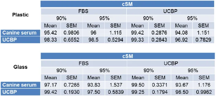
There were two different protein sources used during the thawing process of the cSM samples, canine serum and UCBP. Each sample that was cryopreserved using either 90 or 95% FBS and 90 or 95% UCBP was thawed using canine serum (allogeneic) or UCBP (xenogeneic) as demonstrated in figure 1c).
Those thawed into plastic vials and at 0 hours post-thaw are compared amongst themselves, as well as those thawed into glass vials and at 0 hours post-thaw (table 2). These results are also represented in figure 2.
This was not done for the rSM or cDP samples, those were only thawed with UCBP.
Table 2 - Post-thaw viability means and SEM values of cSM samples thawed using canine serum and UCBP into
15
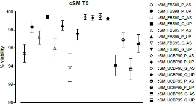
Figure 2 – Plotting of cSM viability values for all samples at 0 hours. Canine synovial mesenchymal stem cells (cSM);
fetal bovine serum (FBS); plastic (P); glass (G); canine serum (AS); human umbilical cord plasma, (UCBP, UP); 90% and 95% (90 and 95 respectively).
When comparing the corresponding viability values directly of the samples thawed using canine serum with those thawed using UCBP into either the plastic vials or the glass vials, there appear to be slightly better results with the samples thawed with UCBP in detriment of those thawed with canine serum, yet there were no statistically significant differences. UCBP viability results remained above 96.50±0.9962% whereas canine serum values went as low as 93.67±1.176%. The highest viability results for UCBP were 99.42±0.1930% whereas canine serum results managed to reach 99.50±0.3371%.
This being the case, canine serum could prove to be an adequate protein source for thawing cryopreserved canine MSCs. In light of these results, it would be useful to preform further thawing assays with canine MSCs from other sources for comparison as well as confirmation. If canine serum does prove to be an adequate protein source, the choice between using canine serum or UCBP would come down to a question of availability and costs. Given the possibility of the use of autologous serum, costs could be reduced since it is more readily available as blood may be collected from the same animal the MSCs are obtained from and at the same moment. Also, serum, contrary to plasma, does not require the addition of anticoagulants thus reducing costs further. Costs are always an issue in laboratory settings as well as costs of the final therapy so wherever they may be reduced is always a plus.
16
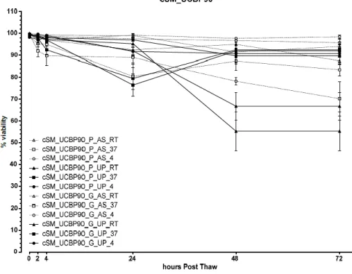
3. Vials and post-thaw preservation of the cSM samples
There are many different vials on the market for use in the laboratory and they may vary significantly in price. Nonetheless, they are all generally made of one of two materials: glass or plastic. Also, depending on their material, their shape may vary making them easier to work with or not.
The purpose of preparing two aliquots in different vial materials of the cSM MSC samples was to establish whether the material the MSCs were preserved in post-thaw interfered with the cellular viability. This was done over a 72 hour period.
This was not done for the rSM or cDP samples, they were thawed into plastic vials only.
The determination of the best temperature at which canine MSCs should be preserved post-thaw, is equally important, since in most clinical settings, especially in veterinary medicine, a laboratory isn’t always handy next to the patient we are trying to treat, nor is it viable to transport the patient to the laboratory so that the treatment may be applied. Therefore, medicine and other therapeutic substances must be conditioned for transportation without a significant alteration of their properties. This is the information we hope to obtain from this trial.
a) Materials and Methods
For the cSM samples, after being thawed with either canine serum or UCBP, six aliquots were prepared, three in glass vials and three in plastic vials and then one vial of each material was placed at each of the following temperatures: 4ºC (refrigeration), 21ºC (room temperature) and 37ºC (incubation).
Readings were taken at 0, 2, 4, 24, 48 and 72 hours post-thaw.
b) Results and discussion
At 0 hours post-thaw, there is no statistical significant difference between the results of the plastic or glass vials, as mentioned above. This is as expected since at 0 hours the vial material did not have the time to interact with the cell suspension and affect viability values. This can be seen in tables 2 and figure 2 above. Nonetheless, there is no apparent or statistical difference in viability throughout the 72 hours when comparing the results of either vial.
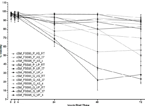
Differences do become apparent in the viabilities of the different temperature settings, particularly from 4 hours onwards. It becomes clear that viability is markedly compromised over time in the samples preserved at 37ºC (incubation) reaching values as low as 22±11.733% at
17
72 hours post-thaw (figure 4). In contrast, samples preserved in refrigeration at 4ºC maintain viability levels higher, only going as low as 83±0.913% at 72 hours post-thaw as can be seen in figure 6 below.
Figure 3 – Viability values (mean±SEM) post-thaw and over 72 hours for the cSM samples cryopreserved with
FBS90. Canine synovial mesenchymal stem cells (cSM); fetal bovine serum (FBS); plastic (P); glass (G); canine serum (AS); human umbilical cord plasma, (UCBP, UP); 90% (90).
Figure 4 – Viability values (mean±SEM) post-thaw and over 72 hours for the cSM samples cryopreserved with
FBS95. Canine synovial mesenchymal stem cells (cSM); fetal bovine serum (FBS); plastic (P); glass (G); canine serum (AS); human umbilical cord plasma, (UCBP, UP); 95% (95).
18
Figure 5 – Viability values (mean±SEM) post-thaw and over 72 hours for the cSM samples cryopreserved with
UCBP90. Canine synovial mesenchymal stem cells (cSM); plastic (P); glass (G); canine serum (AS); human umbilical cord plasma, (UCBP, UP); 90% (90).
Figure 6 – Viability values (mean±SEM) post-thaw and over 72 hours for the cSM samples cryopreserved with
UCBP95. Canine synovial mesenchymal stem cells (cSM); plastic (P); glass (G); canine serum (AS); human umbilical cord plasma, (UCBP, UP); 95% (95).
19
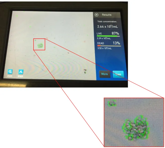
Also, during the study, in the samples thawed with canine serum, agglomerates of viable cells (as those shown in figure 7 below), were seen in more or less quantity and size throughout the samples during the cell viability assay in each reading. This may have rendered the viability and total cell readings inaccurate. They were more present and of larger size in the samples maintained at 37ºC especially after 4 hours which is probably the reason why there was an increase in the SEM value (as seen in figures 3-6), indicating that the readings might not be very precise.
Figure 7 – Agglomerates of viable cells observed while accessing cell viability in cSM samples thawed with canine
serum.
This is important since when samples were evaluated in the previous point (2. Thaw method), at 0 hours, immediately post-thaw, agglomerates we not visible leading one to think of canine serum as an eligible protein source during the thaw process. If focusing only on the statistical data obtained at that time, there would appear to be no important differences between the results from the canine serum and the UCBP immediately post-thaw.
20
Observing the agglomerates it becomes unclear whether the canine serum could be suitable in the thawing process. The reason for the formation of agglomerates must be investigated further since this might hold back the use of a cheap and readily available allogeneic source of protein and an autologous source may be required.
Also, in a previous study by Mitchell and his team in 2015 equine allogeneic serum was used during the cryopreservation stage and similar results to cryopreserving with FBS were obtained. This might be an indication that it is also possible to use allogeneic serum during the thawing process but further experiments must be conducted. There is no mention of the formation of cellular agglomerates so the reason for this to have happened in our study should be explored. A possible reason for this may reside in the fact that Mitchell and his colleagues used commercial serum that might have been subjected to certain processes, eliminating antibodies for example, whereas non-commercial serum collected in house was used here.
Autologous protein sources are a very interesting possibility, even more than allogeneic sources, since it may allow for serum to be collected together with of the collection of the MSCs, from a specific individual. This may serve to reduce costs, allowing for the serum to be stored for use later on in the process, as well as maybe reduce rejection reactions since both MSCs and serum would be from the same origin.
4. Post-thaw preservation of the rSM and cDP samples
As mentioned previously, it is just as imperative as the previous determinations to conclude the optimum temperature at which cells must be conserved after undergoing cryopreservation and subsequent thawing process, and for how long, in order to maintain a high viability and cell number, so that they can be applied at a later time.
This determination will enable calculation of the quantity of viable cells needed to be cryopreserved so that an exact number be recovered after the thawing process and transferred to the animal being treated. Furthermore, it will allow us to determine the best conservation method post-thaw until the cells can be administered to the patient maintaining the highest number of cells and viability possible, increasing the ‘window of opportunity’ to allow for the transportation of therapeutic doses from the preparation facilities to the clinic or application site (in the case of ambulatory application).
Three different temperature settings were tested, refrigeration at 4ºC, room temperature at 21ºC and incubation at 37ºC for the rSM and cDP MSCs samples over a period of 24 hours.
21 a) Materials and Methods
Three aliquots of each rat synovia and canine dental pulp MSCs samples (90 and 95% FBS and 90 and 95% UCBP) were prepared as described previously and then placed at different temperatures: one at 4ºC in refrigeration, another at 21ºC room temperature and the third incubated at 37ºC over a period of 24 hours for each of the samples.
Readings were taken at 0, 2, 4 and 24 hours post-thaw.
b) Results and discussion
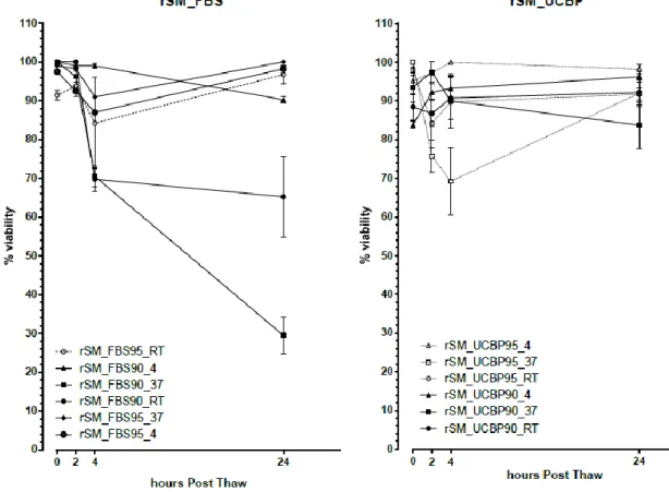
In the rSM samples (figure 8) cryopreserved with FBS at 95%, maintained at room temperature, there is an increase in viability from the 4 hour reading, 91±4.796SEM, to the 24 hour reading 100±0% probably due to the dead, non-viable cells suffering lysis and therefore no longer being taken into account. The same happened with the rSM samples cryopreserved with FBS at 95% maintained at 4ºC and 37ºC. There are a few more inconsistencies of the like with the rSM samples cryopreserved with UCBP.
The rSM samples were the first used in the study and served to establish thawing and counting procedures so their results might be inconsistent and compromised due to that.
Figure 8 – Viability values (mean±SEM) post-thaw and over 24 hours for the rSM samples cryopreserved with
UCBP95. Rat synovial mesenchymal stem cells (rSM); fetal bovine serum (FBS); human umbilical cord plasma, (UCBP, UP); 90 and 95% (90 and 95 respectively).
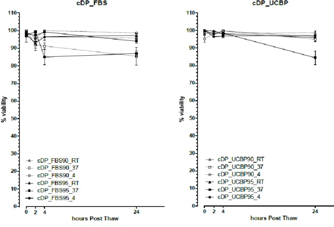
22
Otherwise, in the cDP samples (figure 9), there seems to more consistency in the viability percentage loss in each of the comparable samples where the samples cryopreserved in FBS 90 and 95% preserved at 37ºC had a viability of only 85.5±4.992% and 87±1.472% at 24 hours respectfully whereas the equivalent samples preserved at 4ºC had a viability of 98.75±0.479% and 94±1.354% at the end of the 24 hours. In the same way, the samples cryopreserved in UCBP 90 and 95% preserved at 37ºC had a viability of 95±1.414% and 84.5±3.884% at 24 hours respectfully whereas the equivalent samples preserved at 4ºC had a viability of 96.5±0.957% and 97.250±1.031% at the end of the 24 hours.
Similarly to what happened with the rSM samples, in some cDP samples there was an increase in the mean viability values from the 4 hour readings to the 24 hour readings, but there was also an increase in the SEM.
Figure 9 – Viability values (mean±SEM) post-thaw and over 24 hours for the rSM samples cryopreserved with
UCBP95. Rat synovial mesenchymal stem cells (rSM); fetal bovine serum (FBS); human umbilical cord plasma, (UCBP, UP); 90 and 95% (90 and 95 respectively).
In conclusion, and as shown in figures 3-6, 8 and 9, the viability loss of the samples maintained at 4ºC, over the 24 hour period, was generally lower than that of those maintained at 21ºC and 37ºC being the latter the ones presenting the worst results, rendering statistically significant results.
23 VI.CONCLUSION
It is clear that high and predictable survival rates for cell cultures are important due to the time consuming, costly and difficult preparation processes. Consequently, methods used for cell cryopreservation must ensure the highest viability possible.
There has been a large amount of developmental work undertaken to ensure successful cryopreservation and thawing of cell culture yet the basic principle of successful cryopreservation and thawing continues to be a slow freeze and quick thaw although there are other important factors that influence the rate of success such as the quality and type of reagents used and the work method.
Besides the cryopreservation and thawing methods, other factors that can affect the viability of cryopreserved cells include growth conditions prior to harvesting, the physiological state of the cells and the cell density.
As mentioned before, the purpose of this study was to improve cryopreservation and thawing methods as well as to establish the best preservation temperature of thawed MSCs over time enabling cells to be transported to their location of use without reducing their viability.
It was possible to determine that regarding cryopreservation technique, there is no significant difference in loss of viability between using FBS or UCBP as a serum/protein and that supplementation of up to 95%, and only 5% DMSO may be used not compromising cell viability. During the thawing process, UCBP and canine serum presented similar results in viability leading one to conclude that canine serum might be an adequate protein source for the thawing process but once post-thaw viability was evaluated over time, it became clear that this was not so. The samples thawed with canine serum formed cellular agglomerates compromising them and ultimately one must conclude that UCBP might be a better choice. Nonetheless, there have been previous studies where allogeneic sources of protein source have rendered satisfying results and did not encounter these problems although they were used as a serum supplementation source during the cryopreservation process and not the thawing process. If the formation of agglomerates could be solved (for example by removing antigens) it would give leeway for laboratory choice of protein source depending on accessibility, costs and shelf-life for example. A new protocol could be implemented where, at the same time as MSCs are collected, blood is drawn as to be used in the thaw process of those cells later on (an autologous source of serum) or of MSCs of other individuals of the same species (as an allogeneic source of serum). Further tests can be performed to conclude whether allogeneic serum is an acceptable protein source for this process in all species thus significantly reducing costs of acquisition of commercial reagents.
24
However, it became very apparent that when it comes to preserving cells post-thaw, the best method is to do so is at 4ºC, in refrigeration, or at least the lowest temperature possible since room temperature (21ºC) also yielded better viability results than incubation at 37ºC. This is extremely useful, especially in the veterinary field, where veterinarians must frequently travel to apply the therapies to their patients. It is important to know how long post-thaw cells will remain with an acceptable viability, above a desired percentage, so as to calculate the number of cells needed to be cryopreserved for a given treatment.
This study still leaves room for improvement. A study with clear and separate experiments for each of the objectives of the present study would allow for a more clear and accurate interpretation of the results. As with any study, an increased amount of samples and readings is always beneficial and would allow for a diminution of the SEM value, rendering more accurate means.
Also, each of these experiments done individually with total cells numbers together with viabilities would provide us with a lot of useful information since we may have a sample with 100% viability but a significant reduction in the total cell number by cell lysis for example, or the formation of agglomerates as was the case in this study.
Despite enthusiastic levels of interest in the field of cell therapy where MSCs are presenting to be of great use, the reality is that other than the use of haematopoietic stem cells, many other therapies have yet to be thoroughly proven for their therapeutic benefit and safety in application (Lin et al., 2013).
25 VII.REFERENCES
Afanasyev B. V., Elstner E. E., Zander A. R., (2009) "A. J. Friedenstein, founder of the mesenchymal stem cell concept". In Cellular Therapy and Transplantation, 1, 3, 35-38.
Alge D. L., Zhou D., Adams L. L., Wyss B. K., Shadday M. D., Woods E. J., Gabriel Chu T. M., Goebel W. S., (2010) "Donor-matched comparison of dental pulp stem cells and bone marrow-derived mesenchymal stem cells in a rat model". In J Tissue Eng Regen Med, 4, 1, 73-81. Bhattacharyya S., Khanduja K. L., (2010) "New hope in the horizon: cancer stems cells". In Acta Biochimica et Biophysica Sinica, 42, 4, 237-242.
Bianco P., Robey P. G., Simmons P. J., (2008) "Mesenchymal stem cells: revisiting history, concepts, and assays". In Cell Stem Cell, 2, 4, 313-319.
Caplan A. I., Dennis J. E., (2006) "Mesenchymal stem cells as trophic mediators". In J Cell Biochem, 98, 5, 1076-1084.
Cheng P. H., Snyder B., Fillos D., Ibegbu C. C., Huang A. H., Chan A. W., (2008) "Postnatal stem/progenitor cells derived from the dental pulp of adult chimpanzee". In BMC Cell Biology, 9, 20.
Collart-Dutilleul P.-Y., Chaubron F., De Vos J., Cuisinier F.J., (2015) "Allogenic banking of dental pulp stem cells for innovative therapeutics". In World Journal of Stem Cells, 7, 1010-1021.
Cooper R. L., (1998) "Development of sensory processes during limb regeneration in adult crayfish". In Journal of Experimental Biology, 201, 11, 1745-1752.
Cristofalo V. J., Lorenzini A., Allen R. G., Torres C., Tresini M., (2004) "Replicative senescence: a critical review". In Mechanisms of Ageing and Development, 125, 10–11, 827-848.
de Almeida P. E., Ransohoff J. D., Nahid M. A., Wu J. C., (2013) "Immunogenicity of Pluripotent Stem Cells and Their Derivatives". In Circulation research, 112, 3, 549-561.
De Schauwer C., Goossens K., Piepers S., Hoogewijs M. K., Govaere J. L., Smits K., Meyer E., Van Soom A., Van de Walle G. R., (2014) "Characterization and profiling of immunomodulatory genes of equine mesenchymal stromal cells from non-invasive sources". In Stem Cell Research & Therapy, 5, 1, 6.
DiMarino A., Caplan A., Bonfield T., (2013) "Mesenchymal Stem Cells in Tissue Repair". In Frontiers in Immunology, 4, 201.
Dominici M., Le Blanc K., Mueller I., Slaper-Cortenbach I., Marini F., Krause D., Deans R., Keating A., Prockop D., Horwitz E., (2006) "Minimal criteria for defining multipotent
26
mesenchymal stromal cells. The International Society for Cellular Therapy position statement". In Cytotherapy, 8, 4, 315-317.
El-Hashash A., (2016) "Developmental and Stem Cell Biology in Health and Disease", p. 260. Bentham Science Publishers.
Fortier L. A., Travis A. J., (2011) "Stem cells in veterinary medicine". In Stem Cell Research & Therapy, 2, 1, 9.
Giai Via A., Frizziero A., Oliva F., (2012) "Biological properties of mesenchymal Stem Cells from different sources". In Muscles, Ligaments and Tendons Journal, 2,3, 154-162.
Gurtner G. C., Werner S., Barrandon Y., Longaker M. T., (2008) "Wound repair and regeneration". In Nature, 453, 314-321.
Hatakeyama A., Uchida S., Utsunomiya H., Tsukamoto M., Nakashima H., Nakamura E., Pascual-Garrido C., Sekiya I., Sakai A., (2017) "Isolation and Characterization of Synovial Mesenchymal Stem Cell Derived from Hip Joints: A Comparative Analysis with a Matched Control Knee Group". In Stem Cells International, 2017, 1-13.
Hayflick L., (1965) "The limited in vitro lifetime of human diploid cell strains". In Experimental Cell Research, 37, 3, 614-636.
Hayflick L., Moorhead P. S., (1961) "The serial cultivation of human diploid cell strains". In Experimental Cell Research, 25, 3, 585-621.
Heathman T. R. J., Nienow A. W., McCall M. J., Coopman K., Kara B., Hewitt C. J., (2015) "The translation of cell-based therapies: clinical landscape and manufacturing challenges". In Regenerative Medicine, 10, 1, 49-64.
Hillmann A., Ahrberg A. B., Brehm W., Heller S., Josten C., Paebst F., Burk J., (2016) "Comparative Characterization of Human and Equine Mesenchymal Stromal Cells: A Basis for Translational Studies in the Equine Model". In Cell Transplantation, 25,1, 109-124.
Hou P., Li Y., Zhang X., Liu C., Guan J., Li H., Zhao T., Ye J., Yang W., Liu K., Ge J., Xu J., Zhang Q., Zhao Y., Deng H., (2013) "Pluripotent Stem Cells Induced from Mouse Somatic Cells by Small-Molecule Compounds". In Science, 341,6146, 651-654.
Hunt C. J., (2011) "Cryopreservation of Human Stem Cells for Clinical Application: A Review". In Transfusion Medicine and Hemotherapy, 38,2, 107-123.
Hyun I., (2010) "The bioethics of stem cell research and therapy". In The Journal of Clinical Investigation, 120,1, 71-75.
27
Ikebe C., Suzuki K., (2014) "Mesenchymal Stem Cells for Regenerative Therapy: Optimization of Cell Preparation Protocols". In BioMed Research International, 2014, 11.
Iohara K., Zheng L., Ito M., Tomokiyo A., Matsushita K., Nakashima M., (2006) "Side Population Cells Isolated from Porcine Dental Pulp Tissue with Self-Renewal and Multipotency for Dentinogenesis, Chondrogenesis, Adipogenesis, and Neurogenesis". In STEM CELLS, 24,11, 2493-2503.
Juanes F., Smith L. D., (1995) "The ecological consequences of limb damage and loss in decapod crustaceans: a review and prospectus". In Journal of Experimental Marine Biology and Ecology, 193,1, 197-223.
Lane S. W., Williams D. A., Watt F. M., (2014) "Modulating the stem cell niche for tissue regeneration". In Nature biotechnology, 32,8, 795-803.
Ledesma-Martínez E., Mendoza-Núñez V. M., Santiago-Osorio E., (2016) "Mesenchymal Stem Cells Derived from Dental Pulp: A Review". In Stem Cells International, 2016, 4709572.
Lin H.-T., Otsu M., Nakauchi H., (2013) "Stem cell therapy: an exercise in patience and prudence". In Philosophical Transactions of the Royal Society B: Biological Sciences, 368,1609, 20110334.
Liu Y., Xu X., Ma X., Martin-Rendon E., Watt S., Cui Z., (2010) "Cryopreservation of human bone marrow-derived mesenchymal stem cells with reduced dimethylsulfoxide and well-defined freezing solutions". In Biotechnology Progress, 26,6, 1635-1643.
Mafi R., Hindocha S., Mafi P., Griffin M., Khan W. S., (2011) "Sources of Adult Mesenchymal Stem Cells Applicable for Musculoskeletal Applications - A Systematic Review of the Literature". In The Open Orthopaedics Journal, 5, 242-248.
Maginnis T. L., (2006) "The costs of autotomy and regeneration in animals: a review and framework for future research". In Behavioral Ecology, 17,5, 857-872.
Mao A. S., Mooney D. J., (2015) "Regenerative medicine: Current therapies and future directions". In Proceedings of the National Academy of Sciences of the United States of America, 112,47, 14452-14459.
Martinello T., Bronzini I., Maccatrozzo L., Mollo A., Sampaolesi M., Mascarello F., Decaminada M., Patruno M., (2011) "Canine adipose-derived-mesenchymal stem cells do not lose stem features after a long-term cryopreservation". In Research in Veterinary Science, 91,1, 18-24. Meirelles L. d. S., Chagastelles P. C., Nardi N. B., (2006) "Mesenchymal stem cells reside in virtually all post-natal organs and tissues". In Journal of Cell Science, 119,11, 2204-2213.
28
Melton D., 2014: Chapter 2 - ‘Stemness’: Definitions, Criteria, and Standards. Essentials of Stem Cell Biology, Third Edition, 7-17, Academic Press, Boston.
Metcalfe A. D., Ferguson M. W. J., (2007) "Tissue engineering of replacement skin: the crossroads of biomaterials, wound healing, embryonic development, stem cells and regeneration". In Journal of The Royal Society Interface, 4,14, 413-437.
Mitchell A., Rivas K. A., Smith R., Watts A. E., (2015) "Cryopreservation of equine mesenchymal stem cells in 95 % autologous serum and 5 % DMSO does not alter post-thaw growth or morphology in vitro compared to fetal bovine serum or allogeneic serum at 20 or 95 % and DMSO at 10 or 5 %". In Stem Cell Research & Therapy, 6, 231.
Mrozik K. M., Zilm P. S., Bagley C. J., Hack S., Hoffmann P., Gronthos S., Bartold P. M., (2010) "Proteomic characterization of mesenchymal stem cell-like populations derived from ovine periodontal ligament, dental pulp, and bone marrow: analysis of differentially expressed proteins". In Stem Cells Dev, 19,10, 1485-1499.
Naaldijk Y., Staude M., Fedorova V., Stolzing A., (2012) "Effect of different freezing rates during cryopreservation of rat mesenchymal stem cells using combinations of hydroxyethyl starch and dimethylsulfoxide". In BMC Biotechnology, 12,1, 49.
Nakagawa Y., Muneta T., Kondo S., Mizuno M., Takakuda K., Ichinose S., Tabuchi T., Koga H., Tsuji K., Sekiya I., (2015) "Synovial mesenchymal stem cells promote healing after meniscal repair in microminipigs". In Osteoarthritis and Cartilage, 23,6, 1007-1017.
Nakatsuka R., Nozaki T., Uemura Y., Matsuoka Y., Sasaki Y., Shinohara M., Ohura K., Sonoda Y., (2010) "5-Aza-2'-deoxycytidine treatment induces skeletal myogenic differentiation of mouse dental pulp stem cells". In Archives of Oral Biology, 55,5, 350-357.
Polge C., Smith A. U., Parkes A., (1949) "Revival of spermatozoa after vitrification and dehydration at low temperatures". In Nature, 164,4172, 666.
Potdar P. D., Jethmalani Y. D., (2015) "Human dental pulp stem cells: Applications in future regenerative medicine". In World Journal of Stem Cells, 7,5, 839-851.
Ramalho-Santos M., Willenbring H., (2007) "On the Origin of the Term "Stem Cell"". In Cell Stem Cell, 1,1, 35-38.
Rink J. C., (2013) "Stem cell systems and regeneration in planaria". In Development Genes and Evolution, 223,1-2, 67-84.
Ross C. A., Akimov S. S., (2014) "Human-induced pluripotent stem cells: potential for neurodegenerative diseases". In Human Molecular Genetics, 23,R1, R17-R26.
29
Rouhana L., Tasaki J., (2016) "Epigenetics and Shared Molecular Processes in the Regeneration of Complex Structures". In Stem Cells International, 2016, 9.
Salem H. K., Thiemermann C., (2010) "Mesenchymal Stromal Cells: Current Understanding and Clinical Status". In STEM CELLS, 28,3, 585-596.
Schroeder T., (2008) "Imaging stem-cell-driven regeneration in mammals". In Nature, 453,7193, 345-351.
Squillaro T., Peluso G., Galderisi U., (2016) "Clinical Trials With Mesenchymal Stem Cells: An Update". In Cell Transplant, 25,5, 829-848.
Tang C., Weissman I. L., Drukker M., 2013: Immunogenicity of In Vitro Maintained and Matured Populations: Potential Barriers to Engraftment of Human Pluripotent Stem Cell Derivatives. In: N. Zavazava (ed.), Embryonic Stem Cell Immunobiology: Methods and Protocols. Humana Press, Totowa, NJ.
Taran R., Mamidi M. K., Singh G., Dutta S., Parhar I. S., John J. P., Bhonde R., Pal R., Das A. K., (2014) "In vitro and in vivo neurogenic potential of mesenchymal stem cells isolated from different sources". In Journal of Biosciences, 39,1, 157–169.
Wagner D. E., Wang I. E., Reddien P. W., (2011) "Clonogenic Neoblasts Are Pluripotent Adult Stem Cells That Underlie Planarian Regeneration". In Science, 332,6031, 811-816.
Wei X., Yang X., Han Z.-p., Qu F.-f., Shao L., Shi Y.-f., (2013) "Mesenchymal stem cells: a new trend for cell therapy". In Acta Pharmacol Sin, 34,6, 747-754.