Miguel Pedro Lopes Batista
Degree in Biology
Novel Alginate-Chitosan Aerogel Fibres
For Potential Wound Healing
Applications
Dissertation to obtain Master Degree in Biotechnology
Supervisor: Vanessa Gonçalves, PhD, iBET
Co-supervisor: Frédéric Gaspar, PhD, iBET
Jury
President: Prof. Doutora Susana Filipe Barreiros Arguer: Prof. Doutora Ana Rita Cruz Duarte Supervisor: Doutora Vanessa Gonçalves
Miguel Pedro Lopes Batista
Degree in BiologyNovel Alginate-Chitosan Aerogel Fibres for Potential Wound
Healing Applications
Dissertation to obtain Master Degree in Biotechnology
Supervisor: Vanessa Gonçalves, PhD, iBET
Co-supervisor: Frédéric Gaspar, PhD, iBET
Jury
President: Prof. Doutora Susana Filipe Barreiros Arguer: Prof. Doutora Ana Rita Cruz Duarte Supervisor: Doutora Vanessa Gonçalves
“Copyright”
Novel Alginate-Chitosan Aerogel Fibres for Potential Wound Healing Applications
Miguel Pedro Lopes Batista, FCT/UNL e UNL
VII Acknowledgments
A todos aqueles que direta ou indiretamente contribuíram para que esta etapa fosse alcançada, o meu profundo agradecimento.
Aos meus orientadores, à Vanessa e ao Frédéric, agradeço-vos por toda a confiança depositada e por todo o conhecimento que me transmitiram. A maneira como ambos pensam a ciência e a vossa capacidade multidisciplinar foram sem dúvida a inspiração que me orientou neste trabalho e me irá orientar para o futuro.
À Doutora Ana Matias agradeço a oportunidade de me ter permitido desenvolver este trabalho no grupo Nutraceuticals & Bioactives Process Technology. Deixar também uma palavra de profundo agradecimento a todos os membros deste grupo pela ajuda e companheirismo prestado. Um obrigado especial à Carolina e à Ana Roda pelo vosso apoio. À Doutora Teresa Crespo e a todos os membros dos grupos Food Safety & Microbiology e Membrane Processes pela vossa disponibilidade e boa disposição.
Ao Baixinho e ao Pedro, obrigado pela vossa presença e por todas as felicidades e tristezas partilhadas ao longo desta caminhada. Um profundo obrigado aos amigos de longa data Simão, Diogo, Pimentel, Louro, Salomé, Melo e David pelo vosso apoio e compreensão da minha ausência nos momentos mais atribulados. Aos grandes amigos de Coimbra, que mesmo longe se fizeram sentir sempre perto,
obrigado Costa, Bibiana, Sandro, Cardoso e Daniel. Aos companheiros “lisboetas”, obrigado Sofia,
Serafim, Tiago e Adriana, que me fizeram sentir menos deslocado nesta aventura.
IX Abstract
Aerogels are very interesting materials with high porosity whose wound healing applications are arousing great interest. In particular, aerogels produced from marine polymers are of particular interest due to their attractive properties such as the antimicrobial activity of chitosan or the capacity to provide a moist environment of alginate. The aim of this work was to evaluate the potential for wound healing applications of alginate-chitosan aerogels in the form of fibres. To produce the fibres, a polyelectrolyte complex hydrogel of both polymers was made by the emulsion-gelation method. Through solvent exchange an alcogel was obtained which was then dried with supercritical CO2. Once the fibres were
produced, the characterization of its solid state, biocompatibility, cell migration stimulation and antimicrobial activity were carried out. To characterize the solid state, determination of the fibre’s
chitosan content was first performed. Then, the morphology, its textural properties and the ionic interaction between both polymers was also analysed. Fibres biocompatibility and stimulation of cell migration were evaluated by two in vitro methods, the direct contact method described in ISO 10993-5 and the scratch assay, respectively, using in both methods the mouse fibroblast NCTC clone 929 cell line. The antimicrobial activity was evaluated against Staphylococcus aureus and Klebsiella pneumoniae by two standard methods (dynamic and static) described in ASTM E 2149-01 and in ISO 20743:2013, respectively. In this work, the influence of chitosan’s molecular weight and content on the fibre characteristics was also evaluated. In addition, in order to compare with a consumer product already on the market, cell and antibacterial assays were also performed for a dry calcium-sodium alginate wound dressing. The obtained results suggest that these alginate-chitosan aerogel fibres are good candidates for wound healing applications.
Keywords
XI Resumo
Aerogéis são materiais muito interessantes com elevada porosidade, cujas aplicações em tratamento de feridas estão a despertar interesse. Particularmente, a produção de aerogéis a partir de polímeros marinhos apresentam particular interesse devido às suas propriedades atrativas, tais como a atividade antimicrobiana do quitosano ou a capacidade de proporcionar um ambiente húmido do alginato. O objetivo deste trabalho foi avaliar o potencial de aerogéis de alginato-quitosano na forma de fibras para aplicações em tratamentos de feridas. Para produzir as fibras, produziu-se um complexo polieletrolítico na forma de hidrogel entre os polímeros pelo método de emulsão-gelificação. Através do processo de troca de solvente foi obtido um alcogel o qual foi secado por CO2 supercrítico. Assim que
produzidas, realizou-se a caracterização do estado sólido, biocompatibilidade, estimulação da migração celular e atividade antimicrobiana das fibras. Para caracterizar o estado sólido, começou-se por determinar o conteúdo de quitosano. Em seguida, a morfologia, as propriedades texturais e a interação iónica entre os dois polímeros também foram analisadas. A biocompatibilidade e a estimulação da migração celular foram respetivamente avaliadas através de dois métodos in vitro, o método por contato direto descrito na ISO 10993-5 e o ensaio de migração celular, utilizando em ambos a linha celular de fibroblastos de ratinhos NCTC clone 929. A atividade antimicrobiana foi avaliada em Staphylococcus aureus e Klebsiella pneumoniae por dois métodos padrão (dinâmico e estático) descritos na norma ASTM E 2149-01 e na ISO 20743:2013. Neste trabalho também foi avaliada a influência do peso molecular e do conteúdo de quitosano nas características das fibras. Além disso, de modo a comparar com um produto de consumo já existente no mercado, os ensaios celulares e antibacterianos também foram realizados para um apósito de alginato de cálcio e de sódio. Os resultados obtidos sugerem elevado potencial destas fibras para aplicações em tratamentos de feridas.
Palavras-chave
XIII List of Contents
Acknowledgments ...VII
Abstract ...IX
Keywords ...IX
Resumo ...XI
Palavras-chave ...XI
List of Contents ...XIII
List of Figures ... XV
List of Tables ... XVII
List of Abbreviations... XIX
1. Introduction ... 1
1.1 Aerogels production ... 1
1.2 Aerogels from biopolymers ... 3
1.2.1 Alginate ... 4
1.2.2 Chitosan... 6
1.2.3 Polyelectrolyte complexes ... 8
1.3 Biomedical applications of aerogels ... 10
1.3.1 Wound healing process ... 10
1.3.2 Wound dressings materials ... 11
1.4 Aim of the thesis ... 13
2. Materials and Methods ... 15
2.1 Aerogel fibres production ... 15
2.1.1 Reagents ... 15
2.1.2 Stock solutions ... 15
2.1.3 Sol-Gel process ... 15
2.1.4 Solvent exchange ... 16
2.1.5 Drying with supercritical carbon dioxide ... 16
2.2 Fibres weighing and sterilization ... 17
2.3 Solid-state characterization ... 17
2.3.1 Scanning electron microscopy (SEM) ... 17
2.3.2 Fourier-transform infrared spectroscopy (FTIR)... 17
2.3.3 Brunauer-Emmett-Teller (BET) surface area analysis and Barrett-Joyner-Halenda (BJH) pore size and volume analysis ... 18
2.3.4 Elemental analysis (EA) ... 18
2.4 Cell-based assays ... 18
2.4.1 Reagents ... 18
2.4.2 Cell culture ... 18
XIV
2.4.4 Bioactivity - Scratch assay. ... 19
2.5 Antibacterial activity evaluation assays... 20
2.5.1 Materials ... 20
2.5.2 Bacterial test species, bacterial suspensions and standardized inoculum preparation ... 20
2.5.3 Well diffusion and agar plate methods ... 21
2.5.4 ASTM E 2149-01 standard test method for determining the antimicrobial activity of immobilized antimicrobial agents under dynamic contact conditions. ... 21
2.5.5 ISO 20743:2013(E) Textiles — Determination of antibacterial activity of textile products by absorption method. ... 21
2.5.6 Antimicrobial susceptibility testing by broth microdilution ... 22
2.6 Statistical analysis ... 23
3. Results and Discussion ... 25
3.1 Screening and optimization of assays for evaluation of potential wound healing applications of alginate-chitosan aerogel fibres ... 25
3.1.1 Aerogel fibres production ... 25
3.1.2 Implementation of sterilization method ... 25
3.1.3 Implementation of cell culture ... 25
3.1.4 Optimization of cell-based assays ... 26
3.1.4.1 Biocompatibility – ISO 10993-5 in vitro cytotoxicity assay ... 26
3.1.4.2 Bioactivity - Scratch assay ... 26
3.1.5 Evaluation of antimicrobial activity: screening and assays optimization ... 26
3.1.5.1 Well diffusion and agar plate methods ... 26
3.1.5.2 Static method ... 26
3.1.5.3 Dynamic method ... 27
3.2 Evaluation of alginate-chitosan aerogel fibres for potential wound healing applications ... 27
3.2.1 Influence of chitosan molecular weight ... 27
3.2.2 Influence of chitosan content ... 34
4. Conclusion ... 45
XV List of Figures
Figure 1.1 - Main steps of the production process of an aerogel. ... 2 Figure 1.2 - Pressure versus temperature phase diagram of CO2 indicating the critical point and the sub-
and super-critical regions [5]. ... 2 Figure 1.3 - (a) Scheme of the forces exerted by the surface tension due to the interface of gas-liquid
phases; (b) Effect of gel drying method: gel monolyths of pectin of the same dimensions prepared by thermal gelation dried under supercritical drying (aerogel) and under air drying (xerogel) [4]. 3 Figure 1.4 - Representative alginate structure: G-block, M-block, and alternating MG-blocks in alginate
[13]. ... 5 Figure 1.5 - Structure of chitin and chitosan (reproduced from [22]). ... 7 Figure 1.6 - Schematic representation of polyanion-polycation interactions: mixing of the oppositely
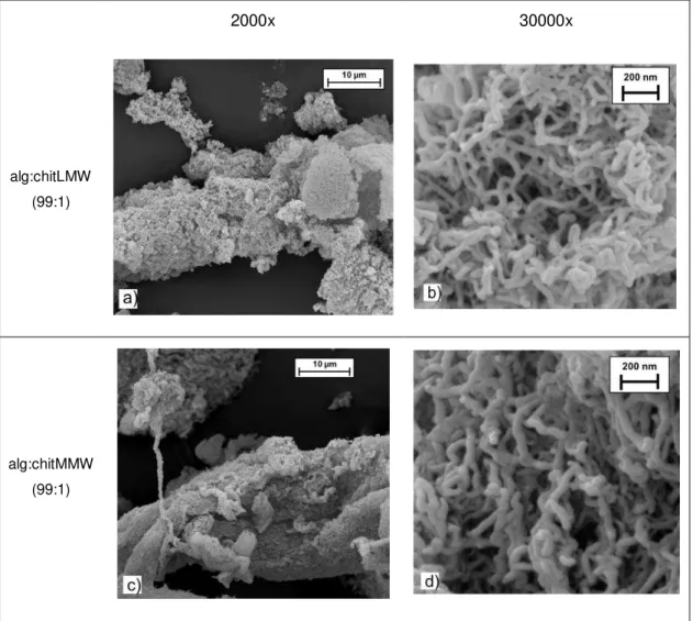
charged polyelectrolytes in aqueous solution leads to formation of a dense phase (PEC) caused by the cooperative electrostatic interactions between the ions [28], [31]. ... 9 Figure 1.7 - Work plan for the present thesis organized in two main tasks. ... 13 Figure 2.1 - Schematic diagram of the supercritical fluid drying apparatus. (Adapted from Waters, 2010) ... 17 Figure 3.1 - Photographs of the aerogel fibres produced with different chitosan MW (low and medium). ... 27 Figure 3.2 - SEM micrographs of alg:chitLMW 99:1 (a,b) and alg:chitMMW 99:1 (c,d) aerogel fibres, at
2000x (a,c) and 30000x (b,d) magnification. ... 29 Figure 3.3 - FTIR spectra of (a) sodium alginate, (b) MMW chitosan and (c) LMW chitosan raw materials;
(d) alg:chitMMW 99:1 and (e) alg:chitLMW 99:1 aerogel fibres. ... 30 Figure 3.4 - BET surface area (a) and BJH pore volume (b) of alg:chitLMW 99:1 and alg:chitMMW 99:1
aerogel fibres. ... 31 Figure 3.5 - Cytotoxicity assay using MTS reagent: samples (fibres and pure compounds) were
incubated, at a concentration of 1.7 mg/mL, in NCTC clone 929 cell line during 24 h at 37ºC and 5% CO2 humidified atmosphere (mean ± SD, n=3; except controls n=6). Solution of 10% (v/v) of
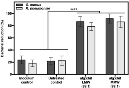
DMSO in cell culture media was used as a positive cytotoxic control. If viability is reduced to <70% of the control, samples have a cytotoxic potential. Statistically significant differences comparing all conditions are indicated by **** (p < 0.0001). ... 32 Figure 3.6 - Percent reduction of S. aureus and K. pneumoniae resulting from contact with samples, at
XVI Figure 3.8 - SEM micrographs of alg:chitLMW 99:1 (a,b), alg:chitLMW 19:1 (c,d) and alg:chitLMW 9:1 (e,f) aerogel fibres, at 2,000x (a,c,e) and 30,000x (b,d,f) magnification. ... 36 Figure 3.9 - FTIR spectra of (a) sodium alginate and (b) LMW chitosan raw materials; (c) alg:chitLMW
9:1, (d) alg:chitLMW 19:1 and (e) alg:chitLMW 99:1 aerogel fibres. ... 37 Figure 3.10 - BET surface area (a) and BJH pore volume (b) of alg:chitLMW 99:1, 19:1 and 9:1 aerogel
fibres... 38 Figure 3.11 - Cytotoxicity assay using MTS reagent: samples were incubated, at a concentration of
1.7 mg/mL, in NCTC clone 929 cell line during 24 h at 37ºC and 5% CO2 humidified atmosphere
(mean ± SD, n=3; except controls n=6). Solution of 10% (v/v) of DMSO in cell culture media was used as a positive cytotoxic control. If viability is reduced to <70% of the control, samples have a cytotoxic potential. Statistically significant differences comparing all conditions are indicated by **** (p < 0,0001). ... 39 Figure 3.12 - Scratch assay: samples were incubated, at a concentration of 1.7 mg/mL, in NCTC clone
929 fibroblasts, during 8h at 37ºC and 5% CO2 humidified atmosphere (mean ± SD, n=4).
Statistically significant differences when compared to control conditions are indicated by **** (p < 0.0001). ... 40 Figure 3.13 - Percent reduction of S. aureus resulting from contact with samples, at a concentration of
0.8 mg/mL, during 2.5 h at 37ºC (mean ± SD, n=3; except controls n=6). Cotton disks were used as untreated control. Statistically significant differences observed between all conditions are
indicated by *** (at least p ≤ 0.0004). ... 41 Figure 3.14 - Percent reduction of K. pneumoniae resulting from contact with samples, at a concentration
of 0.8 mg/mL, during 2.5 h at 37ºC (mean ± SD, n=3; except controls n=6). Cotton disks were used as untreated control. Statistically significant differences observed between all conditions are indicated by **** (p < 0.0001). ... 41 Figure 3.15 - Percent reduction of S. aureus and K. pneumoniae resulting from contact with samples, at
XVII List of Tables
Table 1.1 - Examples of some of the commercial wound dressing materials... 12 Table 2.1 - Mass of alginate and chitosan present in stock solutions used in fibre productions and final
polymer ratio. ... 16 Table 3.1 - Chitosan content of alg:chitLMW (99:1) and alg:chitMMW 99:1 aerogel fibres; and respective
chitosan raw materials used in their production. ... 28 Table 3.2 - Antimicrobial property efficacy of the tested material against S. aureus and K. pneumoniae
from contact with samples, at a concentration of 2 g/mL, during 24 h at 37ºC (mean ± SD, n=3).
Efficacy is defined as significant (for 2 ≤ antibacterial value < 3), strong (for antibacterial value ≥
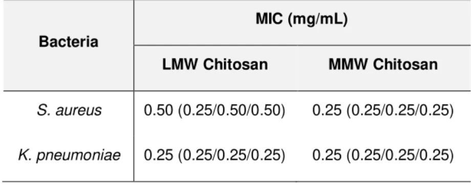
3), or N/A (not applicable for values < 2). ... 33 Table 3.3 – Antimicrobial susceptibility testing of low and medium MW chitosans against S. aureus and
K. pneumoniae. Median and triplicate values of MICs (mg/mL) are presented. ... 34 Table 3.4 - Chitosan content of alg:chitLMW 99:1, 19:1 and 9:1 aerogel fibres; and respective chitosan
raw material used in their production. ... 35 Table 3.5 - Efficacy of antibacterial property of the tested material against S. aureus and K. pneumoniae
from contact with samples, at a concentration of 2 g/mL, during 24 h at 37ºC (mean ± SD, n=3).
Efficacy is defined as significant (2 ≤ antibacterial value < 3), strong (antibacterial value ≥ 3), and
XIX List of Abbreviations
Abbreviation Full form
Alg Alginate
ANOVA Analysis of variance
AST Antimicrobial susceptibility testing BET Brunauer–Emmett–Teller
BJH Barrett-Joyner-Halenda
BPR Back pressure regulator
CAMHB Cation-adjusted Mueller-Hinton broth
Chit Chitosan
CLSI Clinical and Laboratory Standards Institute
DD Degree of deacetylation
DMSO Dimethyl sulfoxide
EA Elemental analysis
EACC European Collection of Authenticated Cell Cultures EDTA Ethylenediaminetetraacetic acid
FBS Fetal bovine serum
FEG-SEM Field emission gun scanning electron microscopy FTIR Fourier-transform infrared spectroscopy
G Guluronate
GRAS Generally recognized as safe
ISO International Organization for Standardization
LMW Low molecular weight
M Mannuronate
MEM Minimum essential medium
MHA Mueller-Hinton agar
MIC Minimal inhibitory concentration
MMW Medium molecular weight
XX
MW Molecular weight
NA Nutrient agar
NB Nutrient broth
NCTC National Collection of Type Cultures
NEAA Non-essential amino acid
PBS Phosphate buffered saline
PEC Polyelectrolyte complex
SC-CO2 Supercritical carbon dioxide
SD Standard deviation
SEM Scanning electron microscopy
TSA Tryptic soy agar
TSB Tryptic soy broth
UV Ultra violet
1 1. Introduction
1.1 Aerogels production
Aerogels are a type of material which interest has increased in recent years. Discovered in the
1930’s by Stephens Kistler, these innovative materials have gained considerable interest due to
the unique combination of its properties. They have high porosity (~80–99.8%), low density (~0.003–0.5 g/cm3) and weight, low thermal conductivity, flexibility, high surface area (~200–
1200 m2/g) and very low dielectric constant that give these materials a huge potential for
applications in a wide spectrum of areas, ranging from construction, chemical engineering and more recently to life sciences and medicine [1]–[3].
An aerogel consists of an amorphous solid material resulting from a hydrogel, where the fluid of this gel is removed and replaced with a gas, keeping the gel structure almost intact, which results in its characteristic significant porosity [3]. It is the type of drying/extraction of the liquid phase of hydrogel that determines the formation of this innovative material. This drying can be performed through supercritical fluid technology, a methodology of compressed fluids that has revolutionized the extraction of various compounds, as well as the production of new materials such as aerogels [2], [4]. Prior to drying step, two steps are important in the production of an aerogel (Figure 1.1). A first step relates to the formation of the hydrogel, usually starting from an aqueous solution. The gelation can be triggered by an action of physical or chemical nature. For example, physical hydrogel may refer to a reversible crosslink formed between polymeric chains under appropriate conditions through weak forces (e.g. hydrogen bonding or ionic interactions). Also, the gel induction through pH or temperature variations are some examples of mechanisms for the preparation of physical hydrogels [3], [4]. On the other hand, a chemical hydrogel may refer to wet gels obtained by establishing covalent bonds between polymeric chains mediated by the addition of chemical cross-linking agents [3], [4]. After obtaining the hydrogel, a second step before drying is performed. Since the drying is mostly done with supercritical carbon dioxide (scCO2), the hydrogel solvent is exchanged for an organic solvent, usually ethanol because of its
affinity for scCO2 [3]. In carrying out this step, it must also be taken into account that the chosen
2
Figure 1.1 - Main steps of the production process of an aerogel.
As previously stated, the drying technique after the solvent exchange step is a critical factor in obtaining an aerogel. So, conventional drying techniques (e.g. air-drying and lyophilization) cannot be adopted since capillary forces from fluid evaporation can lead to the shrinkage of the initial structure, obtaining another type of dry gel much less porous, such as a xerogel. So, supercritical drying technology is used in order to avoid capillary forces, ensuring the structural integrity of the gel from which the aerogel results [3].
A supercritical fluid is understood as a substance that is at a temperature and pressure above its critical point, in which a phase separation between the liquid and gaseous states is not distinguished [3]. Among the various properties of supercritical fluid, its liquid-like density and its gas-like viscosity and diffusivity stands out [5]. Within the supercritical fluids, one of the most used is scCO2. A high number of properties make this gas one of the most used solvents in supercritical
technology. Among them, the fact that it is a very abundant gas, non-toxic, non-flammable, economically cheaper, non-carcinogenic, and easily recyclable [6]. The pressure (73.8 bar) and specifically the temperature (31.1 ºC) required to reach its critical point (Figure 1.2) are relatively close to ambient conditions, making this supercritical fluid one of the safest to operate [4].
3 As previously stated, to produce an aerogel capillary forces must be avoided, so that the initial structure of the gel remains practically the same after solvent removal. This is why scCO2 and
ethanol are usually used in the production of aerogels. In Figure 1.1, a solvent exchange step is carried out in the production of an aerogel. This step is done so that the solvent present in the hydrogel has complete miscibility with scCO2, something that would not be possible with H2O due
to their miscibility gap [7]. The exchanged solvent is usually a low molecular weight (LMW) alcohol, which under certain pressure and temperature conditions achieves complete miscibility in the scCO2. For ethanol, miscibility in scCO2 is assured at operating conditions of 120 bar and
40ºC [2]. This creates the conditions that prevent capillary stress due to the absence of menisci liquid-vapor interface (Figure 1.3a), something that would happen if the gel was dried for example by evaporation, which would lead to shrinkage of the structure, resulting in a much less porous material like a xerogel [3] (Figure 1.3b).
(a) (b)
Figure 1.3 - (a) Scheme of the forces exerted by the surface tension due to the interface of gas-liquid phases; (b) Effect of gel drying method: gel monolyths of pectin of the same dimensions prepared by thermal gelation dried under supercritical drying (aerogel) and under air drying (xerogel) [4].
1.2 Aerogels from biopolymers
4 algae, and chitin present in animals [4]. Within these various compounds, marine polysaccharides such as alginate and chitosan (deacetylated chitin) have gained prominence in aerogels production. There are already several studies where aerogels from these polymers have been produced, as is the case of Robitzer et al. [9] who explored the relationship at nanoscale of the polymer organization of alginate aerogel with its origin gel. Martins et al. [10] studied the potential of biomedical applications of alginate-based aerogels, evidencing the high potential of this materials, particularly for bone regeneration. Besides alginate, aerogels composed of other marine polymers are also emerging, such as the ones described in the work of Ayers et al. [11], who studied the synthesis and properties of chitosan-silica hybrid aerogels, and verified the low cytotoxicity of this material. These biopolymers stand out for their abundant sources, since they can be obtained through waste recovery from the food industry (chitosan) or through algae (alginate), one of the most abundant marine sources of polysaccharides that are easily cropped [12]. As such, there has been an increase in the economic and scientific interest of these biopolymers due to the abundant natural availability and their physicochemical and biological properties, which will be discussed below as well as the interaction between both polysaccharides.
1.2.1Alginate
Alginate is an abundant natural polymer mostly obtained from brown algae, exerting structural function, giving mechanical strength and flexibility. This negatively charged polymer has gained increasing relevance in several areas such as biomedical sciences and engineering. This increase in interest is due to its vast properties such as biocompatibility, ease of gelation, low toxicity and relatively low cost [13], [14]. In an economy increasingly growing, it is important to respond to industrial needs and at the same time ensuring the resources sustainability. Therefore, it can be said that alginate is a resource that can be considered almost inexhaustible, since macroalgae can be easily collected cultivated.
5 marine algae, this type of production may allow the reduction in batch-to-batch variability. The pathway of alginate biosynthesis is generally divided into (1) synthesis of GDP-mannuronic acid precursor, (2) cytoplasmic membrane transfer and polymerization, (3) periplasmic transfer and modification, and (4) export through the outer membrane [16]. With the increasing knowledge of microorganisms’ genetic regulation and the development of areas such as synthetic
biotechnology, it may be possible to produce alginate with more uniform properties and with additional features.
Alginate is a linear copolymer, which is a polymer derived from more than one species of monomer, containing blocks of (1,4)-linked -D-mannuronic acid (M) and -L-guluronic acid (G) residues. The blocks are composed of homopolymeric regions of consecutive G residues (GGGGGG), consecutive M residues (MMMMMM), and alternating M and G residues (GMGMGM), termed G-, M- and MG-blocks respectively [15].
Figure 1.4 - Representative alginate structure: G-block, M-block, and alternating MG-blocks in alginate [13].
As stated above, the alginate composition may vary depending on its source, so the G and M contents of the commercial alginates may vary depending on their extraction source. The composition, sequence, and size of these residue blocks are critical factors that alter the physical properties of alginate [14]. Besides these characteristics, molecular weight (MW), ranging between 32,000 and 400,000 g/mol, is also a determining factor in the physical properties of commercial alginates[13].
6 is widely used in the food industry as a thickener and emulsifier [17]. Alginate also has several properties that make it an excellent candidate for biomedical applications. The fact that it is a biocompatible polymer, does not present toxicity and is biodegradable makes this polymer ideal for human body direct applications [13]. The gelation ability is another interesting property for biomedical applications, since it allows the formation of hydrogels, microspheres, fibres, microcapsules and sponges [18]. These are different types of materials commonly used in biomedical applications such as wound healing, drug delivery and tissue engineering [13]. Another interesting property is that it presents a high bioadhesivity, comparing with other charged polymers, making this polymer an excellent candidate for drug delivery systems [17].
1.2.2Chitosan
Chitin has a structural function similar to cellulose supporting cell and body surfaces and is mostly found in the exoskeleton of crustaceans, such as shrimp, crab and lobsters [19]. Chitosan, a polycationic polysaccharide, is also known as deacetylated chitin, and as such is considered a pseudonatural polymer. This cationic biopolymer presents a set of unique properties like biocompatibility, biodegradability and bioactivity that has greatly increased the interest of this polymer in biomedical applications [20]. Another interesting fact is that this polymer can be obtained through the waste recovery from food industry [19], an important aspect for the contribution to a circular economy, an essential concept for sustainable development.
Briefly, the chitosan obtaining process begins with the processing of the crustacean shells by removing calcium carbonate (decalcification) and proteins (deproteination) that are in large quantities. Chitin is then obtained, deacetylated using an alkaline solution, mainly of sodium hydroxide, for 1h-3h to give chitin with a deacetylation degree in the order of 70%, known as chitosan [21].
7
Figure 1.5 - Structure of chitin and chitosan (reproduced from [22]).
8 1.2.3Polyelectrolyte complexes
Polyelectrolyte complexes (PECs) are the result of electrostatic interactions between polyions of oppositely charged polymers. The formation of PECs is directly related to contact, quantity and charge of both polyanionic and polycationic polymers [28]. Thus, the time, pH and temperature of interaction as well as the concentration and ratio of both polymers are important factors for the formation of these complexes. Another important factor is the distribution and density of positive and negative charges of both polymers, as well as their degrees of ionization [28]. Since electrostatic interactions are established between both polymers without the need of a chemical cross-linker agent, the PECs are generally non-toxic and biocompatible [29]. These complexes, in the form of hydrogels, microparticles or membranes, have gained considerable relevance in areas such as biotechnology and biomedicine, acting as drug delivery systems, biosensors, enzyme immobilizers or materials to aid bone and skin regeneration [28]–[30].
9
Figure 1.6 - Schematic representation of polyanion-polycation interactions: mixing of the oppositely charged polyelectrolytes in aqueous solution leads to formation of a dense phase (PEC) caused by the cooperative electrostatic interactions between the ions [28], [31].
Like for alginate or chitosan-only gels, the complex between both polymers also exhibits interesting physicochemical properties with potential biomedical applications. It is the example of the work of Liao et al. [32] that has demonstrated that the production of a PEC fibre of alginate-chitosan can be an effective drug carrier with potential advantages of high loading level and high encapsulation efficiency. Another study that demonstrated the biomedical potential of these materials was the one from Alsharabasy et al. [30], which demonstrated the preparation of a high quality alginate chitosan complex in the form of a hydrogel film that stimulates cell proliferation and wound closure, demonstrating the potentiality of these materials for wound healing applications.
10 .
1.3 Biomedical applications of aerogels
The unique properties of the aerogels combined with the properties of the polymers that originate them, may offer numerous biomedical and pharmaceutical applications [1], [3]. Biocompatibility and biodegradability, as well as a good relationship between porosity and mechanical strength of aerogels obtained from biopolymers, are important properties for biomedical applications such as substrates for tissue regeneration, drug delivery systems, medical cardiovascular devices and wound care [1]–[3]. Among the various biomedical applications, the use of aerogels in the treatment of wounds has been an area that has also gained relevance.
1.3.1Wound healing process
The skin, the largest organ of the human body, acts as a barrier against possible external aggressions, whether physical, chemical or even biological. It is an essential organ that goes through a very complex and highly regulated healing process when damaged [33]. When a severe aggression occurs, the tissue regeneration process must be highly accompanied by the responsible health professionals. This monitoring must be done in order to avoid complications such as the development of chronic non-healing wounds and serious infections that may even endanger the patient's life. Despite the improvement in medical care, there has been an increase in the development of this complications due to ageing and an increase in elderly population, as well as an increase in the prevalence of chronic diseases such as diabetes that affect the normal process of wound healing [34].
11 collagen (type III) [34]. All cells that are no longer needed, such as endothelial cells from the vascular vessel network, are also removed and scar tissue formation occurs.
Wounds may assume various classifications, with respect to time, depth or type of injury. In relation to time, wounds with less than 6 hours are classified as acute. When the injury occurred more than 6 hours ago, but less than 5 days, are classified as subacute. After 5 days all wounds are chronic. According to the depth, they can be superficial, deep dermal or full thickness wounds. The type of wound may be due to incision, shearing, crushing or burning and consequently each of them can also be classified as sterile or contaminated [36]. The greatest and challenging dilemma of wound healing are the chronic non-healing wounds, as they mean that the normal healing process has been disturbed, leading to very severe complications such as severe infections and great tissue losses [24]. Among the chronic wounds are highlighted as clinically most frequent the pressure ulcers, diabetic ulcers, venous and arterial ulcers [37].
Wound contamination is a factor that requires a lot of concern since it can lead to one of the most serious complications of skin lesions. In a first "phase" the existence of non-replicative microorganisms in the wound is defined as contamination. However, an open wound means that the skin has lost its barrier ability and as such the wounds are colonized, where the adherent microorganisms gain the ability to replicate [38]. Yet, this is a state where there are no tissue losses. Due to a number of factors, such as the microorganism’s concentration, their
pathogenicity and virulence, and the response ability/inability of the host immune system to attack them, favourable conditions to the microorganism growth can be established. This microorganism replicative state may begin to cause damage to the patient's tissue, known as wound infection [38]. From this moment, if the infection is not controlled and eliminated, the spreading can lead to life-threatening conditions such as septicaemia. There are several skin commensal and wound colonizer microorganisms reported as potentially pathogenic, with Staphylococcus aureus and Klebsiella pneumoniae being two examples of commensal and nosocomial microorganisms commonly isolated from noninfected and infected wounds [38], [39].
1.3.2Wound dressings materials
The treatment of wounds is an area that has gained a lot of interest from the scientific, medical and industrial communities. For medical device companies, it has been a market that has increased its attractiveness since it involves a large financial dimension around the world, with an annual market for wound care products estimated at $15.3 billion [40]. An increase in the number of advanced and innovative wound dressings is emerging in the market. Nonetheless, they are still somewhat costly, making them less medically prescribed. However, since they accelerate the healing process and require less dressing changes than conventional products, it could make them less expensive in the future [24].
12 active role in the wound healing process [37]. These advanced wound dressings have emerged as bioactive drug delivery systems, or wound dressings whose own polymeric material is already bioactive, capable of triggering the acceleration of healing and eliminating or hindering the development of infections [37]. In addition, these new wound dressings stand out from the traditional due to the creation of a moist environment (ideal for favouring the wound healing process) and a much less painful and easier of removal [41], [24] There are already several types of commercial wound dressings made up of polymers that can be natural, synthetic or semi-synthetic (Table 1.1). Among the natural ones, some of the most used polymers are alginate, bacterial cellulose and pectin. Polyurethane and polyvinyl alcohol are commonly used synthetic polymers [37]. These types of new polymeric wound dressing materials can be classified regarding formulation or final form. Some of the examples are hydrogels, hydrocolloids and alginates which can be presented in the form of films, foam sheets, membranes or gels. In addition to these advanced wound dressing materials, aerogels are also gaining relevance in this type of applications. The high porosity of these materials presents as prominent property in the maintenance of gaseous exchanges essential for the normal wound healing process. Besides that, aerogels from some polysaccharides, in addition to biocompatibility and biodegradability, also present antimicrobial activity, high water uptake capacity and adhesive nature. Considering the complex process of wound healing, these characteristics make these materials very promising for possible wound care applications [1], [4], [12].
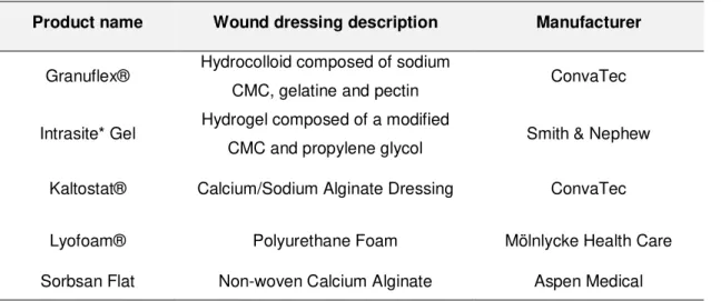
Table 1.1 - Examples of some of the commercial wound dressing materials.
Product name Wound dressing description Manufacturer
Granuflex® Hydrocolloid composed of sodium
CMC, gelatine and pectin ConvaTec
Intrasite* Gel Hydrogel composed of a modified
CMC and propylene glycol Smith & Nephew Kaltostat® Calcium/Sodium Alginate Dressing ConvaTec
Lyofoam® Polyurethane Foam Mölnlycke Health Care
13 1.4 Aim of the thesis
The main objective of the present work was to evaluate the potential of new alginate-chitosan aerogel fibres, for applications in wound treatment.
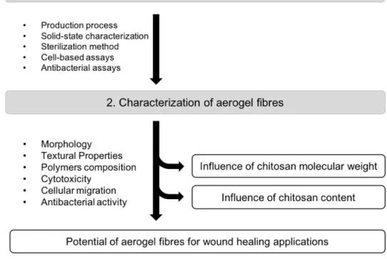
To achieve the proposed aim, the work was divided in two fundamental tasks (Figure 1.7). In a first step the objective was to optimize the fibres production process, and to perform a previous screening of some fibre’s properties. In addition, the purpose of this first step of the work was to optimize and implement the chosen methodologies and techniques to characterize the aerogels. The solid-state, biocompatibility and bioactivity of the fibres were chosen to be characterized. After the first step, the methodologies were implemented to characterize the fibres in terms of chitosan content, morphology, textural properties, ionic interaction between both polymers, cytotoxicity, cell migration stimulation and antimicrobial activity. In addition to evaluating the fibres potential in this biomedical application, another objective was to evaluate the influence of aerogel chitosan content and its MW. In the second step the objective was then to produce the fibres, characterize their chosen properties and thus evaluate the influence of chitosan content and MW on the produced fibres characteristics.
This alignment will allow to achieve the main objective of this work, evaluate the potential of new alginate-chitosan aerogel fibres for wound healing applications.
15 2. Materials and Methods
2.1 Aerogel fibres production
The process described below was the same for all aerogel fibres production, only varying the amount of chitosan stock solution added to the alginate emulsion. Each production was done in duplicate, in two independent assays.
2.1.1Reagents
Alginic acid sodium salt (90.8-106%) and acetic acid (≥99.7%) were purchased from Panreac (Germany). LMW (50-190 kDa, 75-85% deacetylated, viscosity 20-300 cP) and medium molecular weight (MMW) (190-310 kDa, 75-85% deacetylated, viscosity 200-800 cP) chitosans were purchased from Sigma-Aldrich (Steinheim, Germany). Sorbitan monooleate (Span 80) was purchased from Merck (Germany). Paraffin oil was purchased form LabChem (USA). n-Hexane
was purchased from Carlo Erba (Spain) and Ethanol (≥99.8%, absolute) was purchased from
Fisher Chemical (USA). 2.1.2Stock solutions
For the emulsion production, the aqueous phase, an aqueous solution of sodium alginate (3 wt.%), was previously prepared by continuous stirring overnight at room temperature. The oil phase was produced by mixing paraffin oil with surfactant Span 80 (3 wt.%). In addition, acidic solutions of LMW and MMW chitosan (1.5 wt.%) were also prepared by dissolving chitosan in acetic acid (50% v/v) by continuous stirring overnight at room temperature.
2.1.3Sol-Gel process
An Water-in-Oil (W/O) emulsion was made, adding slowly the aqueous phase, sodium alginate stock solution to the oil phase stock solution and then mixing with Ultra-Turrax homogenizer (T25, Ika Works Inc., USA) for 2 min at 24,000 rpm to provide energy to the system, facilitating the dispersion and homogenization [42]. The emulsions were always produced in the same W/O ratio (1:3 w/w).
16
Table 2.1 - Mass of alginate and chitosan present in stock solutions used in fibre productions and final polymer ratio.
Aerogel Fibres (w/w) Alginate (g) Chitosan (g)
alg:LMWchit (99:1) 3 0.03
alg:MMWchit (99:1) 3 0.03
alg:LMWchit (19:1) 3 0.16
alg:LMWchit (9:1) 3 0.33
2.1.4Solvent exchange
After overnight rest period, separation of phases occurred in the emulsion; consequently, top oil phase was removed by aspiration, followed by a centrifugation at 3583 g for 30 min with another removal by aspiration. To remove the remaining paraffin oil from the hydrogel, hexane was added to the centrifugation vial, stirred vigorously and centrifuged at 1593 g for 20 min removing the top liquid layer by aspiration (this step was done twice). For the solvent exchange, remaining suspension was rinsed with ethanol/water mixtures (30, 60, 90 and 100 vol.%) followed by centrifugation (1593 g, 20 min) and aspiration of the supernatant. Finally, to ensure water removal, this step was repeated three times with absolute ethanol. Centrifugations were done using a Beckman Coulter Avanti J-26 XPI centrifuge, a Beckman JA-10 rotor and Beckman 500mL centrifuge tubes. After solvent exchange, the alcogels were placed in filter papers which were stapled and placed in vials immersed in pure ethanol.
2.1.5Drying with supercritical carbon dioxide
The alcogels were dried through scCO2, using the high-pressure laboratory equipment (Thar
Technology, Pittsburgh, PA, USA, model SFE-500F-2-C50), whose scheme is shown in Figure 2.1. Some glass beads were placed on the bottom of the drying vessel to facilitate the contact and diffusion of scCO2 through the alcogel matrix. The coffee filters with the alcogels were placed
inside the extraction vessel with more glass beads, to fully occupy its volume, and pure ethanol to avoid the alcogels drying by atmospheric air before the supercritical CO2 drying process
started. The pressurization of the vessel until 120 bar was performed in the following conditions: vessel heater at 40ºC, heat exchanger at 40ºC, CO2 pump at 40 g/min and back pressure
regulator (BPR) at 120 bar.
When the vessel pressure was reaching 120 bar, the CO2 pump flow rate was decreased, and
the drying was maintained in a CO2 flow range of 8-12 g/min for 4 h. At the end the vessel was
17
Figure 2.1 - Schematic diagram of the supercritical fluid drying apparatus. (Adapted from Waters, 2010)
2.2 Fibres weighing and sterilization
Since the sterility of medical devices used in wound healing treatment is required, aerogel fibres produced in this work were also subjected to sterilization process [43]. This will also guarantee their sterility in all cell and bacterial assays performed. The sterilization was performed using ultraviolet (UV) irradiation as already referenced in the literature [44], with some modifications. The sample mass required for each assay was previously weighed in petri dishes and then sterilized through exposure to the UV lamps of a Biological Safety Cabinets for 4 h (±1 h) at room temperature. After exposure, a sterility test was performed to confirm the efficacy of the procedure. The aerogel fibres were incubated in Tryptic Soy Broth (TSB, Scharlau, Spain) at 37°C for 3 days. After 3 days, to ensure the absence of residual contamination, the suspension was plated in Tryptone Soya Agar (TSA, Oxoid, UK) for 24 h at 37ºC. The material was considered sterile when no turbidity was observed after the TSB incubation and no microbial growth was observed on the TSA plates.
2.3 Solid-state characterization
2.3.1Scanning electron microscopy (SEM)
Field Emission Gun Scanning Electron Microscopy (FEG-SEM) (JEOL, model JSM7001, Japan) was used to examine morphology. The samples were prepared for observation by covering with gold/palladium (Au/Pd), in a sputter coater (Quorum Technologies, model Q150TES). Micrographs of the prepared aliquots were taken at an acceleration voltage of 10 kV.
2.3.2Fourier-transform infrared spectroscopy (FTIR)
The equipment used was a Thermo Scientific FTIR spectrometer (San Jose, USA) Class 1 Laser
Product Nicolet 6100 using ATR accessories with a diamond crystal of 42⁰ for solids. The
18 Corporation). The background spectrum of the air was collected before each sample spectrum acquisition. To clean the crystal, water and acetone were used and after that the crystal was dried with a soft tissue. For the sample spectrum acquisition, the different samples were placed in the corresponding ATR crystal and the spectra were recorded with 32 scans between 4,000-650 cm -1 and with a resolution of 4 cm-1.
2.3.3Brunauer-Emmett-Teller (BET) surface area analysis and Barrett-Joyner-Halenda (BJH) pore size and volume analysis
Specific surface area and pore volume were determined by low temperature nitrogen adsorption desorption analysis (Quantachrome Nova 3000e) using Brunauer–Emmett–Teller (BET) and Barrett Joyner–Halenda (BJH) methods, respectively. Prior to the measurements, the samples were degassed at 348 K for 24 h. The results are presented as the mean values of two independent production processes.
2.3.4Elemental analysis (EA)
The percentage of nitrogen content in the aerogel fibres was quantified by elemental analysis using a Thermo Finnigan Flash EA 1112 CHNS. The samples were weighed 2-3 mg and the total time of analysis was 12 min. The nitrogen content was converted to chitosan content (on a weight percentage basis, wt.%), as described in the literature [45], [46]. Calculated as follows:
𝐶ℎ𝑖𝑡𝑜𝑠𝑎𝑛 (𝑤𝑡. %) =𝑁(𝑤𝑡.%)× 𝐶(𝑔/𝑚𝑜𝑙)𝑁 (𝑔/𝑚𝑜𝑙) (Eq.1)
This calculation is based on the weight percentage of nitrogen in the fibres, N (wt.%), determined by elemental analysis; the average MW of chitosan, C (g/mol), used to produce the fibres (considering the deacetylation degree) and the MW of elemental nitrogen, N (g/mol).
2.4 Cell-based assays
2.4.1Reagents
Minimum essential medium (MEM) with Earle′s balanced salts and 2.0 mM L-glutamine, phosphate buffered saline (PBS) and dimethyl sulfoxide (DMSO) were purchased from Sigma (USA). Non-essential aminoacids (NEAA), foetal bovine serum (FBS) and 0.25% (w/v) Trypsin-EDTA were purchased from Gibco (Life Technologies, USA). AQueous One Solution Cell Proliferation Assay (MTS) was purchased from Promega (USA).
2.4.2Cell culture
Mouse fibroblasts NCTC clone 929 (ECACC 88102702) cells were purchased from European Collection of Authenticated Cell Cultures (EACC, Public Health England, Salisbury, UK). Cells were routinely grown in a standard medium MEM supplemented with 1% NEAA and 10% heat-inactivated FBS. Stock cells were maintained as monolayers in 75 cm2 culture flasks, cultured
every week (seeding 30,000 cells/cm²) and incubated at 37°C in a 5% CO2 humidified
19 using 0.25% trypsin/EDTA at 37°C. The cells were collected, and viability was determined using standard trypan blue staining procedure. Cell counting was performed using a haemocytometer. All cellular assays described below were performed with cells between passages 10 and 25.
2.4.3Biocompatibility – ISO 10993-5 in vitro cytotoxicity assay
In order to mimic the contact of the fibres with skin cells, the aerogel fibres biocompatibility was evaluated by the direct contact method described in ISO 10993-5, a highly sensitive test of medical devices toxicity [47]. Cell viability was quantified through MTS cytotoxicity test, an assay based on the bio-reduction of the MTS tetrazolium compound by viable cells to generate a formazan dye that is soluble in cell culture media. The quantity of formazan dye, directly proportional to the number of living cells in culture, was quantified by measuring the absorbance at 490 nm.
NCTC clone 929 cells were seeded into 24-well plates (volume of 0.6 mL) with a density of 3.0×10⁴ cells/cm2 and maintained in culture for 24 h (~1 doubling period) to form a semi-confluent
monolayer. This incubation period ensures cell recovery, adherence and progression to exponential growth phase. After 24 h, new MEM supplemented with 0.5% FBS was replaced, and following the direct contact method, cells were incubated for 24 h with 1 mg of sample per well, giving a final sample concentration of 1.7 mg/mL. The concentration used in this test was close as possible to the concentration used in the ASTM E 2149 assay, described below. Lastly, the culture media was removed, cells were rinsed with PBS and incubated for 2 h with 0.6 mL of MTS
reagent assay, diluted according to the manufacturer’s information. The absorbance was
recorded at 490 nm using a microplate spectrophotometer (EPOCH, 219 Bio-Tek, USA). Experiments were performed in triplicate in three independent assays. The positive control of cytotoxicity was done with a treatment of 10% (v/v) DMSO solution diluted in MEM.
Results were expressed in terms of percentage of cellular viability (Viab.%) relative to control (cells without aerogel fibres). The lower the Viab.% value, the higher the cytotoxic potential of the test item is. Cytotoxic effect was considered for viability percentage below 70%, according to ISO 10993-5.
2.4.4Bioactivity - Scratch assay.
One of the key steps in the wound healing process is the migration of fibroblasts initiating the proliferative phase [35], and as such, the in vitro scratch assay mimicking cell migration during wound healing was adopted. In this assay an image was captured after conducting a "scratch" in a cell monolayer. After a certain time interval (or regular intervals) the same area is again recorded to compare and quantify the migration rate of cells [48]. The migration assay was performed with the same cell line, seeding and sample concentration of the cytotoxicity assay. The plate wells were previously marked with a pen to help picture acquisition always in the same region.
20 p200 pipette tip the cell monolayer was scratched in a straight-line crossing from one side of the well to the other creating the "scratch". The wells were then rinsed twice with PBS to remove all the suspension cells and freshly new MEM supplemented with 0.5% FBS was replaced. The samples were added and incubated for 8 h at 37ºC in a 5% CO2 humidified atmosphere. The
same scratch area was captured at time point 0 h and 8 h, by microscope (Olympus CKX41) with a 4X objective, equipped with OPTIKAM 4083.B5 microscopy digital USB camera operated with OptikalSview software. Measurement of the wound area was manually calculated using Fiji software (ImageJ) and the results were expressed in terms of percentage (%) of wound recovery, calculated as follows:
𝑊𝑜𝑢𝑛𝑑 𝑟𝑒𝑐𝑜𝑣𝑒𝑟𝑒𝑑 (%) = (𝐴𝑡=0ℎ−𝐴𝑡=8ℎ)
𝐴𝑡=0ℎ × 100 (Eq. 2)
Where,
𝐴𝑡=0ℎ is the mean value of the measured area at t=0 h;
𝐴𝑡=8ℎ is the mean value of the measured area at t=8 h.
2.5 Antibacterial activity evaluation assays
2.5.1Materials
The growth media used in the tests described below were TSB, TSA, Nutrient Broth (NB, Oxoid, UK), Nutrient Agar (NA, Oxoid, UK), cation-adjusted Mueller Hinton Broth (CAMHB, BD Difco, USA) and Mueller Hinton Agar (MHA, BD Difco, USA). Working buffer solution (0.3 mM KH2PO4)
was made dissolving PBS Tablets – Calbiochem (Merck, Germany) in distilled water. Saline solution was made dissolving (0,85% w/v) Sodium Chloride (Panreac, Spain) in distilled water. Kaltostat® was purchased from ConvaTec (UK). McFarland Standard 0.5 (Pro Lab Diagnostics, UK) was used as a reference to adjust the turbidity of bacterial suspensions. Antibiotic test discs (Fisher Scientific, EU) were used as untreated samples. All solutions were heat sterilized at 121°C for 15 min (moist heat sterilization).
2.5.2Bacterial test species, bacterial suspensions and standardized inoculum preparation
21 to a 0.5 McFarland standard (~1.5-3.0×10⁸ CFU/ml). All antimicrobial activity assays described
below were performed with both species.
2.5.3Well diffusion and agar plate methods
The antimicrobial activity of the fibres was tested by two diffusion methods. The well diffusion assay was performed by adapting the Disk Diffusion Test CLSI M02-A11 guidelines [49]. The agar plate method was performed according to AATCC Test Method 90-2016 guidelines [50].
2.5.4ASTM E 2149-01 standard test method for determining the antimicrobial activity of immobilized antimicrobial agents under dynamic contact conditions.
To measure the antimicrobial activity by the Flask‐Shake Method (ASTM E 2149‐01), hereon mentioned as dynamic method, all samples (aerogel fibres, Kaltostat® and the control fabric cotton disks) were prepared by weighing 40 mg and sterilizing as mentioned before (2.2). The working bacterial solution was prepared by diluting a standardized inoculum (2.5.2) into sterile working buffer solution in order to obtain a final concentration of ~1.5-3.0×10⁵ CFU/ml. Briefly,
the working bacterial solution (50 ± 0.1 ml) was added to a sterile 250 ml Erlenmeyer. Once this step was done, the bacterial concentration of each flask was determined by performing serial dilutions and standard plate count techniques, defining the 0 h contact time. The samples were then placed in the flasks and incubated under orbital shaking (180 rpm) for 2.5 h. At the end of this period, the bacterial concentration of each flask was determined as was done for the 0 h contact time subgroup. Each assay included an inoculum only control and an untreated fabric control that were processed in the same way as each sample. The percent reduction of the organisms resulting from contact with the specimen was calculated using the following formula:
𝑅𝑒𝑑𝑢𝑐𝑡𝑖𝑜𝑛, % (𝐶𝐹𝑈/𝑚𝐿) =𝐵−𝐴𝐵 × 100 (Eq. 3)
Were,
A = CFU per millilitre for the flask containing the treated specimen after 2.5 h contact time;
B = CFU per millilitre for the flask used to determine “A” before the addition of the
treated specimen (0 h contact time).
2.5.5ISO 20743:2013(E) Textiles — Determination of antibacterial activity of textile products by absorption method.
22 prepared for both contact time (0 h and 24 h). All vials containing the samples were inoculated with 20 μl of the previously adjusted bacterial suspension (1-3×10⁵ CFU/ml) in order to allow it to
be fully absorbed. Following the inoculation step, to one vial of each sample was added immediately 2 ml of shake-out physiological saline. The concentration of these resulting suspensions was quantified by standard plate count techniques and defined as 0 h contact time. The remaining vials of each sample were incubated at 37ºC for 24 h. After the incubation, the samples contained in the vials were washed out with shake-out physiological saline and plated out as described above, defining the 24 h contact time. All plating was carried out in duplicate. The number of colonies counted was used to calculate the growth values on the control and test samples. The results of this experiment are expressed as mean values of three biological replicates performed.
The antibacterial activity value (A) was calculated using the following formula: 𝐴 = (𝑙𝑔𝐶𝑡− 𝑙𝑔𝐶0) − (𝑙𝑔𝑇𝑡− 𝑙𝑔𝑇0) = 𝐹 − 𝐺 (Eq. 4)
where
A is the antibacterial activity value;
F is the growth value on the control specimen 𝐹 = (𝑙𝑔𝐶𝑡− 𝑙𝑔𝐶0);
G is the growth value on the antibacterial testing specimen 𝐺 = (𝑙𝑔𝑇𝑡− 𝑙𝑔𝑇0); 𝑙𝑔𝐶𝑡 is the common logarithm of the arithmetic average of the numbers of
bacteria obtained from the control specimen after the 24 h incubation; 𝑙𝑔𝐶0 is the common logarithm of the arithmetic average of the numbers of
bacteria obtained from the control specimen immediately after inoculation;
𝑙𝑔𝑇𝑡 is the common logarithm of arithmetic average of the numbers of bacteria
obtained from the antibacterial testing specimens after the 24 h incubation;
𝑙𝑔𝑇0 is the common logarithm of arithmetic average of the numbers of bacteria
obtained from the antibacterial testing specimens immediately after inoculation.
2.5.6Antimicrobial susceptibility testing by broth microdilution
23 in CAMHB to ensure that, after inoculation, each well contained approximately 5×10⁴ CFU. Each
inoculated microtiter plate was incubated under aerobic conditions at 37ºC for 24 h. Minimal inhibitory concentration (MIC) values were read as the lowest chitosan concentration for which visible growth was inhibited after 24 h of incubation. For each chitosan stock solution assayed, a growth control (CAMHB and diluted inoculum), a medium sterility control (CAMHB), and a stock solution sterility control (CAMHB and chitosan stock solution) were also tested. The results of this experiment are expressed as median values of three biological replicates performed.
The highest tested concentrations were 0.5 mg/mL and 0.25 mg/mL for LMW and MMW chitosan, respectively. The concentration of acetic acid present at these tested concentrations was lower than the MIC previously determined for this acid.
2.6 Statistical analysis
All data statistically analysed are expressed as means ± standard errors (SD). Each individual experiment was performed at least in triplicate. The statistical analysis was done using GraphPad Prism 6 (GraphPad Software, Inc., CA). All values were tested for normal distribution and equal variance. When homogeneous variances were confirmed, data were analysed by One Way
25 3. Results and Discussion
3.1 Screening and optimization of assays for evaluation of potential wound healing
applications of alginate-chitosan aerogel fibres
3.1.1Aerogel fibres production
To initiate the work, a first batch of fibres was made according to a methodology already established in the laboratory [52]. Some optimizations were made to the process, namely a vacuum aspiration method was developed, replacing the decantation method, to reduce material losses in the step of solvent exchange. This first batch was produced to be further used in the optimization of protocols and assays used to evaluate the potential of these fibres for wound healing applications.
3.1.2Implementation of sterilization method
Due to the hydrophilicity of the produced fibres, their sterilization by autoclaving with humid heat cannot be done since structural changes, such as fibres contraction or formation of a hydrogel, might occur. One of the most commonly used techniques for sterilization of single-used medical devices is sterilization through radiation, namely ionizing radiation. However, this is a method used by the industry for large-scale productions [53], and not adapted to laboratory scale. Another technique of sterilization by radiation is through UV, a method used in the laboratory for the biological safety cabinet sterilization. This technique, as described by Sabri [44], was adopted for the aerogel fibres sterilization. After the aerogel fibres exposure to UV radiation for 4 h (±1 h), the sterility test showed no growth of microorganisms, validating the method to ensure the fibres required sterilization. Moreover, in order to evaluate if the sterilization process had no influence on physical or chemical aerogel properties, solid-state characterization was performed before and after UV sterilization. The results obtained showed that the radiation had no influence on the characteristics of the fibres (data not shown).
3.1.3Implementation of cell culture
The cell line chosen to perform the cell-based assays was the NCTC clone 929 that is described in ISO 10993-5:2009, which is the standard method chosen to evaluate the fibres biocompatibility. After the purchase of the cell line, the culture methods were implemented in the laboratory. Two growth curves were performed using two different seeding densities referenced by the supplier, 10,000 cells/cm2 and 30,000 cells/cm2, in order to establish the best seeding for the cell culture.
The seeding of 30,000 cells/cm2 was chosen since it showed more linear growth and allowed the
cell subculture twice a week (recommended by the supplier). The seeding of 10,000 cells/cm2
26 3.1.4Optimization of cell-based assays
3.1.4.1 Biocompatibility – ISO 10993-5 in vitro cytotoxicity assay
Some optimizations were made to the method described in the ISO due to the properties of the fibre material and the concentration defined to carry out the tests. The assay was transferred from 96-well to 24-well plates and optimized, with the same seeding, to provide enough area per well ensuring that the entire sample was in homogeneous contact with the cell monolayer. The cytotoxicity evaluation assay was also modified using the MTS assay, which unlike the MTT method described in ISO, produces a water-soluble formazan [54]. The sample concentration defined for this assay (1.7 mg/mL) was as close as possible to the concentration used in the Dynamic method assay (weighing below 1 mg in the available equipment was not precise).
3.1.4.2 Bioactivity - Scratch assay
Unlike biocompatibility, there is no ISO for the evaluation of cell migration. There are several works of scratch assay with fibroblasts reported in the literature, however, there is still some variability between the described methods [55], [56]. The assay was optimized for 12-well plates using the same cell line and cell seeding of the cytotoxicity assay. After performing the scratch, the plates were incubated with three different times: 8 h, 18 h and 24 h. After 18 h and 24 h periods the scratch was totally closed. After the 8 h incubation, a recovery of about 50% was recorded consistently. The assay was then carried out with the 8 h incubation period, where the scratch only with cells and medium was used as control.
3.1.5Evaluation of antimicrobial activity: screening and assays optimization 3.1.5.1 Well diffusion and agar plate methods
To evaluate the antimicrobial activity of the fibres, a first screening was performed using the agar plate method and the well diffusion method. There was no evidence of any type of inhibitory halo in both methods (data not shown). The absence of inhibitory halo suggests that the fibres had no antimicrobial activity detectable by the screening methods performed. However, since both methods rely on diffusion of the samples, the absence of halo could mean that the fibres did not diffuse through the agar, making these methods unsuitable for the evaluation of the material produced [57]. Alternatively, two other methods, described below, were selected to evaluate the antimicrobial activity by contact.
3.1.5.2 Static method
27 3.1.5.3 Dynamic method
The ASTM E 2149 – 01 assay was also adopted because it allows to evaluate the antimicrobial activity by varying the time and the contact dynamics of the samples with the bacterial inoculum, when compared with ISO 20743:2013. For this method, the contact time between the samples and the inoculum is suggested to be 1 h or another contact time specified by the researcher. The time required for the fibres (same sample mass used in Static Method 3.1.5.2) to completely dissolve in the working bacterial dilution was tested and was defined as 2.5 h, so that the fibres were in contact with the whole inoculum in a homogeneous way.
In addition to all optimizations, these tests allowed an initial screening, in which a clear antimicrobial activity of the fibres was verified. As such, the evaluation of the antimicrobial activity was a parameter that was determined and that will be discussed in this work.
3.2 Evaluation of alginate-chitosan aerogel fibres for potential wound healing
applications
3.2.1Influence of chitosan molecular weight
One of the objectives of this work was to evaluate the influence of the chitosan MW on the fibre’s
chemical physical features and potential wound healing properties. In order to understand this influence, fibres were produced varying the chitosan MW, namely fibres with LMW chitosan (alg:chitLMW mass ratio 99:1) and MMW chitosan (alg:chitMMW mass ratio 99:1). These productions were done in duplicate, performing each batch independently.
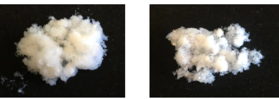
alg:chitLMW (99:1) alg:chitMMW (99:1)
Figure 3.1 - Photographs of the aerogel fibres produced with different chitosan MW (low and medium).
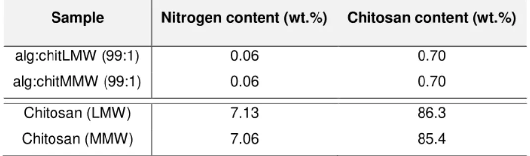
28 totally pure, since they had chitosan contents of 86.3 wt.% and 85.4 wt.% for LMW and MMW, respectively. This may be a reason for the percentages of chitosan content in the aerogels were slightly lower in relation to their theoretical value. Besides the two types of fibres presented similar chitosan contents, also the values between each production batch of both aerogel fibres were similar (data not shown).
Table 3.1 - Chitosan content of alg:chitLMW (99:1) and alg:chitMMW 99:1 aerogel fibres; and respective chitosan raw materials used in their production.
Sample Nitrogen content (wt.%) Chitosan content (wt.%)
alg:chitLMW (99:1) 0.06 0.70
alg:chitMMW (99:1) 0.06 0.70
Chitosan (LMW) 7.13 86.3
Chitosan (MMW) 7.06 85.4
Despite being visually equal, that is, both presenting an ultra-light and cotton-like structure, some characterization techniques were applied in order to characterize the aerogel fibres solid-state. To evaluate the morphology of the produced material, SEM micrographs of the fibres produced with the different chitosan MWs were obtained (Figure 3.2). Pictures at higher magnification (30,000x magnification; Figure 3.2b and Figure 3.2d) allowed to verify that produced aerogels are constituted by a network of nanofibers linked to each other, forming porous structures. As in several research works in which the production of aerogels of alginate or chitosan are described [9], [12], these images indicate the efficacy of the scCO2drying in preserve the structural integrity
29
2000x 30000x
alg:chitLMW (99:1)
alg:chitMMW (99:1)
Figure 3.2 - SEM micrographs of alg:chitLMW 99:1 (a,b) and alg:chitMMW 99:1 (c,d) aerogel fibres, at 2000x (a,c) and 30000x (b,d) magnification.
To complement the aerogels solid-state characterization, Figure 3.3 shows the comparison of FTIR spectra of the produced fibres and their pure origin polymers, chitosan (LMW and MMW) and alginate. The typical bands of alginate, corresponding to the two carboxyl groups (-COOH) of the molecular chain, were observed at 1591 cm-1 and 1400 cm-1 (Figure 3.3a). The FTIR
spectrum of LMW (Figure 3.3c) and MMW (Figure 3.3b) chitosan presented, respectively, characteristic peaks of the Amide I at 1645 cm-1 and 1647 cm-1 and characteristic bands of amino
group (–NH2) at 1572 cm-1 and 1566 cm-1. Therefore, spectra of the pure polymers are consistent
![Figure 1.2 - Pressure versus temperature phase diagram of CO 2 indicating the critical point and the sub- and super-critical regions [5]](https://thumb-eu.123doks.com/thumbv2/123dok_br/16503182.734126/22.892.149.724.115.354/figure-pressure-temperature-diagram-indicating-critical-critical-regions.webp)
![Figure 1.4 - Representative alginate structure: G-block, M-block, and alternating MG-blocks in alginate [13]](https://thumb-eu.123doks.com/thumbv2/123dok_br/16503182.734126/25.892.189.684.479.874/figure-representative-alginate-structure-block-alternating-blocks-alginate.webp)