Joana Grand-Guillaume-Perrenoud Silvestre
Ferreira
Licenciada em Biotecnologia
The role of post-translational
modifications on STAT3 interactions
Dissertação para obtenção do Grau de Mestre em Bioquímica para a Saúde
Orientador: Dr. Federico Herrera, Investigador, ITQB
Joana Grand-Guillaume-Perrenoud Silvestre
Ferreira
Licenciada em Biotecnologia
The role of post-translational
modifications on STAT3 interactions
Dissertação para obtenção do Grau de Mestre em Bioquímica para a Saúde
Orientador: Dr. Federico Herrera, Investigador, ITQB
Presidente: Doutor Pedro Manuel H. M. Matias Arguente: Doutora Joana São José Dias Amaral
Vogal: Doutora Margarida Archer Franco frazão Doutor Federico Herrera García
Instituto de Tecnologia Química e Biológica
T h e r o le o f p o s t-tr a n s la tio n a l m o d if ic a ti o n s o n S T A T 3 i n te ra c tio n s J o a n a F e rr e ir a 2017
“The role of post-translational modifications on STAT3 interactions” Copyright Joana Grand-Guillaume-Perrenoud Silvestre Ferreira, Faculdade de Ciências e Tecnologia, Universidade Nova de Lisboa.
O Instituto de Tecnologia Química e Biológica António Xavier e a Universidade Nova de Lisboa têm o direito, perpétuo e sem limites geográficos, de arquivar e publicar esta dissertação através de exemplares impressos reproduzidos em papel ou de forma digital, ou por qualquer outro meio conhecido ou que venha a ser inventado, e de a divulgar através de repositórios científicos e de admitir a sua cópia e distribuição com objetivos educacionais ou de investigação, não comerciais, desde que seja dado credito ao autor e editor.
I
Acknowledgements
I would like to express my gratitude to all of those involved in the development of this thesis:
First, to my supervisor Dr. Federico Herrera for the opportunity to work in his laboratory, for the dedication, patience and great incentive for the development of this project, thank you. Also thank you for all the transmission of knowledge which encouraged my scientific and personal growth.
I would like to give a special thank you to my labmates, Catarina Almeida, Joana Santos and Ricardo Vilela for all support, patience and encouragement. Thank you for all the conversations and advices.
To Dr. Isabel Pacheco for all the support in the laboratory.
To the Unit of Imaging and Cytometry at IGC for all the technical support and availability. A special thanks to my boyfriend, Miguel Correia, who always manifested his confidence in me and for his huge support in all this project. Thank you for always being by my side and for all help you gave me throughout this important stage of my life. Thank you also for all the good times you gave me when I felt less well. Thank you for all your love.
Last but not least, an enormous thanks to my family, especially my parents and sister, and also my friends for the unlimited support and care. Without them, this thesis would have never been accomplished. A special thanks to my sister, Sarah Ferreira and my friend Raquel Sousa for their unconditional support, patience and all good times and laughs shared, thank you.
III Abstract
The JAK/STAT3 pathway is involved in multiple biological phenomena, mostly related to stress or tissue damage, but also development and cancer. A rate-limiting step of the pathway involves STAT3 dimerization, phosphorylation and translocation to the nucleus. Originally, it was thought that STAT3 dimerized upon phosphorylation by JAKs on Y705. However, current evidence indicates that STAT3 exists as a dimer prior to phosphorylation and activation, and that phosphorylation only induces a change in the conformation of the dimer.
One of the aims of this thesis was to explore the importance of post-translational modifications on STAT3 interactions. To this end we used the BiFC system developed in our lab (Venus-STAT3 BiFC) to create STAT3 mutants on several key residues for acetylation, methylation and phosphorylation: K49, K140, K685, Y705 and S727. Through flow cytometry and fluorescence microscopy, we found that none of the mutants interfere with STAT3 dimerization and intracellular localization.
The other objective was to investigate whether the dimerization of unphosphorylated STAT3 is actually spontaneous or regulated by intracellular pathways. For this, a library of 82 kinase inhibitors was screened for their effect on STAT3 dimerization. We found that PLK-1, Aurora and MEK inhibitors prevented STAT3 spontaneous dimerization. The later steps in the screening were done with the PLK-1 family, which were tested at different concentrations and later against the STAT3 mutants. Our preliminary results indicate that HMN-214 could regulate the dimerization of unphosphorylated STAT3 and that S727 may be involved in this effect.
The relevance of our findings stems from the fact that STAT3 is an important molecule in developmental astrogliogenesis, reactive gliosis during neurodegeneration and brain or spinal cord injury, and some types of cancer.
V Resumo
A via da JAK/STAT3 está envolvida em múltiplos fenómenos biológicos, essencialmente relacionados com stress ou dano tecidular, mas também no desenvolvimento e cancro. Os passos limitantes desta via envolvem a fosforilação, dimerização e translocação para o núcleo do fator de transcrição STAT3. Inicialmente, pensava-se que o STAT3 dimerizava antes de ser fosforilado pelas JAKs no residuo Y705. No entanto, estudos recentes indicam que o STAT3 dimeriza antes de ser fosforilado e ativado e que a fosforilação apenas induz uma alteração conformacional do dímero.
Um dos objetivos da presente tese foi explorar a importância das modificações pós-traducionais nas interações do STAT3. Para este fim, foi usado o sistema BiFC desenvolvido no nosso laboratório (Venus-STAT3 BiFC) para criar mutantes do STAT3 em resíduos-chave para a acetilação, metilação e fosforilação: K49, K140, K685, Y705 e S727. Através de citometria de fluxo e microscopia de fluorescência, observámos que nenhum dos mutantes interfere com a dimerização nem com a localização intracelular do STAT3.
Outro objetivo foi investigar se a dimerização do STAT3 não-fosforilado é espontânea ou regulada por outras vias intracelulares. Para tal foi realizado um rastreio com 82 inibidores de cinases para observar o efeito na dimerização do STAT3, sem que este interfira nos níveis da sua expressão. Através da citometria de fluxo e Western blot, as famílias PLK-1, Aurora e MEK foram consideradas promissoras. Dentro destas, a família PLK-1 foi escolhida para testes com diferentes concentrações e mais tarde em mutantes do STAT3. Os nossos resultados preliminares indicaram que o HMN-214 pode regular a dimerização do STAT3 não-fosforilado e que o resíduo S727 poderá estar envolvido nesta regulação.
A relevância dos nossos resultados advém do facto de o STAT3 ser uma molécula importante no desenvolvimento astrogliogénico, na gliose reativa durante a neurodegeneração e em lesões do cérebro e da espinal medula, e em alguns tipos de cancro.
VII
General Contents
1. Introduction ...1
1.1. The JAK/STAT3 pathway ...1
1.2. Cytokines and receptors ...2
1.3. JAK family ...3
1.4. STAT family ...4
1.4.1. STAT3 ...6
1.4.2. Regulation ...7
1.5. Post-translational modifications ...9
1.5.1. Phosphorylation, methylation and acetylation of STAT3 ...9
2. Aims ... 11
3. Methods ... 13
3.1. Material and reagents ... 13
3.2. Generation of STAT3 mutants... 15
3.3. Cell cultures ... 17 3.4. Flow cytometry ... 17 3.5. Protein extraction ... 18 3.6. Western blot... 19 3.7. Fluorescence microscopy ... 19 4. Results ... 21
4.1. JAK/STAT3 pathway is activated by LIF in HEK cells ... 21
4.2. The Venus-STAT3 BiFC system ... 21
4.3. STAT3 mutants do not affect STAT3 dimerization ... 24
4.4. Polo-like kinase (PLK)-1 inhibitors reduce spontaneous STAT3 dimerization ... 27
4.4.1. HMN-214 (PLK-1 inhibitor 59) reduces spontaneous STAT3 dimerization ... 31
4.4.2. HMN-214 prevents spontaneous STAT3 dimerization on most STAT3 mutants ... 33
5. Discussion ... 35
6. Conclusions ... 39
IX
Index of Images
Figure 1.1 – The JAK/STAT3 pathway. ... 1
Figure 1.2 – IL-6 family of cytokine receptor complexes. ... 3
Figure 1.3 - The structure of JAKs.... 4
Figure 1.4 – The human STAT protein: functional domains and isoforms. ... 5
Figure 1.5 – Negative regulation of STATs by PIAS, SOCS and SHP. ... 8
Figure 3.1 – A BiFC celular model for the visualization of STAT3 dimers in living cells. ... 13
Figure 3.2 – Plasmid maps of the Venus-STAT3 BiFC constructs.... 14
Figure 3.3 – Schematic representation for the generation of the STAT3 K49R mutant construct... 15
Figure 3.4 - Schematic representation of cell transfection and different types of analyses. ... 17
Figure 4.1 – JAK/STAT3 patway is activated by LIF in HEK cells.... 21
Figure 4.2 – The Venus-STAT3 BiFC system in living HEK cells. ... 22
Figure 4.3 – Structure of STAT3 with our target phosphorylation, acetylation and methylation sites. ... 23
Figure 4.4 – Besides from the phosphorylation residues, others are also important for the correct phosphorylation and activation of STAT3. ... 23
Figure 4.5 - STAT3 mutants have no effect on fluorescence levels in HEK cells. ... 25
Figure 4.6 – Mutations of PTM residues have no effect on the intracellular localization of STAT3 dimers. ... 26
Figure 4.7 - Screening with a library of kinase inhibitors.... 29
Figure 4.8 –Kinase inhibitor 28 is autofluorescent. ... 30
Figure 4.9 - Effect of a library of kinase inhibitors on STAT3 expression. ... 31
Figure 4.10 – PLK-1 inhibitors apparently decrease STAT3 dimerization... 32
Figure 4.11 – Inhibitor 59 (HMN-214) at concentration of 0.5 M does not decrease STAT3 expression. ... 32
Figure 4.12 – HMN-214 decrease the dimerization of STAT3.... 33
XI Index of Tables
Table 3.1 – Primers for site-directed mutagenesis. ... 16 Table 4.1 – Combination of the Venus-STAT3 BiFC constructs.... 25
XIII
List of abbreviations A - Alanine
BiFC - Bimolecular fluorescence complementation BMP - Bone morphogenetic protein
BSA - Bovine serum albumin CBP - CREB-binding protein CCD - Coiled-coil domain CDK - Cyclin-dependent kinase CHK - Checkpoint kinase
CIS - Cytokine-inducible SH2-containing protein CNS - Central Nervous System
CNTF - Ciliary neurotrophic factor CON - Control
CT-1 - Cardiotrophin-1 DBD - DNA binding domain
DMEM - Dulbecco’s Modified Eagle’s Medium EGF - Epidermal growth factor
F - Phenylalanine FBS - Fetal bovine serum
GAPDH - Glyceraldehyde-3-phosphate dehydrogenase GFAP - Glial fibrillary acidic protein
Gp130 - Glycoprotein 130
XIV HepG2 - Liver hepatocellular carcinoma cells IL - Interleukin
IFN - Interferon JAK - Janus kinase JH - Jak homology LB - Luria Broth
LIF - Leukemia inhibitory factor
LIFR - Leukemia inhibitory factor receptor K - Lysine
NSC - Neural stem cell NTD - N-terminal domain OSM - Oncostatin M
P/S - Penicillin/Streptomycin solution PBS - Phosphate buffered saline PC3 - Prostate cancer cells PCR - Polymerase chain reaction PI - Propidium iodide
PIAS - Protein inhibitor of activated STAT PLK-1 - Polo-Like Kinase 1
PPIs - Protein-Protein interactions R - Arginine
RA - Retinoic acid
RAR - Retinoic acid receptor SDS - Sodium dodecyl sulphate
XV S - Serine
SH2 - Scr homology 2
SHP - SH2 domain-containing phosphatase STAM - Signal-transducing adaptor molecules
STAT - Signal transducer of activators of transcription StIP - STAT-interacting protein
SOCS - Suppressor of cytokine signaling TAD - Transactivation domain
TBS - Tris-HCL buffer saline
TBS-T - Tris-buffered Saline Tween 20 U3A - Human sarcoma cells
V1 - Venus 1 V2 - Venus 2 WT - Wild-type Y - Tyrosine
The role of post-translational modifications on STAT3 interactions Introduction
1 1. Introduction
1.1. The JAK/STAT3 pathway
Cytokines play a very important role in the regulation of many biological responses, transducing signals from the outside of the cells to the inside through specific receptors.1 The
Janus Kinase (JAK)/ Signal Transducer and Activator of Transcription (STAT) 3 pathway is one of the pathways which can be activated by cytokines as well as hormones and growth factors. This pathway is a conserved signalling cascade, present from invertebrates such as Drosophila to mammalians.2,3
The JAK/STAT3 pathway starts when the ligands associate to their corresponding membrane receptor, promoting a modification on the conformation of the cytoplasmic tail of the receptor, which is coupled to JAKs. These kinases become activated by transphosphorylation, allowing the receptor without intrinsic tyrosine kinase activity to become phosphorylated at specific tyrosine residues on conserved regions.4 These phosphorylated domains will serve as
docking sites for STAT3 dimers. STAT3 dimers are then recruited by means of their own Scr homology 2 (SH2) domains, binding to the receptor and becoming phosphorylated by JAKs on tyrosine (Y) 705. Once activated, phosphorylated STAT3 dimers are released to the cytoplasm and translocate to the nucleus, by a mechanism dependent on importin -5 and the Ran nuclear import pathway. In the nucleus, STAT3 binds specific sequences in the deoxyribonucleic acid (DNA), named STAT3-responsive elements, and regulates the transcription of target genes (Figure 1.1), such as gfap, c-fos, c-myc, sox2, mcl-1 and il-10.2,5–8 After activating gene
transcription, STAT3 is dephosphorylated and returns to the cytoplasm.9
Figure 1.1 – The JAK/STAT3 pathway.
The ligand (cytokine, hormone or growth factor) binds the receptor which is coupled to JAK. JAK phosphorylates specific tyrosine residues in the receptor, providing a docking site for STAT3 dimers. STAT3 is then phosphorylated by JAKs and subsequently translocates to the nucleus to bind the DNA at specific sequences, leading to the transcription of target genes.
JA K JAK P P Ligand Receptor ST AT 3 ST AT 3 ST AT 3 ST AT 3 ST AT 3 ST AT 3 P P P P Cytoplasm Nucleus Activation of target genes
The role of post-translational modifications on STAT3 interactions Introduction
2
This pathway is involved in many biological events and, when its regulation is disrupted, a number of pathological occurrences can appear including immune disorders, cancer and cardiovascular diseases.2,10
The JAK/STAT3 pathway is associated with gene regulation during development, hormone release, inflammation and tumorigenesis. STAT3 expression in injured axons is increased as a specific response to initiate neuronal regeneration.10 Also, STAT3 is required for upregulation
of the glial fibrillary acidic protein (GFAP), astrogliosis and glial scar formation after spinal cord injury, as well as for reducing inflammation after central nervous system (CNS) injury.2,10
Addition of ciliary neurotrophic factor (CNTF) and retinoic acid (RA) to neural precursor cells from embryos in different developmental stages promotes either neurogenesis or astrogliogenesis, both through the activation of the JAK/STAT3 pathway.11,12
Recently, JAK/STAT3 has been implicated in several inflammation-related pathologies such as cancer and obesity. This may bring several questions such as: what is really the role of the JAK/STAT3 pathway in inflammation? Is it anti-inflammatory or pro-inflammatory? Although the JAK/STAT3 pathway works towards reducing inflammation following CNS injury, interleukin (IL)-6 (a pro-infammatory cytokine) mediated-activation of JAK/STAT3 seems to work in the opposite direction, promoting inflammation. This is especially true in many types of inflammation-mediated cancers, such as gastric cancers. Increased and persistently activated inflammation mainly mediated by IL-6 and IL-11 in the intestine affects normal proliferation of epithelial cells, eventually leading to tumorigenesis. Leukemia inhibitory factor (LIF) could also be a relevant player in tumorigenesis, as its overactivates the JAK/STAT3 pathway and promotes inflammation in many types of cancer.13
1.2. Cytokines and receptors
The STAT3 can be activated through various classes of receptors: receptors with intrinsic protein tyrosine kinase activity (epidermal growth factor (EGF) receptor and fibroblast growth factor receptor, for example); receptors without intrinsic protein tyrosine kinase activity and G-protein-coupled receptors (such as the macrophage inflammatory protein receptor and RANTES receptor).5 The receptors without intrinsic protein tyrosine kinase activity are especially
interesting to us because some of them are part of the canonical activation pathway for JAK/STAT3 in the CNS. They are subdivided in smaller groups according to their structure and their differential affinity for particular cytokines and growth factors: interferon (IFN) family (IFN-/, IFN-, IL-10, IL-19, IL-20, IL-22), glycoprotein (gp) 130 family (such as IL-6, IL-11, oncostatin M (OSM), LIF, cardiotrophin-1 (CT-1), granulocyte colony-stimulating factor, IL-12, IL-23, leptin), C family ( IL-2, IL-4; IL-7, IL-9, IL-15, IL-21) and single chain family (erythropoietin, growth hormone, prolactin, thrombopoietin).6
The role of post-translational modifications on STAT3 interactions Introduction
3
In this project we focused on the JAK/STAT3 pathway activated by the IL-6 family of cytokines through the gp130 receptor, which are considered very powerful physiological activators of this pathway in a wide range of biological conditions, described above.7
The JAK/STAT3 pathway can be activated by various members of the IL-6 family, including IL-6, IL-11, CNTF, LIF, OSM, and CT-1.14 Here, we are only focused on activation
of the JAK/STAT3 pathway by LIF, which was the cytokine that worked better in our cell lines. The activation of this pathway by LIF involves the activation of the gp130 receptor which is known to be the signal transducing subunit in IL-6 cytokine receptors.4,15 This receptor can
homo- or heterodimerize with LIF receptor (LIFR), IL-6 receptor, OSM receptor, IL-11 receptor or CNTF receptor, and transduce different signals depending on the triggering cytokine (Figure 1.2).16 Gp130-knockout in mice leads to lethality at birth or shortly thereafter. Accordingly, the
knockout of most members of this family of receptors leads to severe and mostly lethal phenotypes, such as the LIFR. The lack of LIFR leads to placental defects and loss of motor neurons in LIFR-knockout mice.17
Figure 1.2 – IL-6 family of cytokine receptor complexes.
Gp130 receptor can homo- or heterodimerize with other members of the IL-6 family of cytokine receptors. However, gp130 must always be present for the signal transduction to occur, in this family of receptors. (Heinrich et al., 1998)
1.3. JAK family
In mammalians the JAK family is composed by four tyrosine kinases (JAK1, JAK2, JAK3 and tyrosine kinase 2), discovered in the early 1990s.10,18 The JAK family plays an important
role in various signal transduction pathways, involved in cell proliferation, differentiation, survival and apoptosis.19 This family of proteins can have different sizes, ranging from 120 to
140 kDa and their structure has seven conserved JAK homology (JH) domains (JH1-JH7) (Figure 1.3).20 The JH1 domain is a functional catalytic domain and the JH2 domain seems to be
a docking site for STATs. Both are present on the carboxyl-terminal region. The domains JH3 through JH7 are required for binding to the receptor.6,21,22
The role of post-translational modifications on STAT3 interactions Introduction
4 Figure 1.3 - The structure of JAKs.
(Imada Leonard, 2000)
The regulation of JAK activity must be strict in order to ensure that cytokine- and growth factor-induced responses are normal. Changes in its natural function almost always lead to a pathological event. Its uncontrolled overactivation may lead to tumorigenesis, while the lack of its activity results in immunological deficiencies, such as severe-combined immunodeficiency.19
Through the study of JAK1-knockout mice it was possible to determine that JAK1 is essential in the mediation of biological responses to cytokine receptors and that it also displays an early postnatal lethal phenotype. JAK2-knockout mice have an embryonically lethal phenotype. These mice died at day 12,5 of the gestation due to the failure in erythropoiesis.6 In the CNS,
studies reveal that JAK1 is involved on astrocyte differentiation and JAK2 in neural stem cell (NSC) proliferation.15 JAK3’s expression is limited to hematopoietic cells and mice without
JAK3 can grow normally in pathogen-free environments, even without lymphocytes. Individuals with JAK3 deficiency have a severe combined immunodeficiency, having defects in T cells and normal B cells. Finally, the tyrosine kinase 2, which was the first member of the JAK family to be involved in cytokine signaling, seems to play a very important role in mediating the biological responses to IL-12 and lipopolysaccharides.6
JAKs are present in an inactive form before cell stimulation. When a cytokine binds the receptor, JAKs are recruited and then auto- or hetero-phosphorylate with other JAK kinases or other tyrosine kinase family members.19
1.4. STAT family
While studying the JAK family and its mechanisms, Darnel et al. (1994) described for the first time a group of transcription factors whose activity was induced by IFN. These factors were STATs which are composed by seven transcription factors (STAT 1, 2, 3, 4, 5a, 5b and
6).10,18 Studies following this one placed STATs in the center of many signal transduction
pathways activated by cytokines, growth factors and hormones.19
The main function of the STAT family is to transduce external signals to the nucleus leading to the activation of target genes to modulate transcription. STAT proteins are latent in the cytoplasm until their activation and are present ubiquitously among most cell types. Furthermore, the activation of the different members of the STAT family seems to be dependent
The role of post-translational modifications on STAT3 interactions Introduction
5
on the cell type more than the cytokine or the member of the JAK family.19,23
The first STAT genes to be discovered were STAT1 and STAT2. Since then, five more STATs were discovered in mammalians.5 The different STATs are encoded by different genes:
STAT1 and STAT4 genes are localized in chromosome 2; STAT3, STAT5a and 5b genes in chromosome 17; and STAT2 and STAT6 genes in chromosome 12.5 STATs are composed by
around 700-850 amino acids and have six conserved domains (Figure 1.4). The N-terminal domain (N) is involved in dimerization, nuclear translocation and protein-protein interaction (PPI). The coiled coil domain (CC) is essential for receptor binding, Y phosphorylation and nuclear export. The DNA binding domain (DNA) is involved in DNA binding specificity and also in nuclear import and export. The linker domain (LK) is responsible for maintaining the structural integrity of the DNA-binding motif. The SH2 domain (SH2) is involved in the recruitment to the receptor and is fundamental for the dimerization of STAT monomers. Finally, the transactivation domain (TA) holds the regulatory serine (S) residue in most STATs, except in STAT2 and STAT6 (Figure 1.4). The SH2 domain is highly conserved among kinase substrates in many cell pathways and without it STATs cannot be phosphorylated nor activated.5,9,19,24–28
Figure 1.4 – The human STAT protein: functional domains and isoforms.
N = N terminal domain, CC = coiled coil domain, DNA = DNA binding domain, LK = linker domain, SH2 = SH2 domain, Y = phosphotyrosyl tail domain, TA = transactivation domain. (Lim, C. P. & Cao, X., 2006)
The role of post-translational modifications on STAT3 interactions Introduction
6
STATs can suffer alternative mRNA splicing and post-translational proteolytic processing leading to the formation of shorter isoforms (, and ). For example, STAT3 can be truncated in its carboxyl-terminal domain generating different isoforms resulting from alternative splicing (STAT3 and STAT3). The -isoform contains all naturally-occurring domains but the -isoform is truncated in the TAD domain, missing the phosphorylation site at S727. Other members of the STAT family can also be subject to this process, such as STAT1, STAT4, STAT5a and STAT5b. This modification can affect S phosphorylation in the TAD domain, attenuating the transcriptional activity, but not the critical Y phosphorylation. This way different proteins may arise with different biological functions and transcriptional activity (Figure 1.4).5,29,30
1.4.1. STAT3
The STAT3 member was identified as an acute-phase response factor when activated by IL-6.31 Actually, STAT3 can be activated by various polypeptide ligands such as 7, 10,
IL-20, leptin, granulocyte colony-stimulating factor, EGF, LIF, CNTF and OSM.30,32 Its
dysregulation or constitutive activation is involved in many carcinomas and many tumors such as leukemias, lymphomas, gastric, brain, prostate, breast, lung, head and neck tumors.32
Contrarily to other members of the STAT family (STAT1, STAT2, STAT4, STAT5a, STAT5b and STAT6), STAT3 null mice were embryonically lethal, proving that STAT3 is required for early development.28,33,34 STAT3 has also been found to play important roles in skin
wound healing, liver regeneration and the reduction of mammary glands.5
This member of the STAT family has several regulatory residues, but the most important are those involved in phosphorylation, present in the carboxyl-terminal domain: the Y705 which is essential to its activation, and the S727, which is considered to increase STAT3 transcriptional activity, however this is not consensually believed.31 Most likely, this regulatory
activity by S727 is promoter or cell-dependent, much like STAT3 activity itself can have different roles in different cell types.35
As mentioned briefly, STAT3 is involved in NSC differentiation. NSCs, when exposed to IL-6, activate STAT3 and decide cell fate. Increasing evidence has been produced towards the understanding that the inhibition of STAT3 can prevent astrogliogenesis and promote neurogenesis. Despite many studies being conducted to elucidate the exact mechanisms by which NSC differentiation is decided, it is still not clear.36 Some studies, point toward the
down-regulation of certain genes such as notch1 and notch2 through the deletion of STAT3 in NSC, thus promoting neurogenesis and suppressing astrogliogenesis.37
STAT3 plays a protective role in the heart. Heart myocyte-specific STAT3-knockout rendered mice significantly more susceptible to cardiac injury. Although the mechanism by which STAT3 protects the heart is not clear, authors considered its anti-inflammatory effects as
The role of post-translational modifications on STAT3 interactions Introduction
7 a plausible mechanism.24
STAT3 also seems to play an essential role in T-cells, macrophages, neutrophils, dendritic cells as well as bone marrow, as it has been described in the last years through the specific knockout of STAT3 in the immune system of mice. Its absence in macrophages and neutrophils produces mice phenotypically more sensitive to lipoproteins and endotoxins and may increase the susceptibility to chronic inflammatory diseases. The lack of STAT3 in mice dendritic cells and bone marrow tissue leads to an increased chance of development of inflammatory syndromes such as inflammatory bowel disease and Crohn’s disease-like pathologies.38
In tumors, as well as immune cells, STAT3 also comes to play in the worsening of the disease because of its involvement in the appearance of resistant strains which also have the ability to evade immune cell activity and tumor suppression genes. All this contributes to the problems found in treating STAT3-associated cancers. STAT3 confers a malignant nature to the tumors, promoting phenomena such as the common uncontrolled growth of tumoral cells but also migration to other tissues and the development of metastasis.8 In healthy brains, STAT3 is
not activated, but in human brain tumors phosphorylated STAT3 is overexpressed.10
Furthermore, STAT3 plays an important role in connective tissue cells, which are involved in the progression of the tumor, making it a promising target in cancer therapy.13 Moreover, many
studies demonstrate that STAT3 can be controlled by means of some drugs, like kinase inhibitors, which prevent STAT3 dimerization, inhibit its translocation to the nucleus and inhibits downstream events without inhibiting upstream targets.32
1.4.2. Regulation
The JAK/STAT3 pathway can be regulated at many levels: interfering with the receptor, the JAKs and/or STAT3, or through the activity of different effector or negative-regulating molecules.
Positively, it can be regulated by signal-transducing adaptor molecules (STAM) which enhance the transcriptional efficiency through as yet unknown mechanisms.7 STAT-interacting
proteins (StIP) can also positively regulate STAT3 by acting as scaffolds for the phosphorylation of STAT3 by JAKs.7
Bone morphogenetic proteins (BMPs) and LIF can work synergistically to ultimately increase STAT3 transcriptional activity. When both cytokines bind to their corresponding membrane receptors, STAT3 and a heteromeric complex composed by Smad proteins are activated and translocated to the nucleus. STAT3 and the Smad complex work together through the physical interaction with another family of transcriptional coactivators: the CREB-binding protein (CBP)/p300 family. These interactions form a transcriptional complex which promotes the expression of specific genes, such as those related to astrocyte differentiation.39 The Notch
The role of post-translational modifications on STAT3 interactions Introduction
8
proteins, work as scaffolds for the facilitation of the association between JAK2 and STAT3, thus promoting the latter’s activity and promotion of astrocyte differentiation.40 Furthermore,
RA and its RA receptor (RAR) have also been shown to be part of the canonical pathway of astrogliogenesis activation. This is possible through the binding of the RAR to the STAT3-Smad-CBP/p300 complex and the further potentiation of astrocyte differentiation.41
Negatively, it can be regulated by protein inhibitors of activated STATs (PIAS), tyrosine phosphatases and supressors of cytokine signaling proteins (SOCS).20 PIAS3 is a member of the
PIAS family which specifically interacts with STAT3. This protein directly interferes with the ability of STAT3 to bind DNA by binding itself to the DNA, attenuating transcriptional activity.2,5,20 There are some tyrosine phosphatases which are characteristic for having an SH2
domain, which are able to recognize other SH2 domains and dephosphorylate them, leading to their inhibition. The SH2 domain-containing phosphatase (SHP)-1 and SHP-2 are two examples which can interact directly with the receptors, JAKs and STAT3.2,42 The SOCS family consists
of eight members, including the cytokine-inducible SH2 containing protein (CIS), all of them acting upstream of STAT3 activation. They can operate on three different levels: 1) by interacting directly with JAKs, blocking their activity; 2) by coming between the phosphorylated receptor and JAKs; and 3) by competing with STAT3 for the docking site of the phosphorylated receptors (Figure 1.5).5
Figure 1.5 – Negative regulation of STATs by PIAS, SOCS and SHP.
The role of post-translational modifications on STAT3 interactions Introduction
9 1.5. Post-translational modifications
Proteins can be regulated by post-translational modifications (PTM) of the amino acid sequence, including phosphorylation/dephosphorylation (on tyrosines, serines and threonines), methylation/demethylation (on lysines), acetylation/deacetylation (on lysines), isomerization, ubiquitination (on lysines), proteolytic cleavage and others. Some of them are described to play a role in the activity of STATs in general and STAT3 in particular.2,5 In this project we are
especially interested in STAT3 phosphorylation, methylation and acetylation, because these are the most important modifications related to STAT3 activation and regulation.
1.5.1. Phosphorylation, methylation and acetylation of STAT3
The phosphorylation of STAT3 on Y705 is the most well documented PTM. Mohr et al. (2013) replaced Y705 by a phenylalanine (F) (STAT3 Y705F) on Human Embryonic Kidney (HEK) cells. This mutant cannot be phosphorylated in this residue and has a dominant-negative activity. When cells were stimulated with IL-6, STAT3 Y705F did not translocate to the nucleus.43 On the same note, earlier, Kaptein et al. (1995) also demonstrated on liver
hepatocellular carcinoma (HepG2) cells that STAT3 Y705F mutants were not able to activate and showed low levels of reporter gene expression following stimulation by IL-6.44
The role of S727 phosphorylation remains controversial. It has been demonstrated to be necessary for STAT3 transcriptional activity, but also that it negatively regulates STAT3 activity. Wen et al. (1995) described that S727 phosphorylation is essential for maximal transcriptional activity in human sarcoma U3A cells. This was demonstrated by mutating this residue and replacing it with an alanine (A) (STAT3 S727A). This mutant showed impaired transcriptional activity in luciferase assays, although it seems this residue was not necessary for binding DNA and it enhances STAT3 activity only once it is bound to DNA.45,46 Interestingly,
Hazan-Halevy et al. (2010) have described that chronic lymphocytic leukemia B-cells constitutively phosphorylate S727 and this alone is enough to allow DNA binding, without need for Y705 phosphorylation.47 On the other hand, S727 also seems to be responsible for the
regulation of the duration of STAT3 activity. In HepG2-STAT3-knockdown cells later reconstituted with STAT3 mutants, STAT3 activity is prolonged when S727 was mutated to A and therefore could not be phosphorylated. This process involves the nuclear phosphatase TC45, which dephosphorylates Y705.48
Interestingly, it seems that even unphosphorylated STAT3 has importance in regulation of gene expression. While phosphorylated STAT3, which is constitutively activated in many cancers, activates proliferation, angiogenesis, invasion and suppression of apoptosis, unphosphorylated STAT3 has been related with DNA architectural roles and maintenance of heterochromatin stability. From different recent studies, monomers and dimers of
The role of post-translational modifications on STAT3 interactions Introduction
10
unphosphorylated STAT3 have been found bound to AT-rich DNA sequence sites within negatively supercoiled plasmid DNA, suggesting that unphosphorylated STAT3 may function as a transcriptional activator and a chromatin/genomic organizer.49,50
STAT3 acetylation can occur on the K685 residue and the dimethylation on the K140 residue.51–53 The K49 residue can be either acetylated and dimethylated.54 K685 acetylation is
essential for the dimerization of STAT3 upon stimulation by OSM.55 However, data were not
conclusive and were later disputed by Dasgupta et al. (2014), who specified that this acetylation is only critical for the transcriptional activity of unphosphorylated, but not for Y705-phosphorylated STAT3.52,55 This PTM is mediated by histone acetyltransferase CBP/p300.55
The dimethylation of the K140 residue by H3K4 methyl transferase SET9 occurs in the nucleus when STAT3 is bound to the promoter of target genes, and it plays mainly an inhibitory role in response to IL-6 stimulation.53 Ray et al. (2005) demonstrated that acetylation of K49 is
required for IL-6-dependent STAT3 activation, a process mediated by CBP/p300.51 Later in
2015, Dasgupta et al. added that this residue can also be dimethylated by the histone methyl transferase EZH2, as a response to IL-6-dependent STAT3 activation. This dimethylation is crucial for the expression of most STAT3-regulated genes, at least when STAT3 is activated by IL-6.54
The role of post-translational modifications on STAT3 interactions Aims
11 2. Aims
Our main goal in this thesis is to study the role of PTM on STAT3 phosphorylation and dimerization. To address this question, we have two specific aims:
1- Elucidate the role of specific residues susceptible of PTMs (i.e. K49, K140, K685, Y705 and S727) on STAT3 phosphorylation, dimerization and intracellular localization; and
2- Screen a library of 82 kinase inhibitors to identify the intracellular pathways involved in STAT3 dimerization and activation.
The role of post-translational modifications on STAT3 interactions Aims
The role of post-translational modifications on STAT3 interactions Methods
13 3. Methods
3.1. Material and reagents
HEK293 cells were purchased from ATCC (Barcelona, Spain). Dulbecco’s Modified Eagle’s Medium (DMEM) was obtained from Lonza (Basel, Switzerland), fetal bovine serum (FBS) from Biowest (Nuaillé, France) and Penicillin/streptomycin (P/S) solution from Thermo Fisher Scientific (MA, USA).
The Venus-STAT3 bimolecular fluorescence complementation (BiFC) assay was designed by Ana Maia Rocha (MSc) and optimized by Catarina Almeida (MSc) at our laboratory (Cell Structure and Dynamics, ITQB-NOVA, Oeiras, Portugal) (Figure 3.1). This system includes two constructs carrying STAT3 fused to two complementary fragments of the Venus fluorescent reporter, Venus1(V1)-STAT3 and Venus 2 (V2)-STAT3, which were synthesized by Invitrogen (MA, USA) (Figure 3.2). The Venus halves were fused to the N-terminal region of STAT3 to guarantee that possible post-translational cleavage of the protein would not interfere with the technique. Fluorescence is due to the reconstitution of the fluorophore and is therefore proportional to the amount of dimers. It can be analysed quantitative and qualitatively by flow cytometry and fluorescence microscopy.56
Figure 3.1 – A BiFC celular model for the visualization of STAT3 dimers in living cells.
Lipofectamine 2000 was acquired from Invitrogen (MA, USA), LIF from R&D systems (Minneapolis, USA) and the library of kinase inhibitors from Selleckchem (Houston, USA).
Trypsin was purchased from GE Healthcare Life Sciences (Buckinghasmshire, United Kingdom) and phosphate buffered saline (PBS) without Ca2+ and Mg2+ from Lonza (Basel,
Switzerland).
Venus 1
ST
AT
3
Venus 2
ST
AT
3
Venus
ST
AT
3
ST
AT
3
The role of post-translational modifications on STAT3 interactions Methods
14
The NE-PERTM Nuclear and Cytoplasm Extraction kit was obtained from Thermo Fisher
Scientific (MA, USA) and Bradford from Applichem Panreac (Darmstadt, Germany). The anti-STAT3 rabbit monoclonal antibody, the anti-Phospho-anti-STAT3 (Y705) rabbit polyclonal antibody and the anti-Phospho-STAT3 (S727) rabbit polyclonal antibody were acquired from Cell Signalling (MA, USA). The anti-glyceraldehyde-3-phosphate dehydrogenase (GAPDH) mouse monoclonal antibody was purchased from Ambion (CA, USA) and the Lamin-B goat polyclonal antibody from Santa Cruz Biotechnology (Dallas, USA). Secondary antibodies ECLTM sheep anti-mouse IgG HRP and donkey anti-rabbit IgG HRP were obtained from GE
Healthcare Life Sciences (Buckinghasmshire, United Kingdom) and the donkey anti-goat IgG HRP from Santa Cruz Biotechnology (Dallas, USA). The chemiluminescent HRP substrate was purchased from Millipore (Billerica, USA).
Figure 3.2 – Plasmid maps of the Venus-STAT3 BiFC constructs. Top image: Venus1-STAT3 and lower image: Venus2-STAT3.
The role of post-translational modifications on STAT3 interactions Methods
15 3.2. Generation of STAT3 mutants
The original Venus-STAT3 BiFC system carrying wild-type (WT) STAT3 was used as a template for Polymerase chain reaction (PCR)-based site-directed mutagenesis. We generated five different STAT3 mutant constructs fused to Venus1 (amino acids 1-158) and another five fused to Venus2 (amino acids 159-238). Figure 3.3 outlines the procedure used to perform the mutants.
Figure 3.3 – Schematic representation for the generation of the STAT3 K49R mutant construct. Different primers were designed and then all of the Venus-STAT3 BiFC mutants (K140R, K685R, Y705F and S727A) were made using the same protocol described in the schematic.
Five pairs of mutagenesis primers were designed by means of PrimerX free software (http://www.bioinformatics.org/primerx/) (Table 3.1). The original lysine (K) residues on positions 49, 140 and 685 were replaced by arginine (R) residues, the tyrosine (Y) residue on position 705 by phenylalanine (F) and the serine (S) residue on position 727 by alanine (A). These mutations prevent ubiquitinylation/acetylation/methylation/SUMOylation or phosphorylation of the corresponding residues but preserve the basic structural function of the original amino acids. PCR was carried out using PfuTurbo DNA polymerase from Stratagene (CA, USA) (1.25U), using venus-STAT3 BiFC constructs as templates (10 ng) and the corresponding mutagenesis primers (125 ng) (Table 3.1). PCR conditions were 30 sec at 95C, 16x 30 sec at 95C, 1 min at 55C, 8 min at 68C and finally 10 min at 68C.
The role of post-translational modifications on STAT3 interactions Methods
16 Table 3.1 – Primers for site-directed mutagenesis.
Mutation Primers K49R Fwd: CCCCTTGGATTGGGAGTCAAGATTG Rev: CAATCTTGACTCCCAATCCAAGGGG K140R Fwd: GGTGACGGAGACACAGCAGATGCTG Rev: CAGCATCTGCTGTCTCTCCGTCACC K685R Fwd: GAGGCATTCGGAAGGTATTGTCGGCC Rev: GGCCGACAATACCTTCCGAATGCCTC Y705F Fwd: CAGGTAGCGCTGCCCCATTCCTGAAGACCAAGTTTATC Rev: GATAAACTTGGTCTTCAGGAATGGGGCAGCGCTACCTG S727A Fwd: CATTGACCTGCCGATGGCACCCCGCACTTTAGATTC Rev: GAATCTAAAGTGCGGGGTGCCATCGGCAGGTCAATG
Mutagenesis reactions were incubated with DpnI from Promega (WI, USA) for 2 h at 37C. DpnI is an endonuclease which cleaves only methylated DNA. The plasmid templates have been amplified in bacteria, where they were methylated, but the in vitro PCR generates a new double-stranded DNA molecule without methylation. Therefore, DpnI only digests the template DNA, in principle leaving exclusively the new copies with the desired mutations. Reactions were transformed into thermocompetent Escherichia coli from NZYtech (Lisbon, Portugal). Competent bacteria were mixed with each construct and incubated for 30 min in ice. Then bacteria were heat-shocked for 45 seconds at 42ºC and incubated for 2 min in ice. Three-hundred µL of Luria Broth (LB) medium (1% w/v Tryptone; 0.5% w/v Yeast extract; 171mM NaCl) without antibiotics were added to the bacteria suspension and incubated in agitation at 120 rpm, 37ºC for 1h. Transformed bacteria were seeded on LB agar 1x (1% w/v Tryptone; 0.5% w/v Yeast extract; 171 mM NaCl; 1.5% w/v Agar) petri dishes containing 100 µg/mL of ampicillin and incubated overnight at 37ºC. In order to obtain enough plasmid DNA for extraction, purification and sequencing, each colony was grown in 3 mL of liquid LB medium containing 100 µg/mL of ampicillin at 37ºC, in agitation at 180 rpm overnight. DNA was then extracted and purified by means of the ZymoPURETM Mini Prep Kit from Zymo Research (CA,
USA). Glycerol stocks were also done by adding 500 µL of glycerol from AppliChem Panreac (Darmstadt, Germany) on 500 µL of the bacteria suspension in LB liquid medium, and the stocks were stored at -80ºC. DNA was quantified by means of a Nanodrop 2000c from Thermo Fisher Scientific Inc. (Waltham, United States), and ultrapure water was used as blank. The
The role of post-translational modifications on STAT3 interactions Methods
17
DNA was then sent for sequencing to GATC (GATC Biotech AG, Germany) to confirm the mutations.
3.3. Cell cultures
HEK293 cells were grown in DMEM medium supplemented with 10% FBS, 2 mM glutamine and 1x P/S mixture, under controlled conditions of temperature (37ºC) and CO2 (5%).
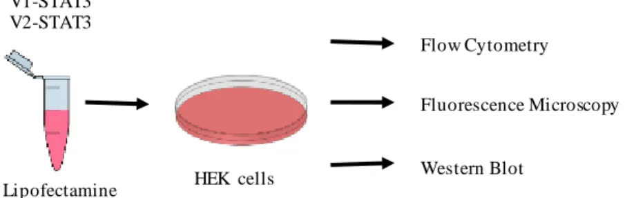
Medium was changed every other day and cells were passed once a week by trypsinization (Trypsin 0.25% w/v) for 5 min at 37ºC. For all the experiments, cells were counted using a Neubauer Chamber from Assistent (Sondheim, Germany) and seeded in different types of dishes according to the type of assay. Transfections were carried out by means of Lipofectamine 2000 in a 1:3 proportion (1 µg of DNA: 3 µL of Lipofectamine), 24 h after seeding. After transfection, STAT3 dimerization and cell death were evaluated by flow cytometry, STAT3 expression and phosphorylation by Western blot and intracellular STAT3 localization by fluorescence microscopy (Figure 3.4).
Figure 3.4 - Schematic representation of cell transfection and different types of analyses.
The transfection mixture was prepared in a microcentrifuge tube and then added to HEK cells. Cells were seeded twenty-four hours earlier in different plates depending on the type of assay. The results were then analyzed by flow cytometry, fluorescence microscopy and Western blot.
3.4. Flow cytometry
Fluorescence intensity of transfected cells was determined by flow cytometry. For this assay, 8x10⁵ cells per well were seeded in 6-well plates from TPP (Trasadingen, Switzerland).
Cells were washed once with PBS and trypsinized (0.05% w/v) at 37ºC for 5 min. Trypsin was neutralized with complete DMEM medium and cells were collected into sterile microcentrifuge tubes. Cells were centrifuged at 300xg for 5 min (VWR micro star 17R centrifuge), the supernatant was discarded and the pellet was resuspended in 500 µL of PBS. Immediately before acquisition, propidium iodide (PI) (1 mg/mL) was added to cells to quantify cell death in our samples, since this staining can cross the membrane of dead cells only.
Ten thousand cells per experimental group were analysed by means of a Cyan ADP flow cytometer from Beckman Coulter (Brea, CA, USA) equipped with a 25mW solid state blue
V1-STAT3 V2-STAT3 Flow Cytometry Fluorescence Microscopy Western Blot HEK cells Lipofectamine
The role of post-translational modifications on STAT3 interactions Methods
18
(488nm) laser, a 50mW solid state violet (405nm) laser, and a 60mW red (643nm) diode. For data analysis and representation, the FlowJo software (Tree Star Inc., Ashland, OR, USA) was used.
3.5. Protein extraction
For Western Blot, 2 x 10⁶ cells were seeded on 60 mm dishes from Thermo Scientific (MA,
USA) and two different protocols were used for protein extraction, depending on the type of sample under analysis.
For total protein extraction, cells were washed once with PBS, lysed with NP-40 lysis buffer (150 mM NaCl, 50 mM Tris-Cl pH 8.0, 1% NP-40) containing cocktail tablets of protease and phosphatase inhibitors from Roche (Basel, Switzerland). Cells were scrapped directly from the plates into microcentrifuge tubes and incubated 10 min in ice. A W-450 D sonicator (Emerson, Danbury, USA) was used for 5 sec at 10% of amplitude. Sonication was essential to disrupt cell membranes and release intracellular proteins, permitting their isolation and detection by Western blot. Cells were then centrifuged at 10000xg for 10 min at 4ºC and the supernatant containing the proteins was collected into a new microcentrifuge tube. Samples were always kept on ice to avoid protein degradation during extraction protocols.
For nuclear and cytoplasmic protein extraction, the NE-PER Nuclear and Cytoplasmic Extraction Reagents kit from Thermo Scientific (MA, USA) was used. Cells were washed once with PBS and trypsinized (0.25% w/v) at 37ºC for 5 min. Trypsin was neutralized with complete DMEM medium and cells were collected into a microcentrifuge tube. Cells were then centrifuged at 300xg for 5 min at room temperature, the supernatant was discarded and the pellet was resuspended in cold cytoplasmic extraction reagent I with protease inhibitors. The suspension was homogenized by vortex (15 seconds) and incubated in ice for 10 min. Cold cytoplasmic extraction reagent II was then added to the mixture, homogenized by vortex and incubated for 1 minute in ice. The mixture was once again vortexed and centrifuged at 16000xg for 5 min. The supernatant, corresponding to the cytoplasmic protein extract, was collected into a new sterile microcentrifuge tube and stored at –20°C. The pellet was resuspended in nuclear extraction reagent with protease inhibitors and homogenized by vortex 4 times every 10 min. Then the sample was centrifuged at 16000xg for 10 min and the supernatant, corresponding to the nuclear protein fraction, was collected into a new sterile microcentrifuge tube and stored at -80°C.
Protein concentration was quantified on a microplate reader from Thermo Scientific (MA, USA) by means of the Bradford method. A standard curve with known concentrations of bovine serum albumin (BSA, 0.125 to 2 µg/µL) was used to determine protein concentration. Samples were incubated with 200 µL of Bradford solution for 5 min and read at 595 nm.
The role of post-translational modifications on STAT3 interactions Methods
19 3.6. Western blot
The same amount of protein (15-20 µg) from each sample was mixed with 4x denaturing loading buffer (0.125 mM Tris pH 6.8; 4% sodium dodecyl sulphate (SDS), 20% glycerol, 10% β-mercaptoethanol, 0.004% bromophenol blue) and boiled for 5 min at 100ºC. Protein samples were loaded and run on 10% w/v SDS-polyacrylamide gel electrophoresis made with Protogel reagents from National Diagnostics (Atlanta, United States). Samples were run in running buffer (25 mM Tris-Base; 3.5 mM SDS; 0.2 M Glycine) at 120V for 1 h and transferred to a nitrocellulose membrane from GE Life Technologies (Boston, USA) at 100V for 1 h in transfer buffer (25 mM Tris-Base; 0.2 M Glycine; 20% methanol).
After electrophoretic transfer, membranes were stained with Ponceau S (0.1% w/v) from Amresco (Solon, United States) to check if protein transfer was efficient. Ponceau solution was removed from the membranes by washing them with Tris-buffered saline Tween 20 (TBS-T) (150 mM NaCl, 50 mM Tris pH 7.4, 0.5% Tween 20). Membranes were then blocked for 1 h in 5% (w/v) non-fat dry milk in TBS at room temperature, washed with TBS-T (10 min, 3 times) and incubated overnight with primary antibodies diluted in 5% BSA in TBS 1x and 0.05% of sodium azide. The primary antibodies used were: anti-phospho-STAT3 Y705 (1:1000, mouse monoclonal), anti-phospho-STAT3 S727 (1:1000, rabbit monoclonal), anti-STAT3 (1:1000, rabbit monoclonal), anti-GAPDH (1:1000 mouse monoclonal) and anti-Lamin-B (1:1000, goat monoclonal). After incubation with primary antibodies, membranes were washed with TBS-T (10 min, 3 times) and incubated for 2 h at 4°C with the appropriate secondary antibody (1:1000 in 5% of non-fat dry milk in PBS), washed with TBS-T (10 min, 3 times) and incubated 1 min with chemiluminescent HRP substrate before imaging in a Chemidoc XRS device from Biorad (CA, USA). GAPDH was used as loading control for cytoplasmic protein extracts and total proteins, and Lamin-B for nuclear protein extracts.
3.7. Fluorescence microscopy
All images of fluorescent living cells were acquired using a Leica DMI6000 fluorescence microscope equipped with a Hamamatsu Flash 4.0 LT sCMOS camera and DAPI + GFP fluorescence filter sets, and controlled by means of the Las X software (MN, USA). For microscopy, 8x10⁵ cells per well were seeded in 35 mm dishes from Ibidi (Planegg, Germany),
coated with poly-L-lysine (20µg/mL) for 1 h at room temperature, for a better adherence of cells. Nuclei were stained with Hoechst 33342 (10µg/mL) from Thermo Fisher Scientific (MA, USA) for 1 min at room temperature. Cells were washed once with PBS and 1 mL of PBS was added to the cells. Pictures of 25-30 cells per experimental group were taken using the 100x objective and then analyzed by means of Fiji free online software (http://fiji.sc/).
The role of post-translational modifications on STAT3 interactions Methods
The role of post-translational modifications on STAT3 interactions Results
21 4. Results
4.1. JAK/STAT3 pathway is activated by LIF in HEK cells
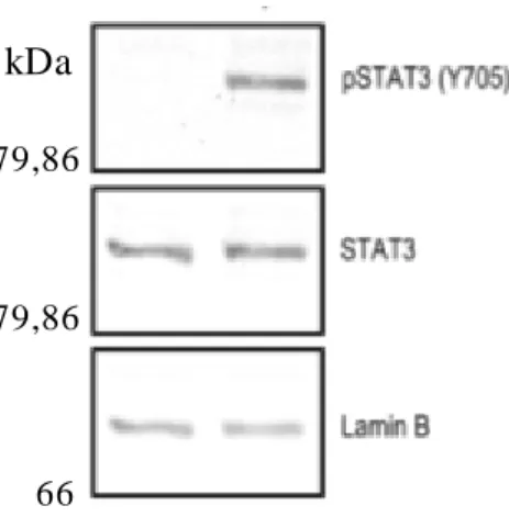
To verify that LIF activates the JAK/STAT3 pathway in HEK cells, these cells were seeded and 19 h later the medium was changed to medium without FBS to allow the activation by LIF. Two hours later, HEK cells were treated with LIF (100 ng/mL) and 2 h later nuclear and cytoplasmic proteins were extracted.
Nuclear proteins were analysed by Western blot to observe endogenous STAT3 phosphorylation. The membrane was incubated with anti-STAT3 phosphorylated on Y705 (pSTAT3 Y705), anti-STAT3 and anti-Lamin-B antibodies (Figure 4.1). Lamin-B levels were used as loading control to make sure that we use the same amount of protein in all of the samples. On the figure below we have cells non-stimulated and stimulated with LIF and we observe that the band of pSTAT3 Y705 only appears when they were stimulated with LIF. The levels of total STAT3 are the same in non-stimulated and stimulated cell.
Figure 4.1 – JAK/STAT3 patway is activated by LIF in HEK cells.
HEK cells were incubated with LIF (100 ng/mL) or the vehicle. Nuclear protein extracts were analysed by Western blot using specific antibodies (pSTAT3 Y705, STAT3 and Lamin-B).
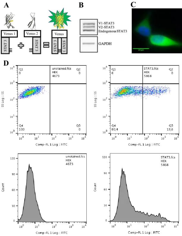
4.2. The Venus-STAT3 BiFC system
Along this project, we are using a BiFC system to study STAT3 dimerization and the effect of its PTM (phosphorylation, acetylation and methylation) in living cells. The BiFC system is composed by two plasmids (V1-STAT3 and V2-STAT3), which are fused to the two halves of the Venus fluorescent protein. When STAT3 dimerizes, a complementation of the two halves of Venus occurs, leading to fluorescence (Figure 4.2A). Protein extracts from HEK cells transfected with the Venus-STAT3 system show three distinct bands upon exposure to STAT3 antibodies. These correspond to V1-STAT3, V2-STAT3 and endogenous STAT3, from the
79,86
kDa
79,86
The role of post-translational modifications on STAT3 interactions Results
22
highest to the lowest molecular weight (Figure 4.2B). In this system, STAT3 effectively dimerizes and produces fluorescence, which is predominantly located in the cytoplasm of HEK cells, as demonstrated by microscopy and flow cytometry analyses (Fig. 4.2C and 4.2D).
Figure 4.2 – The Venus-STAT3 BiFC system in living HEK cells.
A) Representation of the STAT3 BiFC system. HEK cells were transfected with both Venus-STAT3 BiFC plasmids (V1-Venus-STAT3 and V2-Venus-STAT3) and were analyzed 19 h later. B) total protein extracts were analysed by Western blot, and show 3 bands corresponding to V1-STAT3, V2-STAT3 and STAT3 endogenous from top to bottom. C) cells were observed by fluorescence microscopy. Venus-STAT3 is predominantly localized on the cytoplasm (green) and nuclei were stained with Hoechst 33342 (blue). Scale bar: 20 m. D)10000 cells were analysed by flow cytometry, showing a clear signal in the green spectrum (FL-1) in cells transfected with the Venus-STAT3 BiFC constructs.
Venus 1 S T A T 3 Venus 2 S T A T 3 Venus S T A T 3 STA T 3 V1-STAT3 V2-STAT3 Endogenous STAT3
A
B
C
D
GAPDHThe role of post-translational modifications on STAT3 interactions Results
23
As described in section 3.2 (Materials and Methods), using our BiFC constructs as templates, we made 5 different point mutations on STAT3 residues involved in acetylation, methylation and phosphorylation of the protein. Figure 4.3 represents the STAT3 structure with the 5 residues of interest (K49, K140, K685, Y705, S727).
Figure 4.3 – Structure of STAT3 with our target phosphorylation, acetylation and methylation sites.
Several regulation residues in STAT3 were targeted with point mutations to study their purpose in the dimerization and therefore in PPI. (Hendry, L. et al., 2004)
In order to confirm the effects of the different mutations on LIF-stimulated STAT3 phosphorylation on Y705 and S727 residues, nuclear protein extracts were analysed by Western blot by using specific antibodies to phosphorylated STAT3 on Y705 and S727 (Figure 4.4). The reader must be mindful, however, that the results shown herein are not optimal since this Western blot was only done once.
Figure 4.4 – Besides the phosphorylation residues, others are also important for the correct phosphorylation and activation of STAT3.
HEK cells were transfected with mutated or non-mutated Venus-STAT3 BiFC constructs (V1-STAT3 and V2-STAT3). After 19 h medium was changed by medium without FBS and 2 h later cells were stimulated with LIF (100 ng/mL) for 2 h. Nuclear and cytoplasmic protein were extracted and analysed by Western blot. Anti-pSTAT3 Y705, anti-pSTAT3 S727 were used to detect STAT3 phosphorylation. Lamin-B levels were used as loading control.
What is visible in Figure 4.4 is that, firstly, LIF leads to an increase in p-STAT3 levels at both Y705 and S727, confirming once more what was described in section 4.1 (Results).
Acetylation Methylation Methylation Acetylation Phosphorylation Y705 S727 K685 K140 K49 kDa 79,86 66 kDa 79,86 66
The role of post-translational modifications on STAT3 interactions Results
24
Regarding the effects of mutation K49R on the phosphorylation of these two residues, it seems that it does not alter the phosphorylation of Y705, but, on the contrary, seems to slightly reduce the phosphorylation of S727. The increase induced by LIF is so slight it is barely noticeable. This suggests that K49 regulates the phosphorylation of S727, but not Y705.
As for the mutation K140R it appears that not only it affects the phosphorylation of both Y705 and S727 but also seems to interfere with the expression of Lamin-B when cells were stimulated with LIF. In both Western blots, loading controls appeared at considerable lower levels comparing to the control. The combination of the mutation and the activation by LIF should be tested further to assess if K140R does indeed interfere with the phosphorylation, once the loading control problem is solved.
The mutation K685R seems to reduce slightly the phosphorylation of Y705 but significantly reduces S727 phosphorylation. However, it must be taken into account, that the loading controls in the p-STAT3 Y705 gel for the K685R mutation with and without LIF are not very clear, so further studies must be made to confirm these results. Moreover, on the p-STAT3 S727 membrane, the control itself did not show a significant increase in its phosphorylation levels, when cells were stimulated with LIF. This too, adds to the need for more studies and the confirmation of these preliminary results.
Concerning the Y705F mutation, as expected, we see no phosphorylation of this residue. This is evident since tyrosine was replaced by a phenylalanine, a residue which cannot be phosphorylated.43 However, it is still visible that there is phosphorylation of endogenous STAT3
on Y705, corresponding to the third band on the LIF-stimulated lane. This mutation, on the other hand, did not seem to interfere with the S727 phosphorylation.
Finally, regarding the S727A mutation, it seems it is necessary for the correct phosphorylation of Y705. The difference between non-stimulated and stimulated cells seems to be minimal when comparing to the control (WT). As for the phosphorylation levels of S727 itself, once again as expected, they did not increase when serine was replaced by alanine. Interestingly, endogenous STAT3 (lowest band) shows phosphorylation of S727 with and without LIF stimulation.
4.3. STAT3 mutants do not affect STAT3 dimerization
To determine the relative role of the different residues on STAT3 dimerization, HEK cells were transfected with different combinations of mutated and non-mutated Venus-STAT3 BiFC constructs (Table 4.1). Twenty four hours later the samples were analysed by flow cytometry (Figure 4.5) and fluorescence microscopy (Figure 4.6).
The role of post-translational modifications on STAT3 interactions Results
25
Table 4.1 – Combination of the Venus-STAT3 BiFC constructs. V1 V2 K49R K140R K685R Y705F S727A WT K49R 1 K140R 2 7 K685R 3 8 12 Y705F 4 9 13 16 S727A 5 10 14 17 19 WT 6 11 15 18 20 21
The results from flow cytometry show that no combination of the Venus-STAT3 constructs alters significantly the percentage of fluorescence, suggesting that the tested residues do not affect STAT3 dimerization (Figure 4.5).
Figure 4.5 - STAT3 mutants have no effect on fluorescence levels in HEK cells.
HEK cells were treated with the mutants of the Venus-STAT3 BiFC plasmids conjugated with each other. Ten thousand events were analysed by flow cytometry. Percentage of fluorescence of the STAT3 mutants were then compared with the STAT3.
Figure 4.6 shows that STAT3 dimers are predominantly localized on the cytoplasm in all the combinations of STAT3 mutants. However, 4 out of 6 combinations containing the K49R construct (K49R/K49R, K140R/K49R, S727A/K49R, WT/K49R) and also 4 out of 6 combinations with the S727A construct (S727A/K49R, S727A/K140R, S727A/K685R and WT/S727A) show some aggregation. K685R/K140R also showed some aggregates. Given the high incidence of aggregates when K49R and the S727A mutants are present, it is possible that these residues could be relevant for either STAT3 degradation or stability.
1 2 3 4 5 6 7 8 9 1 0 1 1 1 2 1 3 1 4 1 5 1 6 1 7 1 8 1 9 2 0 2 1 0 5 0 1 0 0 1 5 0 % o f fl u o re sc en c e
The role of post-translational modifications on STAT3 interactions Results
26
Figure 4.6 – Mutations of PTM residues have no effect on the intracellular localization of STAT3 dimers.
HEK cells were transfected with Venus-STAT3 BiFC mutants plasmids conjugated with each other and the samples were analysed by fluorescence microscopy. Aggregates are visible as the brighter green spots found in the cytoplasm of the cells. STAT3 was used as control (WT/WT). Scale bar: 20m.