Induced of
Paracoccidioides brasiliensis
Cell Proliferation
in a Ras-Dependent Manner
Ana Eliza Coronel Janu Haniu1, Juliana Terzi Maricato1, Pedro Paulo Moraes Mathias2,
Daniele Gonc¸alves Castilho1, Rodrigo Bernardi Miguel2, Hugo Pequeno Monteiro2, Rosana Puccia1, Wagner Luiz Batista1,3*
1Departamento de Microbiologia, Imunologia e Parasitologia - Universidade Federal de Sa˜o Paulo, Sa˜o Paulo, Brazil,2Departamento de Bioquı´mica/Biologia Molecular – Centro de Terapia Celular e Molecular (CTCMol), Universidade Federal de Sa˜o Paulo, Sa˜o Paulo, Brazil,3Departamento de Cieˆncias Biolo´gicas - Universidade Federal de Sa˜o Paulo/Campus Diadema, Sa˜o Paulo, Brazil
Abstract
Paracoccidioides brasiliensis, a causative agent of paracoccidioidomycosis (PCM), should be able to adapt to dramatic
environmental changes inside the infected host after inhalation of air-borne conidia and transition to pathogenic yeasts. Proteins with antioxidant functions may protect fungal cells against reactive oxygen (ROS) and nitrogen (RNS) species generated by phagocytic cells, thus acting as potential virulence factors. Ras GTPases are involved in stress responses, cell morphology, and differentiation in a range of organisms. Ras, in its activated form, interacts with effector proteins and can initiate a kinase cascade. In lower eukaryotes, Byr2 kinase represents a Ras target. The present study investigated the role of Ras inP. brasiliensisafterin vitrostimulus with ROS or RNS. We have demonstrated that low concentrations of H2O2(0.1 mM) or NO2(0.1–0.25mM) stimulatedP. brasiliensisyeast cell proliferation and that was not observed when yeast cells were
pre-incubated with farnesyltransferase inhibitor. We constructed an expression plasmid containing the Byr2 Ras-binding domain (RBD) fused with GST (RBD-Byr2-GST) to detect the Ras active form. After stimulation with low concentrations of H2O2or NO2, the Ras active form was observed in fungal extracts. Besides, NO2induced a rapid increase inS-nitrosylated Ras levels. This alternative posttranslational modification of Ras, probably in residue Cys123, would lead to an exchange of GDP for GTP and consequent GTPase activation in P. brasiliensis. In conclusion, low concentrations of H2O2 or NO2 stimulated P. brasiliensisproliferation through Ras activation.
Citation:Haniu AECJ, Maricato JT, Mathias PPM, Castilho DG, Miguel RB, et al. (2013) Low Concentrations of Hydrogen Peroxide or Nitrite Induced of
Paracoccidioides brasiliensisCell Proliferation in a Ras-Dependent Manner. PLoS ONE 8(7): e69590. doi:10.1371/journal.pone.0069590 Editor:Robert A. Cramer, Geisel School of Medicine at Dartmouth, United States of America
ReceivedFebruary 17, 2013;AcceptedJune 10, 2013;PublishedJuly 29, 2013
Copyright:ß2013 Haniu et al. This is an open-access article distributed under the terms of the Creative Commons Attribution License, which permits
unrestricted use, distribution, and reproduction in any medium, provided the original author and source are credited.
Funding: This study was supported by Fundac¸a˜o de Amparo a` Pesquisa do Estado de Sa˜o Paulo (grant 2011/14392-2) and Conselho Nacional de Desenvolvimento Cientı´fico e Tecnolo´gico (grant 477764/2009-6) to WLB; and Coordenac¸a˜o Aperfeic¸oamento de Pessoal de Nı´vel Superior (scholarship to AECJH). The funders had no role in study design, data collection and analysis, decision to publish, or preparation of the manuscript.
Competing Interests:The authors have declared that no competing interests exist. * E-mail: [email protected]
Introduction
Paracoccidioides brasiliensis is a thermo-dependent dimorphic fungus responsible for paracoccidioidomycosis (PCM), a systemic mycosis that is prevalent in Latin America. The major protective host immune response againstP. brasiliensisis mediated by cells, as evidenced by granuloma formation [1]. The interaction between
P. brasiliensis and alveolar macrophages is a crucial step for the establishment and progression of infection in susceptible hosts. The yeast pathogenic phase of P. brasiliensis is a facultative intracellular pathogen that is able to survive and replicate within the phagosome of inactivated murine and human macrophages [2]. By contrast, macrophages can engulf this microorganism and confine it within phagosomes where, by action of microbicidal molecules and/or by restriction of essential nutrients, the pathogen can be destroyed. Among the molecules that exert fungicidal action in phagosomes are hydrogen peroxide (H2O2), nitric oxide (NO) and their derivatives. These are generated by NADPH oxidase and inducible nitric oxide synthase (iNOS), respectively
[3,4]. Immune system cells activate the NADPH oxidase complex to generate radical superoxide (O22), which is subsequently converted into H2O2. Macrophages express iNOS with activation of the L-arginine-nitric oxide pathway, and subsequent NO production.
Signaling pathways that control morphological changes, cell proliferation and stress response in P. brasiliensis are largely unknown, but in other dimorphic fungi the involvement of cAMP (cyclic adenosine 39, 59-monophosphate) and MAPK (mitogen-activated protein kinase) is known to be an important factor in this process [8]. Modulation of MAP kinases is an important and highly conserved event in eukaryotes. It is composed of a series of protein kinases, which can sequentially phosphorylate other proteins so that the signal transmissions from the point of origin (typically the cell membrane) to the nucleus can occur [9]. As a result, the target molecules (including transcription factors) are phosphorylated [10,11]. In this context, the Ras protein is prominent in the regulation of signal transduction pathways that mediate adaptive changes. Ras belongs to a large family of low molecular-weight proteins (21 kDa) with GTPase activity. Ras GTPases are molecular switches that are active when GTP-bound and inactive when GDP-bound. Both processes are regulated by enzymatic reactions. Guanine nucleotide exchange factors (GEFs) catalyze the release of GDP from the guanine nucleotide-binding pocket, mediating the exchange of GDP for GTP. The activation state of Ras is self-limited by its intrinsic GTPase activity, which is enhanced to critical regulatory levels by GTPase-activating enzymes (GAPs) [12]. Ras is involved in signal transduction pathways connecting events from many cell surface receptors to intracellular processes [13]. In mammals, depending on the cellular context, Ras activation can stimulate cell division cycle, morphogenesis, differentiation, or apoptosis [13].
In microorganisms, the Ras protein is similarly involved in growth and development processes, morphological changes, and stress responses. In Saccharomyces cerevisiae, Cryptococcus neoformans, and Aspergillus fumigatus, among other fungi, two Ras isoforms (Ras1 and Ras2) have been identified [8]. Waugh et al. [14] demonstrated thatC. neoformansRas proteins share some degree of functional redundancy, since both Ras1 and Ras2 mutants were viable and phenotypically similar to wild type. InTrichoderma reesei, both Ras1 and Ras2 play similar roles in morphogenesis and adjusting cAMP level, but Ras2 is also involved in regulation of cellulase gene expression [15]. Differential functions of Ras1 and Ras2 were also described inBeauveria bassiana[16]. In Schizosacchar-omyces pombeonly Ras1 was identified, which controls two different downstream signaling pathways [17]. However, endomembrane Ras activates a Cdc42 pathway to mediate cell polarity, while plasma membrane Ras selectively regulates a MAP kinase pathway to mediate mating pheromone signalling [18].
Ras proteins are conserved at the N-termini, but differ substantially at the C-termini, where 10–20 amino acids form the hypervariable region. Ras proteins are anchored to the membranes by a series of post-translational modifications occur-ring at theC-terminus. Anchoring of Ras in membranes is believed to be absolutely required for biological activity. Ras proteins contain a CAAX motif at the C-terminus (C = cysteine, A = ali-phatic amino acid, and X = any amino acid), where farnesyl transferase-mediated cysteine farnesylation occurs in the cytosol. This posttranslational modification prompts Ras association with the endoplasmatic reticulum (ER). Farnesylation is followed by the cleavage of the three C-terminal residues (AAX) and subsequent carboxymethylation of the farnesyl-cysteine [19,20].
In P. brasiliensis, two Ras isoforms were characterized with important roles during fungal dimorphism, thermal stress, and in parasite-host interactions [21]. The prenylation site (CAAX) was detected in both isoforms, but with variable sequences (CVIM in Ras1 and CLIL in Ras2) [21]. Nevertheless, nothing is known about GTPases functions inP. brasiliensisfor adaptive responses to oxidative and nitrosative stress. In the present study, we
investigated the importance ofP. brasiliensis Ras GTPase afterin vitrostimulation with ROS and RNS. Using a novel probe that detects activated Ras (Ras-GTP), we showed that low concentra-tions of NO and H2O2mediate cell signaling triggered with the participation of Ras, leading to cell proliferation inP. brasiliensis. Furthermore, we showed that Ras is S-nitrosylated in our test conditions. Therefore, this work shows the beneficial role of Ras activation by low levels of ROS and RNS in P. brasiliensis cell proliferation.
Materials and Methods
2.1. Fungal strain and growth conditions
We usedP. brasiliensis, isolate Pb18, in our experiments. Unless otherwise mentioned, cells were cultured and maintained at 37uC in modified YPD medium (0.5% yeast extract, 0.5% casein peptone, and 1.5% glucose, pH 6.5). CFU count was performed in supplemented BHI plates (Becton Dickinson Company) containing 4% fetal calf serum, 5% spent medium, ampicillin (100 IU/mL) and streptomycin (100mg/mL).
2.2. NO and H2O2stimulation and quantification of P.
brasiliensis CFU
In experiments involving oxidative and nitrosative stress, P. brasiliensis cells were cultivated in modified YPD for 5 days at 37uC. Yeast cells (16105) were seeded in a 6-well culture plate subjected to a 24-h period of starvation with F12 medium, to reduce or stop fungal growth until starting the treatment. This strategy was used to verify the role of H2O2or NO2stimulus on fungal growth and cell signaling. Yeast cells were exposed to different concentrations of H2O2or NaNO2(in culture medium mildly acidic, pH 5.5; in this condition NaNO2releases NO), for 5 h at 37uC [22,23,24]. Then yeast cells were washed and incubated for 24 h at 37uC under shaking in fresh culture media. Finally, 100mL were plated in supplemented BHI plates for 7 days
at 37uC. The experiment was repeated three times. Cell proliferation was evaluated by colony formation unit counts (CFU).
Growth curves were performed by evaluating fungal counts during different days of growth. For that, yeast cell suspension aliquots (100ml) were stained with equal volumes of Trypan Blue
vital dye and counted in a Neubauer chamber (for 4, 8 and 12 days), where viable cells did not stain by the vital dye.
2.3. Plasmid construction
The Ras Binding Domain (RBD) coding sequence of P. brasiliensis Byr2 kinase (GenBank accession number EEH46080, Ste11S. cereviseae homolog) was obtained by PCR, as described previously [25]. Briefly, the RBD region was synthetized by using the primer sense, 59 CCCTTCCTCCAAATTGGCC 39, con-taining anEcoRI site, and the downstream primer anti-sense, 59
2.4. Ras activation in P. brasiliensis
Ras activation was determined using the RBD(Byr2)-GST fusion protein, which tightly binds to the GTP-associated Ras form. After stimulus, yeast cells were collected by centrifugation, washed (3 times), and disrupted with glass beads in ice-cold lysis buffer (25 mM HEPES, pH 7.5, 150 mM NaCl, 1% (w/v) Nonidet P-40, 0.25% (w/v) sodium deoxycholate, 10% (w/v) glycerol, 25 mM NaF, 10 mM MgCl2, 1 mM EDTA, 1 mM sodium vanadate, one tablet of Protease Inhibitor, Roche Diagnostic, Mannheim, Germany, in 50 mL of extraction medium). The solubilized extract was centrifuged at 14,0006g for 15 min, and the supernatant was used in pull-down assays. The protein content of the cell extract was determined with Bradford reagent (Bio-Rad, Hercules, CA, USA). A protein sample (1 mg) was incubated with glutathione-Sepharose beads associated with RBD(Byr2)-GST for 3 h with gentle rocking. The samples were spun at 7,2006gfor 10–20 sec, and the resin was washed three times with lysis/binding/wash buffer (7,2006gfor 30 sec). The final pull-down was assayed by Western blot probed with mouse monoclonal anti-Ras antibody (Oncogene, Research Products). The remaining lysate was probed with the same antibody to determine the levels of total and endogenous Ras. The ratio between Ras signal intensity bound to RBD(Byr2)-GST beads and that obtained from total Ras, determined by densitometry, is proportional to Ras activity [26,27].
A control for probe RBD(Byr2)-GST specificity was performed as described Colombo et al. [27], with some modifications. Pb18 total extracts (1 mg) were incubated in PBS containing protease inhibitor (one tablet of Protease Inhibitor, Roche Diagnostic, Mannheim, Germany, in 50 mL of extraction medium) with 1 mM GTP (Sigma) or GDP (Sigma) at room temperature for 1 h with gentle rocking. Samples (bound either to GTP or to GDP) were incubated with glutathione-Sepharose beads containing cross-linked RBD(Byr2)-GST for 3 h with gentle rocking and detected by Western blotting using anti-Ras antibodies.
2.5. Western blotting
For Western blotting, proteins (25–50mg) were separated in 10
or 12% polyacrylamide gels and transferred to nitrocellulose membranes. After blocking, the membranes were incubated overnight at 4uC with primary anti-Ras antibody. A secondary antibody (anti-mouse) conjugated with horseradish peroxidase was used in the second step of the procedure (room temperature incubation for 1 h). Immunoblots were developed using the Super SignalH(Pierce, Rockford, USA) system.
2.6. Detection of Ras S-nitrosylation
The biotin switch technique (BST) was performed to detect Ras
S-nitrosylation, as described by Forrester et al. [28]. To detect Ras
S-nitrosylation in P. brasiliensis after H2O2 or nitrite treatment, yeasts were cultured in F12 medium for 24 h (starvation), then treated with H2O2or nitrite for increasing time periods, and then yeasts were disrupted with glass beads in buffer containing 25 mM HEPES, 50 mM NaCl, 0.1 mM EDTA, 1% NP-40, 0.5 mM PMSF, and protease inhibitors (Roche Diagnostic, Mannheim, Germany), pH 7.4. Cell debris was removed by centrifugation, and samples (1 – 0.6 mg protein extract) were diluted to 1.8 mL with HEN buffer (100 mM Hepes, 1 mM EDTA, 0.1 mM neocuproine, pH 8.0); SDS and MMTS were added to final concentrations of 2.5 and 0.1%, respectively. Following frequent vortex and incubation at 50uC in the dark for 20 min, lysates were precipitated with 3 volumes of acetone at220uC for 1 h. Proteins were centrifuged at 2,0006gfor 15 min, and the protein pellet was gently washed with 70% acetone (four times). The pellets were
suspended in 240mL HENS (HEN buffer added 1% SDS).
Samples were further incubated with 30mL biotin-HPDP
(2.5 mg/ml) in the presence or absence of 20 mM ascorbate at room temperature, in the dark for 1 h. After acetone precipitation, proteins were resuspended in 250mL HENS, followed by addition
of 750mL of neutralization buffer (25 mM HEPES, 100 mM
NaCl, 1 mM EDTA, 0.5% Triton X-100, pH 7.5). Fifty microliters of streptavidin-agarose beads (pre-washed) were added to each sample and incubated overnight at 4uC. Beads were washed with washing buffer (neutralization buffer with 600 mM NaCl) four times. To detectS-nitrosylated proteins, 50mL of 26 SDS sample buffer were added to the beads and tested by immunoblotting with anti-Ras antibody.
2.7. Statistical analysis
Data are expressed as means6SD. The statistical analysis of significance was assessed by one-way analysis of variance using the Student’st-test for comparison.p,0.05 was considered statistically significant.
Results
3.1. Low concentrations of ROS and RNS induced cell proliferation in P. brasiliensis
It is known that different levels of H2O2 or NO can induce distinct responses within a cell [29]. For example, different transcriptional responses are induced by low (sub-toxic) or high (toxic) levels of H2O2or NO in mammalian cells,S. cerevisiae, and
S. pombe[30,31]. To better understand this type of stimulus inP. brasiliensis, logarithmic growing yeast cells, were cultured in F12 medium for 24 h and subsequently treatedin vitro with different concentrations of H2O2(0.05–30 mM) and NO2(0.1–1000mM)
for 5 h at 37uC and the CFU count was assessed (Figure 1). For cells pre-incubated for 5 h with higher concentrations of H2O2 (10, 15 and 30 mM) and NO2(1, 10, 100 and 1000mM), a typical
dose-response curve with decreased cell viability was observed (Figure 1). For intermediate concentrations of H2O2 (0.5 and 1 mM) and NO2 (0.5mM) there was no change in cell viability
(Figure 1). However, yeast cells pre-incubated with low concen-trations H2O2 (0.1 mM) and NO2 (0.25mM) responded with significant cell proliferation (3.760.166103 and 1.86 0.176103CFU, respectively) when compared to unstimulated controls (2.1360.196103and 1.3560.786103CFU, respectively). Maximum stimulation of fungal proliferation was observed after incubation with 0.1 mM H2O2 and 0.25mM NO2. These data suggested thatP. brasiliensismay benefit from low concentrations of ROS and RNS to proliferate.
3.2. Ras participates in P. brasiliensis proliferation dependent on low concentrations of ROS or RNS
We also tested the effect of low concentrations of H2O2 and NO2 during fungal growth. P. brasiliensis was incubated with or without stimulus in the presence or absence of Ras inhibitor. We observed significant increase in the number of cells growing in the presence of 0.1 mM H2O2(5.83760.8256106cells) and 0.25mM of NO2 (3.6160.082610
6
cells) after 4 days when compared to controls (260.836106 cells and 1.6360.2886106 cells, respec-tively) (Figure 2B), however this effect was abolished in the presence of FPT III. We observed similar results after 8 days of growth (data not shown). However, on the twelfth day there was no statistical difference between the experimental samples and controls (data not shown). Therefore, these results confirm that low concentrations (sub-toxic) of ROS and RNS can lead to P. brasiliensiscell proliferation and that Ras farnesylation is required for this event.
3.3. Low concentrations of ROS or RNS promote Ras activation in P. brasiliensis
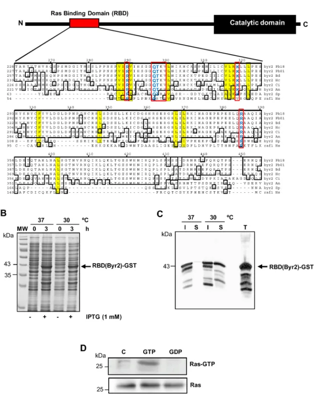
To understand the contribution of Ras onP. brasiliensis redox-dependent proliferation, we evaluated its activation after stimula-tion with low concentrastimula-tions of H2O2 and NO2. In order to do that, we constructed a probe that detects Ras active form (Ras-GTP). In fungi, the serine/threonine kinase Byr2 (Ste11 homo-logue inS. cereviseae) is known to be responsible for the interaction with activated Ras. InP. brasiliensis, the Byr2 gene is composed of four exons separated by three introns. BYR2 has 2,685 bp and encodes a protein of 894 amino acids. The Ras Binding Domain (RBD) shows conserved Ras interaction regions (Figure 3A), as observed in the alignment of Byr2-RBD with its fungal homo-logues fromA. niger,S. cerevisiaeandS. pombe, and with human
Raf-1. The RBD is located in a regulatory region at theN-terminal, between amino acids 216 and 395. According to Scheffzek et al. [36], the Byr2-RBD amino acid residues fromS. pomberesponsible for the interaction with Ras would be Arg74, Lys101, Ala84, Arg83, Thr82, Gln81, and Arg160. All residues are preserved inP. brasiliensis Byr2-RBD, suggesting that the intermolecular interac-tion with activated Ras may also occur (Figure 3A).
The use of a RBD-GST probe in studies on cell signaling is widespread to detect Ras activity in mammalian cells [33,37,38]; however, in fungi there are only few studies using this strategy [27,39,18]. We constructed an expression plasmid containing approximately 537 pb of the Byr2-RBD gene fragment subcloned intoEcoRI andXhoI sites of the pGEX-4T-2 vector. The cloned fragment corresponds to Byr2 amino acids 216–395 (179 amino acids) that include the whole RBD. For expression of the recombinant protein (RBD-GST probe), the expression vector was introduced intoE. coliBL21 and induced with 1 mM of IPTG at 37uC or 30uC. After 3 hours of induction in both temperatures a component of approximately 43 kDa was expressed, which is consistent with the expected molecular mass of the recombinant protein (Figure 3B). The solubility of recombinant RBD(Byr2)-GST was evaluated and we observed that at 37uC most of the fusion protein was expressed as insoluble inclusion bodies (Figure 3C). However, at 30uC we observed that a greater proportion of the RBD(Byr2)-GST recombinant protein was found in the bacterial extract soluble fraction (Figure 3C). To determine specificity of the probe RBD(Byr2)-GST in detecting Ras-GTP we performed in vitro exchange experiments. We incubated Pb18 total extract with high amounts of GTP or GDP, probed it with RBD(Byr2)-GST and immunoblotted the
Figure 1. Low concentrations of H2O2or nitrite promoted cell proliferation inP. brasiliensis.Pb18 yeast cells were seeded in a 6-well
culture plate subjected to a 24-h period of starvation with F12 medium and treated with different concentrations of H2O2(A) or nitrite (B) at pH 5.6 for 5 h at 37uC. Treated cells were plated in BHI and incubated at 37uC for 7–10 days (n = 6 at each point). The graph shows the means6SD of total CFU before and after treatment with H2O2or nitrite for each concentration. Statistically significant samples are indicated (p,0.05). The results are representative of three independent experiments.
precipitate with anti-Ras. Only activated Ras (Ras-GTP) was able to bind to the probe RBD(Byr2)-GST (Figure 3D), indicating that the assay is specific for Ras-GTP.
We next used the RBD(Byr2)-GST probe to assess the ability of low concentrations of ROS and RNS to activate Ras. Ras activity was determined in P. brasiliensis yeasts exposed to 0.1 mM H2O2 and 0.25mM NO2for increasing periods of time. Early Ras activation was observed after 15 min of stimulation with ROS and RNS (Figure 4A). Maximum Ras activation was observed after 60 min of incubation (Figure 4A), and at latter timepoints (180 and 300 min) we observed decrease of Ras activation. Also, the fungal protein extract was incubated with Glutathione-Sepharose beads alone (negative control) and analyzed by Western blot, but no reaction was observed (data not shown). The protein loading of the samples used in the Ras activity assay observed in SDS-PAGE gel stained Coomassie blue proved to be fairly homogeneous (data not shown). We also evaluated by RT-PCR the transcription levels ofRAS1andRAS2inP. brasiliensis
after incubation with 0.1 mM H2O2 or 0.25mM NO2, but no significant changes were observed (data not shown). Therefore, low concentrations of ROS and RNS promoted guanine nucleotide exchanges in the critical cellular signaling protein Ras.
We also investigated the inhibitory effect of farnesyl transferase in Ras activity. Yeast cells were cultivated for 24 h in the presence or absence of 250mM FPT III, followed by treatment for 1 h with
0.1 mM H2O2or 0.25mM nitrite. As shown in Figure 4B, FPT III
substantially inhibited Ras activation in yeast cells ofP. brasiliensis
treated with low concentration of ROS or RNS, as compared with controls without inhibitor.
3.4. NO2promotes Ras S-nitrosylation in P. brasiliensis
It has been shown that NO stimulates guanine nucleotide exchange in Ras and that this event is dependent on S -nitrosylation, which occurs in the active thiol group Cys118 [40]. Initially we checked whether nitrosylable Cys was conserved inP. brasiliensis. By aligning a small fragment of the C-terminal region from different fungi and human Ras, we observed that inP. brasiliensisRas1 there is a Cys homologue at position 123, whereas in Ras2 there is a serine at this position (Figure 5A). Then we used a computer program (http://dbSNO.mbc.nctu.edu.tw, [41]) to predict putative S-nitrosylation sites in P. brasiliensis Ras and detected that Cys123 would be a likely S-nitrosylation site with 95% prediction specificity (Figure 5B). Cys123 is localized in the Ras GTP binding and interaction site (G3) (Figure 5A). Ras2 also showed probable S-nitrosylation sites at Cys55 and Cys176; however, these sites are located elsewhere in the molecule (data not shown). We also evaluated whether Cys123 would be located in Ras hydrophobic region. According to Hess et al. [6] local hydrophobicity might promote specificS-nitrosylation. Analysis of the deducedP. brasiliensisRas1 and Ras2 sequences showed that Cys123 is inserted in Ras1 hydrophobic domain with low surface-probability in the protein (Figure 5C). Moreover, this feature was not observed in the equivalent Ras2 region. These results suggested
Figure 2. Cell proliferation ofP. brasiliensisstimulated by low concentrations of H2O2or nitrite is suppressed in the presence of FPT III inhibitor.(A) Pb18 yeast cells were seeded in a 6-well culture plate subjected to a 24-h period of starvation with F12 medium and pretreated with 250mM FPT III and stimulated with 0.1 mM H2O2or 0.25mM nitrite at pH 5.6 for 5 h at 37uC. Cells were plated in BHI medium at 37uC for 10 days (n = 6 at each point) and CFU were counted. (B) Same as in (A), but cells (1.56105) were cultured in the YPD medium (n = 4 each point) and after 4
days the fungal growth was determined by counting in a Neubauer chamber. The graphs show the mean CFU or number of cells6SD for each sample. The results represent three independent experiments. Statistically significant samples are indicated (p,0.01 or 0.05).
Figure 3. Plasmid construction and production of the RBD(Byr2)-GST probe.(A) Schematic representation of Byr2 showing the localization of conserved domains, highlighting in red the localization of the Ras Binding Domain (RBD). RBD sequences fromP. brasiliensis(Pb – EEH46080), Blastomyces dermatitidis(Bd – EGE86103),H. capsulatum(Hc – EGC48892),Coccidioides immitis(Ci – XP 001242119),S. cerevisiae(Sc – AAB67571), and RBD-Raf-1 ofHomo sapiens(Hs – AGC09606) were aligned with ClustalW (module MegAlign, DNAstar Inc). Conserved sequences are boxed, the residues directly involved in the interaction with activated Ras (Ras-GTP) are indicated in red boxes and key amino acids involved in the interaction with Ras-GTP are shown in yellow. (B) Coomassie blue-stained 10% SDS-PAGE gels showing total bacterial extracts from recombinant bacteria expressing RBD(Byr2)-GST (arrow) stimulated (3 h) or not (0 h) with 1 mM IPTG. (C) Ten microliters of total (T), soluble (S) and insoluble (I) fractions were assayed in Western blots probed with anti-GST antibody. The migration of molecular mass standards (MW) is shown on the left. (D) Pb18 total extract (1 mg) was bound either to GTP (1 mM) or GDP (1 mM) and incubated with RBD(Byr2)-GST fusion protein linked to glutathione-Sepharose. Ras-GTP and total Ras (50mg total protein) eluted with SDS-PAGE sample buffer were loaded in an SDS-PAGE gel. Ras was detected by immunoblotting with anti-Ras antibody.
that Ras1 Cys123 bears S-nitrosylation features. Therefore, we verified if Ras1 was possiblyS-nitrosylated by the biotin switch test [28], which introduces a biotin molecule in theS-nitrosylated Cys (Figure 6A). The detection ofS-nitrosylated protein by the biotin switch method is dependent on treatment with ascorbate, consistent with the reduction dependent on ascorbate from nitrosothiol bonds (Figure 6A). For that, we studied whether lower concentrations of NO2(0.25mM) or H2O2(0.1 mM) would be able to induce Ras1S -nitrosylation. We found that NO2 induced rapid increase in S -nitrosylated Ras levels, with a peak at 30 min (Figure 6B). On the other hand, no change in the level ofS-nitrosylation was observed after treatment with H2O2(Figure 6B). These findings suggested that NO probably plays an important role in the process of Ras activation through itsS-nitrosylation.
Discussion
The ability of pathogenic fungi to resist the deleterious effects of ROS and/or RNS is foreseen as an important virulence
mechanism, particularly in relation to its contact with phagocytic host cells. We have here demonstrated that sub-toxic concentra-tions of ROS and RNS are capable of stimulatingP. brasiliensiscell proliferation in a Ras GTPase activation-dependent manner. In addition, we observed that stimulation with NO2evoked RasS -nitrosylation, which has not been observed after stimulation with H2O2. Cell proliferation upon ROS and RNS stimulation have not been reported before in fungi; however, it remains to be clarified if this phenomenon would occur inP. brasiliensisisolates from other phylogenetic groups [42], or even in isolates of the recently separated speciesP. lutzii[43], apart from virulent Pb18 presently tested. Pb18 represents the main S1 phylogenetic P. brasiliensisgroup [42].
It has frequently been observed that high concentrations of both ROS and RNS are cytotoxic to fungal cells, causing cell death, and this is an effector mechanism of the immune system cells [44]. However, NADPH oxidase knockout mice (deficient for ROS production) showed decreased fungal spread when intratracheally infected with C. neoformans, and were also protected against
Figure 4. Low concentrations of H2O2or nitrite induce Ras activation.(A) Pb18 yeast cells were cultivated in modified YPD for 5 days at
37uC, subjected to a 24-h period of starvation with F12 medium and incubated with 0.1 mM H2O2or 0.25mM NO2at pH 5.6 for different timepoints at 37uC. (B) Pb18 yeast cells were cultivated in modified YPD for 5 days at 37uC, subjected to a 24-h period of starvation with F12 medium and cultured with or without 250mM FPT III inhibitor followed by treatment with 0.1 mM H2O2or 0.25mM NO2at pH 5.6 for 1 h at 37uC. After yeast cells lysis (A) and (B), Ras activation was determined using the GST-RBD(Byr2) fusion protein, which binds with high affinity to the GTP-associated form of Ras. Ras-GTP (active) and total Ras (50mg total protein) were assayed by western blots probed with anti-Ras antibody. Relative densitometric values of bands are shown in the bar graphs.
pulmonary infection [45]. This event, which seems to contradict the literature, may suggest that the microorganisms could somehow benefit from ROS/RNS derived from immune system cells. Our data suggests that one possible mechanism would be by direct contact of the fungus with sub-toxic concentrations of NO2 and H2O2, but that remains to be tested inC. neoformans. Despite the proliferation induced by low doses of ROS and RNS having been previously demonstrated in mammalian cells [32,33,46], this is the first report of stimulation of fungal cell growth by ROS/
RNS. Recently, Srinivasa et al. [47] showed that both low (,1 mM) and high (4–10 mM) concentrations of exogenous H2O2 induce filamentous growth with distinct cell morphology and growth rate inC. albicans, which suggests a differential transcrip-tional response. The authors demonstrated that sub-toxic doses of H2O2induce the formation of pseudohyphae.
In experimental PCM, NO has dual roles. Brummer et al. [1] have demonstrated that activation of mouse (BALB/c) peritoneal macrophages by IFN-c enhances the fungicidal activity. In
Figure 5.P. brasiliensisRas1 has a putative nitrosilable Cys.(A) Ras1 and Ras2 sequences fromP. brasiliensis(Pb – EEH22637 and EEH22450),B. dermatitidis(Bd – XP002628159 and EEQ83443),H. capsulatum(Hc – EEH06649 and EEH07767),C. posadasii(Cp – XP 001246878 and XP 001247157), S. cerevisiae(Sc – AAA34958 and CAA95974) andH. sapiens(Hs – AAH14261) were aligned by ClustalW (module MegAlign, DNAstar Inc). (B) Analysis of putative motifs of S-nitrosylation in Pb18 Ras1 using the computer program dbSNO (http://dbSNO.mbc.nctu.edu.tw, [37]). (C) Kyte-Doolittle hydrophilicity and Emini Surface Probability plots (Protean module; DNAstar Inc.) of Pb18 Ras1 and Ras2. Red boxes indicate the location of Ras1 Cys123 and Ras2 Ser144, respectively.
contrast, macrophages from A/J mice were poorly activated by low doses of IFN-c, and secreted only low amounts of IL-12, NO, thus showing poor fungicidal ability [48]. Nascimento et al. [49] showed that NO is essential for host resistance to infection byP. brasiliensis. The authors found that mice genetically deficient for iNOS (inducible nitric oxide synthase) were susceptible to infection by the fungus. On the other hand, the persistent production of NO was correlated with increased infection byP. brasiliensis[49]. Our
in vitrodata suggests that one possible alternatively mechanism of fungal proliferationin vivowould be by direct contact of the fungus with sub-toxic concentrations of NO2and H2O2.
In our study we observed that there was decrease in Ras activity after incubation with FPT III (Fig. 4B). Previous results with theP. brasiliensis showed that farnesylation blockage interfered with vegetative growth of yeast cells and stimulated germinative tube production even at 37uC [21]. Since the farnesylation inhibitor is not specific for Ras incorporation into the membrane, the effects observed in our work could be result from the inhibition of the other Ras-related proteins that are also farnesylated [50]. However, several studies reported the strong involvement of Ras protein in fungal growth and differentiation after different stimuli [17,51,52]. Furthermore, it is known that ROS and RNS are involved in Ras-dependent cell proliferation [32,33,46,53] and fungal conidiation [52]. Thus, it is possible speculate that the FPT III effect on fungal growth and Ras activity were dependent on decreasing Ras activity in the same cell (Figure 4A and 4B).
By using a probe developed to detect active Ras, we also demonstrated that, as in mammal cells, P. brasiliensis Ras may participate in cell proliferation in a redox-dependent manner. Thus, GTPase Ras is a highly preserved signaling protein that
transmits receptor signals from the cell surface to a variety of effectors, thereby regulating important physiological processes such as growth, morphology, and survival in eukaryotes from yeast to humans. Moreover, it can regulate virulence in human pathogenic fungi [54,55,56]. Ras can be activated by different stimuli, and it is known that ROS and RNS can induce this process [33,46,53]. It is known, for example, that NO, a free radical with signaling properties [57], stimulates human Ras activity by S-nitrosylation of the Cys118 residue [40]. S -nitrosylation is a reversible posttranslational modification derived from the interaction of NO with the thiol group of specific cysteines [58]. Redox regulation of Ras GTPases occurs in a redox-active cysteine (X) present in a conserved NKXD motif [59]. This Cys homologue was identified only inP. brasiliensisRas1 (Cys123), which was also identified as a putativeS-nitrosylation site. GTPases with a redox-sensitive NKCD motif can be activated by NO2and other RIs (reactive intermediates). Several cell-based andin vitrostudies have shown that NO2reacts with Ras through Cys118 to promote nucleotide exchange and Ras activation [60]. Raines et al. [61] speculated that NO2-mediated guanine nucleotide release occurs through a radical propagation mecha-nism involving Ras thiyl radical conversion to a Ras-GDP guanine radical. The guanine base is particularly sensitive to reaction with free radicals [59] and formation of a guanine radical is likely to alter interactions with Ras, resulting in the release of Ras-bound GDP [62]. Thus, NO can increase Ras downstream signaling through the mitogen-activated kinase pathway [63].
On the basis of the above observations, we believe that low levels of NO could lead to RasS-nitrosylation (probably in residue Cys123), which would lead to exchange of GDP for GTP, and
Figure 6. Low concentrations of nitrite induce RasS-nitrosylation inP. brasiliensis.(A) Schematic diagram of theS-nitrosylation assay. A cysteine is indicated with a free thiol, disulfide, or nitrosothiol conformation. The free thiols are made unreactive by methylthiolation with MMTS. Next step, nitrosothiols are selectively reduced with ascorbate to reform the thiol, which then reacts with the thiol-modifying reagent biotin-HPDP. (B)In vitro S-nitrosylation ofP. brasiliensisextracts after stimulus with 0.1 mM H2O2or 0.25mM NO2at pH 5.6 for increasing periods at 37uC. Cell extracts were analyzed by the biotin-switch technique (see the Materials and Methods sections for details), and Western blots were probed with anti-Ras antibody.
consequent GTPase activation inP. brasiliensis. That would trigger the Ras-dependent downstream cell-signalling pathway. Recently, proteinS-nitrosylation has drawn attention as an event capable of inducing biological responses in microorganisms. Seth et al. [64] demonstrated that the transcription factor OxyR from E. coli, which is activated by oxidation under aerobic conditions, can also be activated by S-nitrosylation under anaerobic conditions, thus inducing gene expression through alternative PTM.
On the other hand, it is known that oxidative modifications trigged by H2O2leads to Ras activation [65], and this radical is involved in Ras signal transduction to the nucleus, mediating Ras-induced cell cycle progression [66,67]. However, the mechanisms by which ROS carries the Ras signal to the nucleus are still unclear.
Testing Ras activity was only possible because of the use of a RBD(Byr2)-GST probe, which detects Ras active form (Ras-GTP). Byr2 fromP. brasiliensis(in Pb18 this gene was annotated as a dual-specificity mitogen-activated protein kinase - dSOR1) is also known as Ste11, homologous to mammalian Raf1, and it is a target for regulating Ras [36,68]. InS. pombe, the structure of Ras-Byr2-RBD complex revealed that Ras-Byr2-RBD shows essentially the same folding structure as that verified in the RBD of Raf-1 [36]. When comparing the Byr2-RBD region ofP. brasiliensiswith that of S. pombe, we observed that all major amino acid residues that participate in the intermolecular interaction with Ras are preserved (Figure 3A). We also observed that the residues of interaction with Byr2 corresponding to amino acids Asp33, Glu37, Asp38, Ser39, Arg41 and Asp54, which were detected by Scheffzek et al. [36], are preserved in the Ras1 and Ras2 from
P. brasiliensis. Thus, we believe that the Byr2-RBD protein fromP.
brasiliensiswould be able to interact with the activated Ras form (Ras1 and Ras2); that was actually confirmed by pull-down testing. Our probe has become an important tool to verify Ras activity and it may be further used in other conditions and even with other fungal systems.
In summary, this study demonstrated that inP. brasiliensislow concentrations of ROS and RNS can switch the Ras-GDP for Ras-GTP with consequent activation and triggering of a mitogenic signal transduction. This event is interesting because it has been shown that resistance to ROS and RNS is an important virulence factor in pathogenic fungi. In our model, the fungus used sub-toxic concentrations of ROS and RNS to proliferate. In these conditions, in addition to survive, we believe the fungus would grow and develop within low ROS-producer macrophages. It has been proposed that the intracellular parasitism would be an important event for the establishment and progression of PCM in a susceptible host [69]. Thus, a more detailed characterization of these signaling cascades that allow microorganisms to cause disease would be crucial to understand fungal pathogenesis.
Acknowledgments
We are thankful to Alison Felipe Alencar Chaves and Larissa Valle Guilhen Longo for generous and competent technical assistance.
Author Contributions
Conceived and designed the experiments: WLB. Performed the experi-ments: AECJH JTM PPMM DGC RBM. Analyzed the data: AECJH HPM RP WLB. Contributed reagents/materials/analysis tools: HPM WLB. Wrote the paper: RP WLB.
References
1. Brummer E, Hanson LH, Restrepo A, Stevens DA. (1989) Intracellular multiplication of Paracoccidioides brasiliensis in macrophages – killing and restriction of multiplication by activated macrophages. Infection and Immunity
57:2289–2294.
2. Calvi SA, Peracoli MT, Mendes RP, Marcondes-Machado J, Fecchio D, et al. (2003) Effect of cytokines on the in vitro fungicidal activity of monocytes from paracoccidioidomycosis patients. Microbes Infect 5:107–113.
3. Romani L. (2004) Immunity to fungal infections. Nature Review Immunology 4:1–23.
4. Nathan C. (2006) Neutrophilis and immunity: challenges and opportunities. Nature Review Immunology 6:173–182.
5. Tavares AH, Simoneide SS, Dantas A, Campos EG, Andrade RV, et al. (2007) Early transcriptional response ofParacoccidioides brasiliensisupon internalization by murine macrophages. Microbes and Infection 9:583–590.
6. Hess DT, Matsumoto A, Kim SO, Marshall HE, Stamler JS. (2005) Protein S-nitrosylation: Purview and parameters. Nature Rev Mol Cell Biol 6: 150–166. 7. Monteiro HP, Arai RJ, Travassos LR. (2008) Protein tyrosine phosphorylation and protein tyrosine nitration in redox signaling. Antioxidant Redox Signal 10(5):843–889.
8. Lengeler KB, Davidson RC, D’souza C, Harashima T, Shen WC, et al. (2000) Signal transduction cascades regulating fungal development and virulence. Microbiol Mol Biol Rev 64(4):746–785.
9. Cano E, Mahadevan LC. (1995) Parallel signal processing among mammalian MAPKs. Trends Biochem Sci 20(3):117–122.
10. De´rijard B, Hibi M, Wu IH, Barrett T, Su B, et al. (1994) JNK1: a protein kinase stimulated by UV light and Ha-Ras that binds and phosphorylates the c-Jun activation domain. Cell 76(6):1025–1037.
11. Elion EA, Satterberg B, Kranz JE. (1993) FUS3 phosphorylates multiple components of the mating signal transduction cascade: evidence for STE12 and FAR1. Mol Biol Cell 4(5):495–510.
12. Takai Y, Sasaki T, and Matozaki T. (2001) Small GTP-binding proteins. Physiol Rev 81: 153–208.
13. Karnoub AE, Weinberg RA. (2008) Ras oncogene: split personalities. Nature Rev Mol Cell Biol 9, 517–531.
14. Waugh MS, Nichols CB, De Cesare CM, Cox GM, Heitman J, et al. (2002) Ras1 and Ras2 contribute shared and unique roles in physiology and virulence ofCryptococcus neoformans. Microbiology 148(Pt 1):191–201.
15. Zhang J, Zhang Y, Zhong Y, Qu Y, Wang T. (2012) Ras GTPases modulate morphogenesis, sporulation and cellulase gene expression in the cellulolytic fungusTrichoderma reesei. PLoS One 7(11):e48786.
16. Xie XQ, Guan Y, Ying SH, Feng MG. (2013) Differentiated functions of Ras1 and Ras2 proteins in regulating the germination, growth, conidiation, multi-stress tolerance and virulence ofBeauveria bassiana. Environ Microbiol 15(2):447– 462.
17. Tamanoi F. Ras signaling in yeast. (2011) Genes Cancer 2(3):210–215. 18. Onken B, Wiener H, Philips MR, Chang EC. (2006) Compartmentalized
signaling of Ras in fission yeast. Proc Natl Acad Sci 103(24):9045–9050. 19. Fehrenbacher N, Bar-Sagi D, Philips M. (2009) Ras/MAPK signaling from
endomembranes. Mol Oncol 3(4):297–307.
20. Chang EC, Philips MR. (2006) Spatial segregation of Ras signaling: new evidence from fission yeast. Cell Cycle 5(17):1936–1939.
21. Fernandes L, Paes HC, Tavares AH, Silva SS, Dantas A, et al. (2008) Transcriptional profile of ras1 and ras2 and the potential role of farnesylation in the dimorphism of the human pathogenParacoccidioides brasiliensis. FEMS Yeast Res 8(2):300–310.
22. Schnappinger D, Ehrt S, Voskuil MI, Liu Y, Mangan JA, et al. (2003) Transcriptional Adaptation of Mycobacterium tuberculosis within Macrophages: Insights into the Phagosomal Environment. J Exp Med 198(5):693–704. 23. Rhee KY, Erdjument-Bromage H, Tempst P, Nathan CF. (2005)S-nitroso
proteome ofMycobacterium tuberculosis: Enzymes of intermediary metabolism and antioxidant defense. Proc Natl Acad Sci U S A 102(2):467–472.
24. Nathan C, Shiloh MU (2000) Reactive oxygen and nitrogen intermediates in the relationship between mammalian hosts and microbial pathogens. Proc Natl Acad Sci U S A 97(16):8841–8848.
25. Batista WL, Matsuo AL, Ganiko L, Barros TF, Veiga TR et al. (2006) The PbMDJ1 gene belongs to a conserved MDJ1/LON locus in thermodimorphic pathogenic fungi and encodes a heat shock protein that localizes to both the mitochondria and cell wall ofParacoccidioides brasiliensis. Eukaryot Cell 5(2):379– 390.
26. van Triest M, de Rooij J, and Bos JL. (2001) Measurement of GTP-bound Ras-like GTPases by activation-specific probes. Methods Enzymol 333:343–348. 27. Colombo S, Ronchetti D, Thevelein JM, Winderickx J, Martegani E. (2004)
Activation state of the Ras2 protein and glucose-induced signaling in Saccharomyces cerevisiae. J Biol Chem 279(45):46715–46722.
28. Forrester MT, Foster MW, Benhar M, Stamler JS. (2011) Detection of ProteinS -Nitrosylation with the Biotin Switch Technique. Free Radic Biol Med 46(2): 119–126.
30. Quinn J, Findlay VJ, Dawson K, Millar JB, Jones N, et al. (2002) Distinct regulatory proteins control the graded transcriptional response to increasing H(2)O(2) levels in fission yeastSchizosaccharomyces pombe. Mol Biol Cell 13(3):805– 816.
31. Vivancos AP, Castillo EA, Jones N, Ayte J, Hidalgo E. (2004) Activation of the redox sensor Pap1 by hydrogen peroxide requires modulation of the intracellular oxidant concentration. Mol Microbiol 52:1427–1435.
32. Villalobo A. (2006) Nitric oxide and cell proliferation. FEBS J 273(11):2329– 2344.
33. Oliveira CJ, Curcio MF, Moraes MS, Tsujita M, Travassos LR, et al. (2008) The low molecular weight S-nitrosothiol, S-nitroso-N-acetylpenicillamine, promotes cell cycle progression in rabbit aortic endothelial cells. Nitric Oxide 18(4):241– 255.
34. Takai Y, Sasaki T, Matozaki T. (2001) Small GTP-binding proteins. Physiol Rev 81(1):153–208.
35. Reuter CW, Morgan MA, Bergmann L. (2000) Targeting the Ras signaling pathway: a rational, mechanism-based treatment for hematologic malignancies? Blood 96(5):1655–1669.
36. Scheffzek K, Gru¨newald P, Wohlgemuth S, Kabsch W, Tu H, et al. (2001)The Ras-Byr2RBD complex: structural basis for Ras effector recognition in yeast. Structure 9(11):1043–1050.
37. Randhawa PK, Rylova S, Heinz JY, Kiser S, Fried JH, et al. (2011) The Ras activator RasGRP3 mediates diabetes-induced embryonic defects and affects endothelial cell migration. Circ Res 108(10):1199–1208.
38. Santibanez JF, Pe´rez-Go´mez E, Fernandez-L A, Garrido-Martin EM, Carnero A, et al. (2010) The TGF-beta co-receptor endoglin modulates the expression and transforming potential of H-Ras. Carcinogenesis 31(12):2145–2154. 39. Rudoni S, Colombo S, Coccetti P, Martegani E. (2001) Role of guanine
nucleotides in the regulation of the Ras/cAMP pathway inSaccharomyces cerevisiae. Biochim Biophys Acta 1538(2–3):181–189.
40. Lander HM, Hajjar DP, Hempstead BL, Mirza UA, Chait BT, et al. (1997) A molecular redox switch on p21(ras) - Structural basis for the nitric oxide-p21(ras) interaction. J Biol Chem 272, 4323–4326.
41. Lee TY, Chen YJ, Lu CT, Ching WC, Teng YC, et al. (2012) dbSNO: a database of cysteineS-nitrosylation. Bioinformatics 28(17):2293–2295. 42. Matute DR, Sepulveda VE, Quesada LM, Goldman GH, Taylor JW, et al.
(2006) Microsatellite Analysis of Three Phylogenetic Species ofParacoccidioides brasiliensis.J Clin Microbiol 44(6): 2153–2157.
43. Teixeira MM, Theodoro RC, Carvalho MJ, Fernandes L, Paes HC, et al. (2009) Phylogenetic analysis reveals a high level of speciation in theParacoccidioides genus. Mol Phylogenet Evol 52:273–283.
44. Calich VL, da Costa TA, Felonato M, Arruda C, Bernardino S, et al. (2008) Innate immunity toParacoccidioides brasiliensisinfection. Mycopathologia 165(4– 5):223–236.
45. Snelgrove RJ, Edwards L, Willians AE, Rae AJ, Hussell T. (2006) In the absence of reactive oxygen species, T cells default to a Th1 phenotype and mediate protection against pulmonary Cryptococcus neofomans infection. J immunol 177:5509–5516.
46. Batista WL, Ogata FT, Curcio MF, Miguel RB, Arai RJ, et al. (2013) S -Nitrosoglutathione and Endothelial Nitric Oxide Synthase-Derived Nitric Oxide Regulate Compartmentalized RasS-Nitrosylation and Stimulate Cell Prolifer-ation. Antioxid Redox Signal 18(3): 221–238.
47. Srinivasa K, Kim J, Yee S, Kim W, Choi W. (2012) A MAP kinase pathway is implicated in the pseudohyphal induction by hydrogen peroxide inCandica albicans. Mol Cells 33(2):183–193.
48. Pina A, Bernardino S, Calich VL. (2008) Alveolar macrophages from susceptible mice are more competent than those of resistant mice to control initial Paracoccidioides brasiliensisinfection. J Leukoc Biol 83(5):1088–1099.
49. Nascimento FR, Calich VL, Rodrı´guez D, Russo M. (2002) Dual role for nitric oxide in paracoccidioidomycosis: essential for resistance, but overproduction associated with susceptibility. J Immunol 168(9):4593–4600.
50. Caponigro F, Casale M, Bryce J. (2003) Farnesyl transferase inhibitors in clinical development. Expert Opin Investig Drugs 12(6):943–954.
51. Cullen PJ, Sprague GF Jr. (2012) The regulation of filamentous growth in yeast. Genetics 190(1):23–49.
52. Belden WJ, Larrondo LF, Froehlich AC, Shi M, Chen CH, et al. (2007) The band mutation inNeurospora crassais a dominant allele of ras-1 implicating RAS signaling in circadian output. Genes Dev 21(12):1494–1505.
53. Pervin S, Singh R, Hernandez E, Wu G, Chaudhuri G. (2007) Nitric oxide in physiologic concentrations targets the translational machinery to increase the proliferation of human breast cancer cells: involvement of mammalian target of rapamycin/eIF4E pathway. Cancer Res 67(1):289–299.
54. Fortwendel JR, Zhao W, Bhabhra R, Park S, Perlin DS, et al. (2005) A fungus-specific ras homolog contributes to the hyphal growth and virulence ofAspergillus fumigatus. Eukaryot Cell 4(12):1982–1989.
55. Leberer E, Harcus D, Dignard D, Johnson L, Ushinsky S, et al. (2001) Ras links cellular morphogenesis to virulence by regulation of the MAP kinase and cAMP signaling pathways in the pathogenic fungusCandida albicans. Mol Microbiol 42: 673–687.
56. Nichols CB, Ferreyra J, Ballou ER, Alspaugh JA. (2009) Subcellular localization directs signaling specificity of theCryptococcus neoformansRas1 protein. Eukaryot Cell 8(2):181–189.
57. Monteiro HP, Stern A. (1996) Redox modulation of tyrosine phosphorylation-dependent signal transduction pathways. Free Rad Biol & Med 21: 323–333. 58. Evangelista AM, Kohr MJ, Murphy E. (2013) S-Nitrosylation: Specificity,
Occupancy, and Interaction with Other Post-Translational Modifications. Antioxid Redox Signal (in press)
59. Mitchell L, Hobbs GA, Aghajanian A, Campbell SL. (2013) Redox Regulation of Ras and Rho GTPases: Mechanism and Function. Antioxid Redox Signal 18(3): 250–258.
60. Davis MF, Vigil D, Campbell SL. Regulation of Ras proteins by reactive nitrogen species. Free radical biology & medicine 51: 565–75, 2011. 61. Raines KW, Bonini MG, Campbell SL. Nitric oxide cell signaling: S-nitrosation
of Ras superfamily GTPases. (2007) Cardiovasc Res 75(2):229–239. 62. Heo J, Prutzman KC, Mocanu V, Campbell SL. (2005) Mechanism of free
radical nitric oxide-mediated Ras guanine nucleotide dissociation. J Mol Biol 346: 1423–1440.
63. Lander HM, Jacovina AT, Davis RJ, Tauras JM. (1996) Differential activation of mitogen-induced protein kinases by nitric-oxide-related species. J Biol Chem 271:19705–19709.
64. Seth D, Hausladen A, Wang YJ, Stamler JS. (2012) Endogenous protein S-Nitrosylation inE. coli: regulation by OxyR. Science 336(6080):470–473. 65. Lander HM, Ogiste JS, Pearce SF, Levi R, Novogrodsky A. (1995) Nitric
oxide-stimulated guanine nucleotide exchange on p21ras. J Biol Chem 270:7017– 7020.
66. Adachi T, Pimentel DR, Heibeck T, Hou X, Lee YJ, et al. (2004) S -glutathiolation of Ras mediates redox-sensitive signaling by angiotensin II in vascular smooth muscle cells. J Biol Chem 279:29857–29862.
67. Bhunia AK, Han H, Snowden A, Chatterjee S. (1997) Redox-regulated signaling by lactosylceramide in the proliferation of human aortic smooth muscle cells. J Biol Chem 272:15642–15649.
68. Tu H, Barr M, Dong DL, Wigler M. (1997) Multiple regulatory domains on the Byr2 protein kinase. Mol Cell Biol 17(10):5876–5887.