Arquivos Brasileiros de Cardiologia - Volume 86, Nº 4, April 2006
Case Report
Case Report
302
Atrogenic Pseudoaneurysm of Axillary Artery
Amélia Cristina Seidel, Fausto Miranda Jr., Leandro V. Fregadolli
Universidade Estadual de Maringá, Universidade Federal de São Paulo - Maringá, PR - São Paulo, SP - Brazil
M a i l i n g A d d r e s s : A m é l i a C r i s t i n a S e i d e l • R u a D r . G e r a r d o B r a g a , 1 1 8 – 8 7 0 5 0 - 6 1 0 – M a r i n g á , P R - B r a z i l E-mail: [email protected] Received on 06/29/05 • Accepted on 07/25/05
Pseudoaneur ysms can be caused by several mechanisms, such as infection, trauma, surgical procedures and interventional radiology following percutaneous catheterization by transfemoral or transbrachial approach1.
With the increasing use of interventional and invasive diagnostic methods to treat cardiovascular diseases, it is important to be aware of the types and incidence of potential complications associated with these procedures.
According to medical literature, the incidence of pseudoaneurysms ranges from 0.1 to 6%2 and up to
0.5 to 9% depending on the diagnostic or therapeutic procedure performed3. However, the number of upper limb
pseudoaneurysms is signifi cantly lower than those that affect the lower limbs (less than 2% of all lesions) 4. There
are only a few reports of axillary aneurysms caused by trauma with shoulder dislocations or gunshot injuries.
The objective of this paper is to illustrate a potential complication associated with interventional procedures and also indicate diagnosis and treatment options.
C
ASE REPORTA 76-year-old male patient was admitted to the hemodynamics and cardiac surgery department with clinical symptoms consistent with angina pectoris. His symptoms of chest pain on exertion and dyspnea were rapidly worsening. The diagnostic procedures aimed to clarify the symptoms.
Although the patient was not obese, his medical history showed that he did not follow a strict lipid-controlled diet. He denied smoking, alcohol consumption and did not have diabetes mellitus.
On physical examination made at the admission, the patient had BP 200x120 mmHg, mildly pale skin and no adventitious sounds on lung auscultation. The cardiac auscultation indicated hypophonetic heart
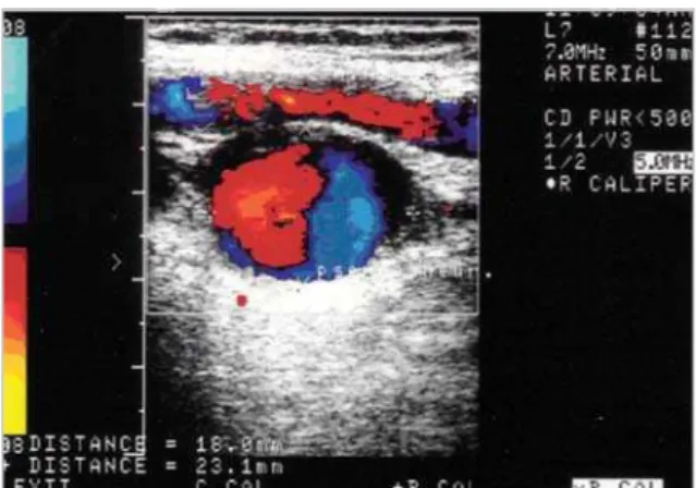
Fig. 1 – Photography of colour Doppler result showing right axillary artery pseudoaneurysm
sounds, frequent extrasystoles and no murmurs. All upper limb pulses were palpable, whereas in the lower limbs only femoral pulses were palpable bilaterally; distal pulses were absent, even with the “blind” Doppler test.
Ancillary [laboratory] tests showed normal enzyme levels (troponins and CK-MB), but increased cholesterol and triglyceride levels. The chest X-ray revealed an enlargement of the cardiac area, and the electrocardiogram showed sinus rhythm and atrial extrasystoles.
The patient underwent coronar y angiography performed by dissection of the right brachial artery. The introduction of the catheter was somewhat diffi cult at the beginning, but after that the procedure went smoothly. At the end of that day, the patient complained of mild paresthesia in the forearm, which was only kept under observation. On the following day, there was a pulsatile mass in his right axillary area with a systolic murmur and slightly increased local temperature, muscle weakness and reduced sensitivity in the right upper limb.
Arquivos Brasileiros de Cardiologia - Volume 86, Nº 4, April 2006
303
The presence of neurological symptoms demanded immediate surgical treatment. An arteriotomy was performed, exposing an atheromatous plaque detached from the endothelium and the pseudoaneurysm orifi ce. Endarterectomy and lateral suture of the artery were performed (Figure 2).
be performed to confi rm the diagnosis6.
The differential diagnosis with parietal hematoma and transmission of arterial pulse must be made immediately in order to start the most appropriate treatment for each case.
Contrary to what happens in the case of arterial injuries, the natural course of pseudoaneurysms is benign and some of them even occlude spontaneously. When this doesn’t happen, other kinds of treatment can be started, such as: ultrasound-guided local compression, thrombin injection, coil embolization or surgical treatment in case none of the above options succeed or complications arise1,3,7.
Therapeutic practice in the management of iatrogenic pseudoaneurysms has undergone dramatic changes over the last decade, and surgery is rarely the option of choice4.
In modern treatment, ultrasound-guided compression is preferable as an initial choice; pseudoaneurysm size does not seem to affect the favorable result of this kind of treatment.
Percutaneous injection of thrombin into the aneurysmal sac is considered a promising and minimally invasive method to treat iatrogenic pseudoaneurysms when the neck is narrow5,8. When there is a branch, or a
wide-necked aneurysm, and provided there is no arteriovenous fi stula, the insuffl ation of a balloon through the aneurysm neck is recommended2.
Short term results of stenting in vascular lesions indicate that this is a low-risk and less invasive procedure than surgery, although mid-term and long-term results are not yet available9. Otkar et al inserted
a 6 mm Hemobahn stent into a pseudoaneurysm in the axillary artery of a 24-year-old female gun-shot victim. After 5 years, the patient developed stenosis due to intimal hyperplasia, and was subsequently treated by balloon angioplasty10.
Julia et al reported that, despite the fact that the endovascular treatment of these injuries may seem attractive, they preferred the open-surgery technique which allows treatment of concomitant injuries and decompression of the axillary fossa caused by the hematoma.
Other authors also agree that surgical treatment is the best option in cases of large hematomas, bleeding, native vessel injury, arterial or venous compression, neurological deficits, or infections2. Even when the diagnosis is
delayed, surgery should be performed immediately. The prognosis depends on ready identifi cation of the injury and timely surgical intervention.
In this case, the patient underwent surgical treatment to remove the hematoma, decompress the brachial plexus and restore the native vessel.
Fig. 2 – Photography of colour Doppler result showing the right axillary artery after surgical correction
The patient’s clinical progress was satisfactory with an immediate improvement of the neurological symptoms. However, complete recovery took place only 30 days post-surgery, after daily motor physical therapy of the limb.
D
ISCUSSIONThe iatrogenic pseudoaneurysm presents early typical clinical fi ndings, and it is often diagnosed within the fi rst hour following puncture while it is still in the hematoma stage. Nevertheless, in this case the rather slow clinical progression allowed a diagnosis to be made only 24 hours after the procedure.
The risk factors to be considered are age, female gender and clinical evidence of peripheral vascular disease. In this case, the peripheral vascular disease was the determinant factor in the occurrence of the pseudoaneurysm reported, since it occurred exactly at the site of an atheromatous plaque that ruptured in the axillary artery. Other factors to be weighed are the inexperience of the professional performing the puncture, a catheter size greater than 8F, and concomitant anticoagulation therapy3.
Despite their small incidence, these aneurysms present a significantly greater potential for more severe complications. An early diagnosis and immediate intervention in order to reduce the high rate of complications and severe long term sequelae are mandatory4.
Medical literature refers to colour Doppler as the technique of choice to evaluate potential complications of brachial or femoral artery catheterization4,5. The technique
is considered as the one that provides the fastest and most reliable diagnosis1, as was the case in this report.
Other studies recommend that an angiogram should also
Arquivos Brasileiros de Cardiologia - Volume 86, Nº 4, April 2006
304
In comparing the three kinds of treatment cited, Stone et al (2003) concluded that the ultrasound-guided injection of thrombin should be the initial treatment of choice, leaving the surgical option for cases when the above treatment fails7.
Delay in identifying these injuries may cause permanent
neurological defi cits, despite the appropriate repair having been made in the axillary artery10.
The prevention of pseudoaneurysms depends on atraumatic arterial puncture, adequate compression after sheath withdrawal, and use of devices for percutaneous artery closure.
R
EFERENCESATROGENIC PSEUDOANEURYSM OF AXILLARY ARTERY
1. Rosa RFC, González IG, García RC. Case report. Pseudoaneurism of the femoral artery after cardiac catheterization. Rev Sanid Mil 2000;54 (5):244-8.
2. Görge G, Kunz T, Kirstein M. Non-surgical therapy of iatrogenic false aneurysms. Dtsch Med Wochenschr 2003;128(1-2):36-40. 3. Righini M, Quéré I, Laroche JP. Prise en charge des faux anévrismes
artériels post-cathétérisme. J Mal Vasc 2004;29(2):63-72. 4. Szendro G, Golcman L, Klimov A et al. Arterial false aneurysm and
their modern management. Isr Med Assoc J 2001;3(1):5-8. 5. Ghersin E, Karram T, Gaitini D et al. Percutaneous ultrasonographically
guided thrombin injection of iatrogenic pseudoaneurysms in unusual sites. J Ultrasound Med 2003;22(8): 809-16.
6. Julia J, Lozano P, Gomez F, Corominas C. Traumatic pseudoaneurysm of the axillary artery following anterior dislocation of the shoulder. Case
report. J Cardiovasc Surg 1998; 39(2):167-9.
7. Stone P, Lohan JA, Copeland SE, Hamrick RE, Tiley EH, Flaherty SK. Iatrogenic pseudoaneur ysms: comparison of treatment modalities, including duplex-guided thrombin injection. W V Med J 2003;99(6):230-2.
8. Undseth RM, Klow NE, Hoffmann P. Treatment of pseudoaneurysms after vascular inter ventions. Tidsskr Nor Laegeforen 2004; 124(10):1384-6.
9. Baltacioglu F, Cimsit NC, Cil B, Cekirge S, Ispir S. Endovascular stent-graft applications in latrogenic vascular injuries. Cardiovasc Intervent Radiol 2003;26(5):434-9.