ISSN: 2067-533X
INTERNATIONAL JOURNAL
OF
CONSERVATION SCIENCE
Volume 4, Issue 3, July-September 2013: 283-294 www.ijcs.uaic.ro
THE USE OF GAMMA IRRADIATION IN THE STERILIZATION
OF
STREPTOMYCES
COLONIZING THE TEMPRA PAINTINGS IN
ANCIENT EGYPTIAN TOMBS
Akmal Ali SAKR 1,2*, Mohamed Farouk GHALY1, Mona Foad ALI3
1 Botany Department, Faculty of Science, Zagazig University, Zagazig, Egypt
2 Conservation Department, National Museum of the Egyptian Civilization, Cairo, Egypt 3 Conservation Department, Faculty of Archaeology, Cairo University, Cairo, Egypt
Abstract
Eight out of forty six Streptomyces strains from mural paintings at the Tell Basta and Tanis tombs were exposed to increasing doses (5, 10, 15, 20, 25kGy) of gamma irradiation. These strains varied in their resistance profile. S. canarius was the most resistant to gamma irradiation doses, as it was totally eliminated at 25kGy, whereas S. chibaensis and S. albidofuscus resisted to 20kGy and S. ambofaciens resisted 15kGy. The other strains under investigation showed a lower resistance to gamma irradiation. Tricyclazole (5, 7, 10 µg/mL) inhibited melanin production after gamma irradiation at doses lower than lethal dose. Gamma irradiation with the previous doses enhanced the chitinease activity of irradiated Streptomyces strains, but S. canarius was the exception. No color change was observed either for pigments or for binding media, after gamma irradiation at the same doses.
Keywords: gamma irradiation; melanin; mural paintings; streptomyces; tricyclazole
Introduction
Most of the paintings in ancient Egyptian tombs were carried out by using the tempera technique, where pigments were mixed with binding media, such as arabic gum, animal glue and egg yolk [1]. Mural paintings suffer from chromatic alteration and disfiguration with biopigments, due to the growth and colonization of Streptomyces [2]. Due to the healthy and environmental hazards imposed by application of biocides and other chemical substances to both conservators and treated cultural heritage objects, so gamma irradiation could be used as a safe and clean alternative agent in eliminating or reducing the number of Streptomyces deteriorating cultural heritage objects in general [3].
Gamma irradiation was used since the 1960s for the sterilization of archives without reducing the tensile strength of paper or causing color change [4-7]. Gamma irradiation has many advantages qualifying it to be a good alternative means in the sterilization of deteriorated cultural heritage objects. It has as high penetrating power inside monuments, for several mms where Streptomyces develop, so it can reduce or eliminate any pathogenic microorganisms colonizing mural paintings. On the other hand, it produces no hazardous traces or secondary radioactivity [8-11], attractive in cost [12], has a short intervention time and is a non-toxic practice for conservators [13].
*
The efficacy of sterilization, or its reducing the number of pathogens colonizing cultural heritage objects by using gamma irradiation
,
should depend on the determination of the appropriate doses or lethal dose (LD) [14], because using lower doses may cause Streptomyces to produce more metabolites as acids, enzymes and pigments, which may harm the irradiated cultural heritage objects. Moreover, using higher doses may destroy the molecules of those objects [15].Determination (LD) is an important parameter for using gamma irradiation in preserving cultural heritage, as is a useful tool for quantitative evaluation of the growth activity of microbial cells, and to prevent occurrence of color change of paintings. This (LD) depends on the initial level of contamination, the radio sensitivity of contaminating flora and age of colonizing Streptomyces [16].
The effect of gamma irradiation on the colonizing Streptomyces should depended on several factors, such as the number and type of microorganisms in the microbial community, their ability for irradiation resistance and the water content of the irradiated objects [16]. Schoneman and Dickson [17] used a 25kGy gamma irradiation dose in the sterilization of deteriorated wood artifacts, but that dose significantly affected the humid wood chemistry, which did not happen with the dry samples. In that regard, Mc Namara et al. [18] reported that lower doses of radiation in wet soils eliminated microbial population more than in other soils, due to the releasing of free radicals that have an inhibitory effect on irradiated Streptomyces isolates, due to the synergetic effect of both direct and indirect effect of gamma irradiation.
Conventionally, radiation effects have been explained using target theory, and according to this, lethal effects of ionizing radiation, are expressed in the surviving irradiated cells due to direct absorption of radiation energy [16], but mortality action of gamma irradiation may attribute to the indirect action through releasing the free radicals that contribute to the destruction of microbial cells as following [14]:
H2O → H2O+ + e- (primary cation)
H2O+→ H+ + HO⋅ (free radical)
e- + H+→ H⋅ (free radical atom)
Many publications report that the appropriate dose for the sterilization of Streptomyces colonizing cultural heritage objects ranges from 20 to 25kGy. Katušin-Ražemet al. [19] used gamma irradiation in the sterilization of Streptomyces sp. colonizing a wooden statue and found that a dose of 25kGy was the common dose for the sterilization of most streptomyces isolated from cultural heritage objects, without any change in appearance, but Petushkova et al. [20] irradiated Streptomyces and Arthrobacter isolated from the Church of the Virgin’s Birth, Ferapontovo, Russia and found that the lethal dose (LD) for most of these microorganisms was 17kGy, without any color change in paintings.
In addition to that, gamma irradiation has some disadvantages, since it is not suitable for the treatment of large and immovable objects, due to the lack of facilities, its short term efficacy and it can not prevent Streptomyces from recolonizoffing the irradiated objects. To overcome this problem, gamma irradiation should be used in combination with biocides, as it was reported that gamma irradiation in combination with biocides, such as Catamine AB and Sulfonol, enhanced the antimicrobial activity of gamma irradiation considerably, due to the synergetic effect of both gamma irradiation and biocides [21].
Moreover, gamma irradiation may be used in combination with antibiotics, as Abel Haleim et al. [22] did by using gamma irradiation in combination with commercial antibiotics and found that gentamycin and spiramycin were the most effective in killing most of tested Streptomyces.
Gamma irradiation caused irradiated cells to produce a melanin pigment as a protective pigment, a defense mechanism, thus causing the foxing of irradiated cultural heritage objects. A melanin inhibitor, 1,8 - dihydroxynaphthalene (DHN), in ethanol was used to reduce or to block the melanin production caused by sterilization by using gamma irradiation [22].
The purpose of this work is to isolate and identify Streptomyces samples taken from mural paintings in ancient Egyptian tombs, by using the 16S rDNA sequence method and to determine the appropriate doses of gamma irradiation for the sterilization of these strains,without causing color change of pigments or chromatic alteration of the irradiated objects.
Materials and Methods
Isolation and Identification of Streptomyces
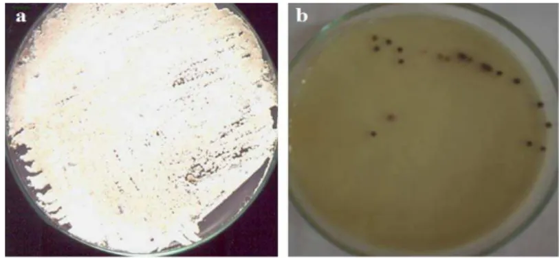
Eight out of forty-six samples were collected from Tell Basta and Tanis, 80 km southeast of Cairo, in June 2008, by using sterile cotton swap, from yellow, red, black, blue parts of paintings and from the stone surface in the investigated tombs (Fig. 1). These samples represented different deterioration symptoms of color change and scaling. Samples were cultured onto starch-nitrate-agar plates (agar 20g; starch 20g; KH2PO4 1g; MgSO4 0.5g; NaCl
0.5g; KNO3 2g and CaCO3 3g / L distilled water) and incubated for 45 days at 30°C.
Fig. 1. Isolation of Streptomyces locations, (a) Ceiling of tomb Oserkon II. Tanis (b) Eastern wall of cnh h3f., Tell Basta (c) Eastern wall of tomb of Ihy, Tell Basta. (d) Eastern wall of tomb Cnh m b3st, T.B. (e) blue color from tomb of Ihy, Tell Basta (f) Limestone saturated
Streptomyces samples were identified morphologically and biochemically, according to the identification keys devised by Kämpfer [23] and confirmed by the 16S rDNA sequence method.
16S rDNA Sequencing
Total DNA was extracted from eight isolated colonies of Streptomyces [24]. The gene coding for 16S rRNA was amplified from each colony by PCR, with universal primers: forward primer (F27), AGAGTTTGATCCTGGCTCAG-3' and reverse primer (R1492), 5'-GGTTACCTTGTTACGACTT-3') [25, 26]. These primers bind to universally conserved regions and permit the amplification of an approximately 1500bp fragment. The PCR amplification was carried out in a Gene-Amp PCR system 9600 thermocycler (Perkin Elmer). The amplification conditions were as follows: 94°C for 10min and 35 cycles of denaturation at 95°C for 30s, annealing-extension at 56°C for 1min, 72°C for 1min and an extension at 72°C for 10min. Presence and yield of specific PCR products (16S rRNA gene) were monitored by running 1% agarose gels. Then PCR product was cleaned up by using a GeneJET™ PCR Purification Kit (Fermentas).
Amplified DNA fragments were partially sequenced at GATC Biotech AG (Konstanz, Germany) by using an ABI 3730xl DNA sequencer, using forward primer (F27). The 16S rDNA sequences which were determined in the present study were stored on the NCBI web server (www.ncbi.nlm.nih.gov). Sequence analysis and comparison to published sequences was made by using the Basic Local Alignment Search Tool (BLAST) program (http://www.ncbi.nlm.nih.gov/blast) [27].
Determination of theAppropriate Gamma Irradiation
To investigate the resistance profile of the Streptomyces samples to gamma irradiation, three replicate spore suspensions in saline solution (0.9% NaCl), from freshly prepared cultures of Streptomyces, were exposed to radiation. The spore suspensions (105spores/mL) were subjected to increasing doses of gamma-irradiation (5, 10, 15, 20, and 25kGy) inside the irradiation chamber of a gamma cell 220 equipment (National Center for Irradiation Researches, Nisr City, Cairo). A Cobalt-60 source, with an average dose rate of 1.4Gy/s was used as radiation source [28]. The irradiated spores were plated onto starch-nitrate-agar plates and incubated for 7 days at 30°C. The D10 values were calculated, as the survival of the cells was
quantified by determining the number of colony forming units (cfu) at different dilutions [29]. The data were normalized to the survival of control samples.
The Effect of Gamma Irradiation on the Chitinease Activity of Irradiated Streptomyces
To study the enzymatic activity of Streptomyces before and after exposure to increasing gamma irradiation doses (5, 10, 15, 20 and 25kGy), Streptomyces isolates were cultured onto starch-nitrate-agar plates, free carbon source and incubated for 7 days at 30°C and the biofilm of Aspergillus japonicus, the most dominant fungi in the investigated tombs, was used as sole carbon source and incubated at 30°C for the same period.
Using Melanin Production Inhibitors
To overcome the problem of melanin production during and after irradiation, melanin inhibitors, such as tricyclazole, were used. Streptomyces samples were cultured on liquid starch-nitrate agar medium . Erlenmeyer flasks, containing 250mL, were prepared, each with 50mL of medium supplemented with tricyclazole, (5-Methyl-1,2,4-triazolo[3,4-b]benzothiazole) provided by Sigma, in three concentrations (5, 7, 10µg/mL), dissolved in dimethylsulfoxide (DMSO) and incubated at 30°C for 7 days [30].
The Effect of Gamma Irradiation on Different Pigments
of gamma irradiation. The comparisons of all pigments before and after irradiation were expressed as values in the grey scale. Visual observations were made to document other phenomena such as cracks, blisters, and scaling or flaking of the paint.
Results
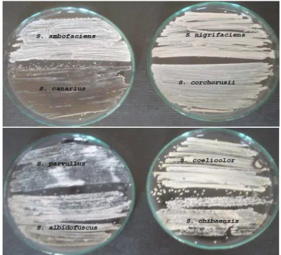
The rDNA sequences of the Streptomyces samples identified in this study with similarity more than 98% and could be closely related to species of S. albidofuscus, S. ambofaciens, S. canaries, S. chibaensis, S. coelicolor, S. corchorusii, S. nigrifaciens, S. parvullus and the accession numbers were included in Table 1.
Streptomyces varied in their resistance profile to doses of gamma irradiation. 5kGy did not affect all Streptomyces isolates, but S. coelicolor, S. corchorusii, S. nigrifaciens, S. parvullus were killed totally at doses of more than 5kGy. Nevertheless, S. ambofaciens could withsatnd up to 15kGy, but S. albidofuscus and S. chibaensis could survive up to 20kGy, Furthermore, S. canarius was an exception and could resist up to 25kGy (Fig. 2). Thus, the doses from 20-25kGy were suitable for the sterilization of most Streptomyces colonizing cultural heritage objects.
Table 1. Phylogenetic affiliation of inoculated strains (Homology of 16S r DNA and similarity in comparison with NCBI Data) N. Location Homology
approximately
Similarity enter genes 16S r DNA
(%)
Authorsۥ accession number
4 Azurite blue, tomb of Ihi, Tell Basta. S. albidofuscus 99 Later name is S. pyridomyceticus
BankIt1507621 JQ625331 7 Yellow color of Southern wall of
Ankh h3 f tomb.
S. ambofaciens 99 BankIt1507642 JQ625332 9 Blue color, ceiling burial tomb of
Oserkon II, Tanis.
S. canaries 99 BankIt1507650 JQ625337 11 Black color, tomb Ankh h.f, Tell
Basta.
S. chibaensis 100 BankIt1507649 JQ625336 46 Red color, tomb Ankh m b3st, Tell
Basta.
S. coelicolor 99 BankIt1507648 JQ625335 10 Limestone, tomb of Oserkon II, Tanis. S. corchorusii 98 BankIt1507647 JQ625334 12 North wall, tomb of Ist, Tell Basta. S. nigrifaciens 98 Later name is S. flavovirens
BankIt1507149 JQ625330 8 Yellow color of Southern wall of
Ankh h3 ftomb.
S. parvullus 99 BankIt1507645 JQ625333
0 0.5 1 1.5 2 2.5 S. a lbido fusc us S. a mbof acie ns
S. c ana rius S. c hib aens is
S. c oelic
olor
S. c orc
horu sii
S. n igrif
acie ns
S. p arvu llus E ff icacy o f g am m a i rr ad at io n d o ses 5kGy 10 kGy 15 kGy 20 kGy 25 kGY
Our results indicated that an exposure of S. albidofuscus to 20kGy of gamma irradiation caused the morphogenesis of irradiated spores (Fig. 3) and irradiation changed the aerial mycelium of S. chibaensis (Fig. 4), whereas the exposure of streptomyces to gamma irradiation doses lower than the lethal dose (LD) caused morphological changes in the architecture of spores and induced mutation. In figure 5 we can observe that the exposure of S. canarius to gamma irradiation enhanced the production of the protective pigment of melanin.
Fig. 3. Morphogenesis S.albidofuscus by gamma irradiation. (a) control, (b) irradiated
Fig. 4. change of color of aerial mycelium of S. chibaensis (a) control, (b) after irradiation dose 20kGy.
Fig. 5. Reduction of colonies and melanin production of S. canaries after gamma irradiation: (a) control, (b) irradiated cells.
used as a melanin inhibitor. Our results indicated that using tricyclazole in alcoholic solution with different concentrations (5, 7, 10µg/mL) inhibited the melanin production of Streptomyces colonizing irradiated cultural heritage objects.
We found that gamma irradiation at doses lower than the lethal dose (LD) (5-10kGy) enhanced the chitinease enzyme activity of the Streptomyces strains (Fig. 6), so the irradiated Streptomyces could decompose the melanized biofilm of Aspergillus japonicus better than the non irradiated cells.
Furthermore, the irradiation of pigments (limonite, hematite, malachite, azurite, vermillion, calcium carbonate, lamp black, lead carbonate and gypsum plaster) with the lower dose (5kGy) and the higher dose (25kGy) caused neither chromatic alteration nor scaling of those pigments (Fig. 7). No effect on the different binding media of animal glue, arabic gum or egg yolk was noticed.
Finally, it was observed that test tubes containing spore suspension and exposed to the previous doses were turned into grey color.
Fig. 6. Chitenolytic activity of isolated Streptomyces on biofilm of Aspergillus japonicus
Discussions
All identified isolates were attributed to the Streptomyces genus, whereas Streptomyces represent the highest percentage among the microbiota of microorganisms colonizing cultural heritage objects, the typical and first colonizers and considered an indicator for advanced stage of deterioration [20].
Streptomyces varied in their resistance profile to doses of gamma irradiation, since a dose 5kGy did not affect all Streptomyces samples, but S. coelicolor, S. corchorusii, S. nigrifaciens, S. parvullus were killed totally at doses above 5kGy. Nevertheless, S. ambofaciens could resist up to 15kGy, but S. albidofuscus and S. chibaensis could survive up to 20kGy. Furthermore, S. canarius was an exception and could survive up to 25kGy. Thus, we concluded that doses from 20-25kGy were suitable for the sterilization most Streptomyces colonizing cultural heritage objects. That was in agreement with the results obtained by Pointing et al. [31] who used a dose of 20kGy for the inactivation of wood biodeterioration agents. Also, Katušin-Ražem and Braun [19], reported that the dose of 25kGy was commonly used for the sterilization or reduction of all viable forms of bacterial cells. Furthermore, it was pointed out that a dose above 20kGy was required for the inactivation of microorganisms colonizing wood objects [32]. Moreover, Severiano et al. [11], mentioned that doses of 15-20kGy were used in the disinfection of microorganisms colonizing deteriorated wood without any remarkable change in appearance and the same dose was used in the sterilization of microorganisms contaminating some Peruvian paintings dated back to the 17th century [33]. Our results indicated that Streptomyces varied in their resistance profile to gamma irradiation even between closely related strains, as different strains had different levels of radiosensitivity to gamma irradiation [34].
We found that the exposure of S. albidofuscus to 20kGy of gamma irradiation caused the morphogenesis of the irradiated spores and changed the aerial mycelium of S. chibaensis, whereas the exposure of streptomyces isolates to gamma irradiation doses lower than lethal dose (LD) caused morphological changes in the architecture of spores and induced mutation. These results confirm the results of prevous relevant studies [35].
From the current results we could observe that lethal dose of the most Streptomyces isolates was high (15-20kGy), this in agreement with Sweiha [36] since Streptomyces as the most resistant genus of actinomycetes due to sporulation as another defense mechanism.
Gamma irradiation has an inhibitory effect on irradiated streptomyces cells, after exposure to doses above 15kGy and it was reported that gamma irradiation caused damages to the DNA of cells, through ionization, by inducing mutation, single and double strand DNA breakages, and even the death of the cell. This effect was a result of the radiolysis of the cellular water in the irradiated objects, which generated active oxygen species, free radicals and peroxides [29, 35, 37]. This damage may have occurred as a direct effect of gamma irradiation through the absorbance of the high electromagnetic energy of gamma radiation by the targeted microorganisms [11].
Our results indicated that the exposure of S. canarius to gamma irradiation lower than the lethal dose enhanced melanin production, which acts as a protective agent against adverse environmental conditions, such as hypersalinity, radiation and heavy metals, because melanin acts as a shield between the cell and its often hostile surroundings, so the surface of the hyphae exposed to radiation becomes highly pigmented [38, 39].
with different concentrations (5, 7, 10µg/mL) inhibited the melanin production of Streptomyces, as these concentrations inhibited the melanin production of most fungi colonizing limestone and marble slabs [30]. Thus, melanin production by Streptomyces after gamma irradiation could be inhibited by treating the deteriorated cultural heritage objects before irradiation with alcoholic solution of tricyclazole, because tricyclazole interrupts the melanin synthesis pathway, without being toxic to the irradiated Streptomyces, thus the resistance of Streptomyces to gamma irradiation is reduced without killing these isolates [40]. With the pass of time, the efficacy of tricyclazole was reduced, since it was reported that during log phase growth of Streptomyces, a detoxification mechanism may be in place that tricyclazole becomes less effective in older cultures [40], so the tricyclazole must be freshly applied.
Our findings indicated that gamma irradiation at doses lower than lethal dose (LD) (5-10kGy) enhanced chitinease enzyme activity of Streptomyces strains, so the irradiated Streptomyces could decompose the melanized biofilm of Aspergillus japonicus better than the non irradiated cells, because decomposition of melanized spores of microorganisms is more difficult than of non melanized ones [39]. Our results are similar to those obtained by Mahrous [14] who performed the irradiation of Streptomyces sp. and Thermoactinomycetes vulgaris isolated from the mummy of the King Sequenenrc, Egyptian Museum, Cairo, with doses lower than the lethal dose (lower than 20kGy), which enhanced the proteolytic activity of these irradiated cells with more than 60%, so the enzymatic decomposition of irrradiated cultural heritage objects would be higher [42].
Furthermore, the irradiation of pigments (limonite, hematite, malachite, azurite, vermillion, calcium carbonate, lamp black, lead carbonate and gypsum plaster) with the lower dose (5kGy) and the higher dose (25kGy) caused neither chromatic alteration nor scaling of the irradiated paintings. No effect on different binding media of animal glue, arabic gum or egg yolk was noticed. The current results are similar to results obtained by Manea et al. [43], who tested the different doses of gamma irradiation (11 and 24.5kGy) on different pigments [red lead Pb3O4; lead white (PbCO3)2•Pb(OH)2; chrome yellow (PbCrO4) and hematite (Fe2O3•H2O)].
Nevertheless, Negut et al. [44], mentioned that the FT/IR patterns of irradiated and non irradiated pigment samples indicated that the exposure to increasing doses lead to chromatic alteration in red lead, because the interaction of gamma irradiation with pigments may change its chemical and physical properties. Moreover, it was reported that pigments on parchment documents in Portugal were not changed when exposed to gamma irradation at doses of 20-25kGy and there was no color change for collagen after irradiation at the same doses, but they did not point out the specific types of pigment that did not change [3].
Moreover, the previous studies pointed out that exposure of cerussite (lead carbonate) and HgS (vermillion) to gamma irradiation caused blackening of these pigments due to transformation to β-HgS by gamma irradiation [45, 46]. On the other hand, it was reported that blackening of lead carbonate is a temporary effect, when the irradiation is stopped, the black stains of lead carbonate progressively fades out and disappeared [47, 48].
Conclusions
Acknowledgements
The authors would like to thank Dr. Aza Shahein, Professor of Irradiation, National Centre for Irradiation Researches, Nasr City, Cairo, Egypt, for performing the irradiation procedure and for her helpful discussions.
References
[1] R. Newman, M. Serpico, Adhesives and binders, Ancient Egyptian Materials and
Technology, (Editors: Nicholson, P., and Shaw, I.), Cambridge University Press, 2000, pp. 475-493.
[2] A.A. Gourbushina, K. Petersen, Distribution of microorganisms on ancient wall paintings as related to associated faunal elements, International Biodeterioration and Biodegradation, 46, 2000, pp. 277-284.
[3] I. Nunes, N. Mesquita, S.C. Verde, M.J. Trigo, A. Ferreira, M.M. Carolino, A. Portugal, M.L. Botelho, Gamma radiation effects on physical properties of parchment documents assessment of Dmax, Radiation Physics and Chemistry, 81, 2012, pp. 1943–1946.
[4] L.A. Belyakova, Gamma irradiation as a means of disinfection of books against spores of mould fungi, Microbiology, 29, 1960, pp. 762-765.
[5] P. Sico, The use of Gamma Rays in book conservation, Nuclear News, 2000, pp. 38-40. [6] M. Gonzalez, A. Calvo, E. Kairiyama, Gamma radiation for preservation of biologically
damaged paper, RadiationPhysics & Chemistry, 63, 2002, pp. 263-265.
[7] G. Magaudda, The recovery of biodeteriorated books and archive documents through gamma radiation: some considerations on the results achieved, Journal of Cultural Heritage, 5, 2004, pp. 113–118.
[8] J. Urban, P. Justa, Conservation by gamma radiation, Museum, 151, 1986, pp. 165-167. [9] F. Butterfield, The potential long term effects of Gamma Irradiation on paper, Studiesin
Conservation, 32, 1987, pp. 181-187.
[10] C.C. Ponta, Irradiation conservation of cutlural heritage, Nuclear Physics News, 18, 2008, pp. 22-24.
[11] L.C. Severiano, F.A. Lahr, Bardi, A.C. Santos, L.B. Machado, Influence of gamma radiation on properties of common Brazilian wood species used in artwork, Progress in Nuclear Energy, 52, 2010, pp. 730-734.
[12] D.C. Negut, C.C. Ponta, M. Georgescu, G. Niculescu, M. Lupu, Effects of gamma irradiation on the color of pigments, Proc. SPIE 6618, 2007, 66180R.
[13] M.M. Manea, C.D. Negut, IR. Stanculescu, C.C. Ponta, Irradiation effects on canvas oil painting: Spectroscopic observations, Radiation Physics and Chemistry, 81, 2012, pp. 1595–1599.
[14] A.M. Mahrous, Application of gamma radiation for the preservation of Ancient Egyptian cultural objects, M Sc Thesis, Women’s College, Ain Shams University, 1988, p.20. [15] J.I. Choi, M. Yoon, D. Kim, Gamma irradiation of cultural heritage artifacts for
disinfection using Monte Carlo Simulations, Applied Radiation and Isotopes, 70 (11), 2012, pp. 2564-2568.
[16] S. Wirkner, K. Takahash, M. Furuta, & T. Hayashi, Calorimetric study on the effect of 60Co g-rays on the growth of microorganisms, Radiation Physics and Chemistry 63,
2002, pp. 327–330.
[16] P. Mora, L. Mora, P. Philippot, Conservation of Wall Paintings, Butterworths, London, 1984.
[17] M. Schoneman, D. Dickson, Growth of Aurobsidium pullumans on lignin breakdown products at weathered wood surfaces, Mycologist, 11(2), 1997, pp. 168-172.
[18] N.P. Mc Namara, H.J. Black, N.A. Bereford, N.R. Parekh, Effects of acute gamma irradiation on chemical, physical and biological proprieties of soils, Applies Soil Ecology,
[19] B. Katušin-Ražem, M. Braun, Irradiation treatment for the protection and conservation of cultural heritage artefacts in Croatia, RadiationPhysics and Chemistry, 78, 2009, pp. 729–731.
[20] J.P. Petushkova, N.N. Lyalikova, F.G Nichiporov, Effect of ionizing radiation on monument deteriorating microorganisms, Radioanalytical and Nuclear Chemistry,
125(2), 1988, pp. 367-371.
[21] F. Cappitelli, C. Sorlini, From papyrus to compact disc: The microbial deterioration of documentary heritage, CriticalReviewMicrobiology, 31, 2005, pp. 1–10.
[22] M.E.F. Abdel-Haleim, M.F. Ali, M.F. Ghaly, A.A. Sakr, Efficiency of antibiotics and gamma irradiation in eliminating Streptomyces strains isolated from paintings of ancient Egyptian tombs, JournalofCulturalHeritage, 14(1), 2013, pp. 45-50.
[23] P. Kämpfer, The Prokaryotes - A Hand Book on the Biology of Bacteria. Vol. 3, The family Streptomycetaceae, Part I, Taxonomy, 3rd ed., (Editors: Dworkin, M., Falkow, S., Rosenberg, E., Schleifer, K-H., and Stackebrandt, E.), Springer-Verlag, Berlin, 2006, pp. 538-604.
[24] J. Sambrook, D. Russel, Molecular Cloning: A Laboratory Manual, 3rd ed., Cold Springs Harbour Press, 2001.
[25] D. Chénbey, L. Philippot, A. Hartmann, C. Hénalut, J.C. Germon, 16S rDNA analysis for characterization of denitrifying bacterial isolated from three agricultural soils, FEMS Microbiology Ecology, 24, 2000, pp. 121–128.
[26] L.W. Enquist, S.G. Bradley, Characterization of deoxyribonucletoide acid from Streptomyces venezuelae, Dev. IndiaMicrobiology, 12, 1971, pp. 225-236.
[27] S.F. Altschul, T.L. Madden, A.A. Schäffer, J. Zhang, Z. Zhang, W. Miller, D.J. Lipman, Gapped BLAST and PSI-BLAST: a new generation of protein database search programs,
Nucleic Acids Research, 17, 1997, pp. 389–402.
[28] N. Aziz, M. El Fouly, M. Abou Shady, L. Moussa, Effect of gamma radiation on the survival of fungal and actinomycetal florae contaminating medicinal plants, Applied Radiation and Isotopes,48, 1997, pp. 71-76.
[29] J.I. Choi, Y.J. Chung, D.I. Kang, K.S. Lee, J.W. Lee, Effect or irradiation on disinfection and mechanical properties of Korean traditional paper Hanji, Radiation Physics and Chemistry, 81, 2012, pp. 1051-1054.
[30] E. Diakumaku, A. Gourbushina, W. Krumbein, L. Panina, S. Soukharjevski, Black fungi in marble and limestones- an aesthetical, chemical and physical problem for the conservation of monument, The Science of the Total Environment, 167, 1995, pp. 295-304.
[31] S. Pointing, E. Jones, A. Jones, Decay prevention in waterlogged archaeological wood using gamma irradiation, International Biodeterioration and Biodgredation,42, 1998, pp. 17-24.
[32] M. Yoon, Y. Chung, D. Kang, I. Kim, Y. Lee, J. Lee, J. Choi, Study of the optimal gamma irradiation for the control of fungi on wood cultural property, Journal of Conservation Science, 27, 2011, pp. 127-134.
[33] M. Rizzo, L. Machado, S. Borrely, M. Sampa, Effects of gamma rays on a restored painting from the XVIIth century, Radiation Physics and Chemistry, 63, 2002, pp. 259– 262.
[34] P. Normand, The families Frankiaceae, Geodermatophilaceae, Acidothermaceae and Sporichthyaceae, Prokaryotes, 3, 2006, pp. 669-681.
[35] H.E. Sweiha, Effect of gamma irradiation on certain soil inhabitant actinomycetes, M Sc Thesis, Faculty of Science, Ain Shams University, 1984, p.168.
[36] L. Moussa, F. Mansour, M. Serage, S. Abou El Nour, Effect of Gamma Radiation on the Physiological Properties and Genetic Materials of Streptomyces albaduncus and S. erythogresius, International Journal of Agriculture and Biology,7, 2005, pp. 197–202. [37] M. Gtari, I. Essoussi, R. Maaoui, H. Sghaier, R. Boujmil, J. Gury, P. Pujic, L. Brusetti, B.
[38] T. Warscheid, W. Krumbein, General aspects and selected cases, Microbially Influenced Corrosion of Materials, (edited by Heitz et al.), Springer-Verlag, Berlin, 1996, pp. 274–295.
[39] M. Butler, A. Day, J. Henson, N. Monery, Pathogenic properties of fungal melanin,
Mycologia, 93, 2001, pp. 1-8.
[40] M.E. Young, H.L. Alakomi, I. Fortune, A. Gourbushina, W. Krumbein, I. Maxwell, C. McCullagh, P. Robertson, M. Saarela, J. Valero, M. Vendrell, Development of biocidal treatment regime to inhibit biological growth on cultural heritage: BIODAM,
Environmental Geology, 56, 2008, pp. 631-641.
[41] G. Lazarovits, A.N. Starratt, H.C. Huang, The effect of tricyclazole and culture medium on production of melanin precursor 1,8-Dihydoxynaphthalene by Sclerotinia sclerotiorum isolate SS7, Pesticide Biochemistry and Physiology,67, 2000, pp.54-62.
[42] J.P. Voets, M. Dedeken, E. Bessems, The behavior of some amino acids in gamma irradiated soils, Naturwissenschaften, 52, 1965, p. 467.
[43] M.M. Manea, I.V. Moise, M. Virgolici, C.D. Negut, O.-H. Barbu, M. Cutrubinis, V. Fugaru, I.R. Stanculescu, C.C. Ponta, Spectroscopic evaluation of painted layer structural changes induced by gamma radiation in experimental models, Radiation Physics and Chemistry, 81, 2012, pp. 160–167.
[44] C.D. Negut, V. Bercu, O.G. Duliu, Defects induced by gamma irradiation in historical pigments, Journal of Cultural Heritage, 13, 2012, pp. 397– 403.
[45] K. Nassau, Gamma ray irradiation induced changes in the color of Tourmalines,
AmericanMineralogist 60, 1975, pp.710-713.
[46] M. Radepont, Understanding of chemical reactions involved in pigment discoloration, in particular in mercury sulfide (HgS) blackening, PhD Thesis, Universiteit Antwerpen, France, 2013, pp.28-30.
[47] E. Kotulanová, P. Bezdicka, D. Hradil, J. Hradilová, S. Svarcová, T. Grygar, Degradation of lead-based pigments by salt solutions, Journal of Cultural Heritage, 10, 2009, pp. 367– 378.
[48] J. Absil, H.P. Garnir, D. Strivay, C. Oger, G. Weber, Study of color centres induced by PIEX irradiation, Nuclear Instruments and Methods in Physics Research B, 198, 2002, pp.90-97.