as a Plausible Substrate for Guiding Sensory Transfer
across the Thalamic Gateway
Se´bastien Be´huret1*, Charlotte Deleuze1, Leonel Gomez1,2, Yves Fre´gnac1, Thierry Bal1*
1Unite´ de Neurosciences, Information et Complexite´ (UNIC), CNRS UPR-3293, Gif-sur-Yvette, France,2Laboratorio de Neurociencias, Facultad de Ciencias, Universidad de la Repu´blica Oriental del Uruguay, Montevideo, Uruguay
Abstract
The thalamus is the primary gateway that relays sensory information to the cerebral cortex. While a single recipient cortical cell receives the convergence of many principal relay cells of the thalamus, each thalamic cell in turn integrates a dense and distributed synaptic feedback from the cortex. During sensory processing, the influence of this functional loop remains largely ignored. Using dynamic-clamp techniques in thalamic slicesin vitro, we combined theoretical and experimental approaches to implement a realistic hybrid retino-thalamo-cortical pathway mixing biological cells and simulated circuits. The synaptic bombardment of cortical origin was mimicked through the injection of a stochastic mixture of excitatory and inhibitory conductances, resulting in a gradable correlation level of afferent activity shared by thalamic cells. The study of the impact of the simulated cortical input on the global retinocortical signal transfer efficiency revealed a novel control mechanism resulting from the collective resonance of all thalamic relay neurons. We show here that the transfer efficiency of sensory input transmission depends on three key features: i) the number of thalamocortical cells involved in the many-to-one convergence from thalamus to cortex, ii) the statistics of the corticothalamic synaptic bombardment and iii) the level of correlation imposed between converging thalamic relay cells. In particular, our results demonstrate counterintuitively that the retinocortical signal transfer efficiency increases when the level of correlation across thalamic cells decreases. This suggests that the transfer efficiency of relay cells could be selectively amplified when they become simultaneously desynchronized by the cortical feedback. When applied to the intact brain, this network regulation mechanism could direct an attentional focus to specific thalamic subassemblies and select the appropriate input lines to the cortex according to the descending influence of cortically-defined ‘‘priors’’.
Citation:Be´huret S, Deleuze C, Gomez L, Fre´gnac Y, Bal T (2013) Cortically-Controlled Population Stochastic Facilitation as a Plausible Substrate for Guiding Sensory Transfer across the Thalamic Gateway. PLoS Comput Biol 9(12): e1003401. doi:10.1371/journal.pcbi.1003401
Editor:Diego Contreras, University of Pennsylvania, United States of America
ReceivedFebruary 12, 2013;AcceptedNovember 4, 2013;PublishedDecember 26, 2013
Copyright:ß2013 Be´huret et al. This is an open-access article distributed under the terms of the Creative Commons Attribution License, which permits unrestricted use, distribution, and reproduction in any medium, provided the original author and source are credited.
Funding:SB was supported by the French DGA (Direction Ge´ne´rale de l’Armement). This work was supported by the CNRS, the Agence Nationale de la Recherche (ANR-10-BLAN-1402: V1-Complex) and EC contracts (Facets (FP6-2004-IST-FETPI 15879), Brain-i-nets 2009-ICT-FET 243914) and BrainScales (FP7-269921)). The funders had no role in study design, data collection and analysis, decision to publish, or preparation of the manuscript.
Competing Interests:The authors have declared that no competing interests exist. * E-mail: [email protected] (SB); [email protected] (TB)
Introduction
The thalamus is the major sensory gateway to the cerebral cortex. Forming the output of the retina, axons of ganglion cells diverge to connect a small number of thalamocortical (TC) neurons in the dorsolateral geniculate nucleus (dLGN); likewise several ganglion cells send convergent connections to individual TC neurons [1]. In turn, a sizable number of TC neurons (ranging from 15 to 125 in the cat [2]) converge onto individual recipient cortical neurons [3]. However, in spite of the fact that it is often described and modeled as a pure feedforward relay, the thalamus receives a massive corticofugal feedback. The functional interac-tions between the feedforward thalamocortical converging stream and the corticothalamic (CT) feedback are not known, and yet likely plays a key role in the control of the global gain and filtering features of the sensory thalamic relays.
Despite the fact that the function and mechanisms of the CT input have attracted much interest they are still a matter of discussion [4,5]. A first accepted view is that the cortical feedback influences the transfer of sensory information by TC cells [6–8]
and may participate to modulate visual responses during attention and awareness [9]. A second and well-publicized hypothesis endows the CT feedback and the thalamic nucleus reticularis (NRT) with a searchlight function [10] or focal attention [11] by enhancing selectively the receptivity of targeted TC neuron populations to attended sensory features. Others envision the thalamus as an ‘‘active blackboard’’ onto which the cortex could write down the results of its computation [12].
Nevertheless, the cellular mechanisms underlying the functional impact of the CT feedback are poorly understood despite a few experimental studies pointing to the spatial sharpening of thalamic receptive field and its ON-OFF antagonism [13], the facilitation of lateral geniculate nucleus (LGN) activity in the awake cat [14] or attentive monkey [15], the synchronizing action on thalamic neurons involved in the detection of co-aligned elements in the visual field [16,17] or the enhancement of the surround antagonism during motion processing [18].
the transfer efficiency of TC neurons through the contextual synaptic bombardment originating from the CT input [19].
In the primary visual cortex (V1; areas 17 and 18 in the cat), layer 6 is the source of the CT synaptic feedback to thalamus. Activity patterns originating from projections of cortical layer 6 remain a major mystery although recent studies suggest behavioral circumstances in which the feedback from corticothalamic neurons could be engaged [20]. In the mouse, layer 6 neurons projecting to thalamus are spontaneously active and their activity increases during unspecific full-field visual stimulation (Fig. 1D in [21]). However, the fine-scale activity of layer 6 circuits during naturalistic vision is unknown. A detailed modeling of the activity of layer 6 seems presently an unreachable target, since it would require including interrelations with all other cortical layers and other related cortical areas (see [20,22]). Instead, our strategy has been to model the top-down cortical input as a configurable activity pattern transmitted by excitatory and inhibitory synapses for which we have fully explored the parameter space.
At the single-cell level, the efficiency of the transfer via the thalamus was established by measuring the spiking probability function of individual TC relay cells, and shown to depend on the statistical context of the synaptic bombardment [19]. When considering the whole population level, the question we want to address is whether cortically-induced modulation of the thalamic transfer efficiency can be deduced solely from the interactions observed at the single-cell level or if it emerges from higher order interactions within the network. In other words, in terms of the global information transfer between retina and cortex, is the combined effect of changes operating in individual cells equivalent to the modulation of the thalamic population as a whole? Our working hypothesis is that the CT synaptic bombardment is able to modulate the transfer efficiency of specific TC neurons, not only at the single-cell level by impacting on the input-output gain [19], but also at the population-level by controlling the contextual correlations in membrane potential fluctuations within subgroups of TC relay cells. We present here a new approach in the study of the sensory transfer gating mechanisms in the thalamus by
exploring the functional impact of higher order interactions arising between multiple TC neurons, both in computer models and in the slice.
Results
The aim of our experimental plan was to combine in computo models of the retino-thalamo-cortical (RTC) pathway and top-down corticothalamic inputs within vitromeasures of information transfer at different points of the circuit. The results are organized consequently to describe the global circuit model and its various implementations, present parametric studies of the dependency on the model on various structural and activity-dependent features, and quantify their functional impact on global information transfer efficiency between retina and cortex.
More specifically, the first part of the results and the methods present the implementation of the circuit model (Fig. 1A) and biological iteratively constructed networks (BICNs)in vitro(Fig. 1B). In the second and third parts, respectively, we tested critical structural parameters of the thalamocortical and retinothalamic circuits topologies (Fig. 1A, i and ii). The fourth part shows the dependency of the model behavior on CT synaptic bombardment statistics (Fig. 1A, iii). In the final parts, we implemented various contextual patterns in the thalamic layer, including membrane potential fluctuation correlation across TC cells imposed via the CT input, in both topologically optimized BICNs and model networks (Fig. 1A, iv).
In all simulations, mutual information analysis (Eq. 19) was carried out to estimate the efficiency of the global information transfer between the retinal input and the cortical response (later referred as ‘‘transfer efficiency’’ (TE); see Methods) [23,24]. This theoretical tool quantifies the non-linear statistical dependencies of specific features between two spike trains such as spike events, absence of spikes or any combinations of these two events in a given time window (see Figure S1 for comparison with other methods).
The thalamocortical convergence circuit model
In our model, the topology of the feedforward retino-thalamo-cortical circuitry (Fig. 1A) is highly schematic, but constrained with detailed biophysical measurements taken from the available literature. It is composed by an ordered layout of populations of thalamocortical neurons in the dLGN converging to a single layer 4 pyramidal neuron of the primary visual cortex (see Methods for details). Circuits were either built from collections of Hodgkin Huxley type model neurons (Eq. 1–3) or reproduced in anin vitro slice preparation of the rat thalamus using an iterative procedure [25] implemented in dynamic-clamp [26–28]. Synapses were conductance-based (Fig. 1, inset; see Methods) and mimicked AMPA (a-amino-3-hydroxy-5-methyl-4-isoxazolepropionic acid) and GABAA(gamma-aminobutyric acid type A) mediated current
flows (Eq. 5 and 6). We based our circuit reconstructions on direct estimates of the structure and size of the elementary thalamic microcircuitry found in the literature. The topology of the circuit was parametrized to test the sensitivity of information transfer on the structural constraints. We varied in the model simulations (Fig. 1A, i) the degree of convergence and weight of TC synapses onto a single target cortical neuron and (Fig. 1A, ii) the divergence/convergence configuration of the retinogeniculate axons and retinal input synchronization.
In order to reproduce the main components of the thalamic input, each artificial or biological TC neuron was fed with an artificial retinal input pattern (Eq. 10) and received cortical inputs simulated by stochastically fluctuating conductances composed of
Author Summary
Most of the sensory information in the early visual system is relayed from the retina to the primary visual cortex through principal relay cells in the thalamus. While relay cells receive,7–16% of their synapses from retina, they
mixed excitatory and inhibitory inputs (see Eq. 7 and 8). Exploring the effects of the CT inputs requires modeling an artificial synaptic signal whose statistical signature can be experimentally controlled. With this approach it is possible to generate a large range of artificial CT activity patterns and explore their effects by stimulating recipient neurons with the resulting contextual
synaptic bombardments. It should be emphasized that we simulate here the contextual synaptic noise in an open-loop fashion. In other words, it does not depend on the activity of the model cortical cell but rather is controlled by a set of statistical parameters. This contextual synaptic noise produced background membrane voltage fluctuations in TC neurons and was designed
such as to mimic the dynamics of the direct AMPA synapses and the disynaptic GABAergic input originating from local interneu-rons and neuinterneu-rons from the NRT in the wake state where the cortical input is presumably irregular. A distinctive feature of this paradigm is that each one of the TC neurons can be either modulated by a neuron-specific pattern of synaptic fluctuations or share common synaptic inputs with neighboring cells. In order to control the functional impact of the CT input, we analyzed critical factors of the statistics of this synaptic noise such as the ratio of inhibitory versus excitatory conductances, the amplitude of the conductance fluctuations and the level of coherence of the noise shared by TC neurons. Relevant model parameters are summa-rized in Table 1.
We used BICNs in vitro and computer modeling (Fig. 1B) to reproduce and explore systematically the voltage dynamics of neural circuit existing in the intact brain. The activity patterns of biologically recorded relay TC cells were replayed to simulate the synaptic convergence activity of the thalamic layer and stimulate the modeled cortical cell in the primary visual cortex, similar to that described above in the model circuit. A BICN hybrid thalamic layer thus consisted of a population of a parametrized set of pseudo-neurons, whose output trains replayed simultaneously individual response patterns recorded sequentially in biological TC neurons (see Methods).
Figures 1C and 1D show examples of voltage traces of TC neurons chosen among a larger population for both BICNs (in vitro) and model (in computo) circuits. The fluctuating voltage recordings illustrated for the TC cells are the result of different synaptic bombardment sequences for each trace. The synaptic bombard-ment has been optimized in order to maximize the transfer efficiency according to paradigms explored in later sections.
Parametric dependency on thalamocortical convergence and synaptic weight
In this first set of simulations, the statistics of the corticothalamic input were uniform across the whole thalamic population but the individual time patterns were chosen to be independent between each of the TC cells. The entire thalamic population was connected by a unique retinal cell mimicking the discharge pattern of an ON-center Y cell (30 Hz gamma 3 distribution, [29,30]; see Eq. 10). Hence, the TC cells input differed only in their individual corticothalamic synaptic noise pattern.
We adjusted the synaptic weight (see Eq. 6 and inset in Fig. 1) of the thalamocortical synapse to a biologically realistic value (,2.3 nS, [31]) and varied the population size (Fig. 2A, black curve). The TE was shown to reach a maximum for a convergence ratio of 80–100 cells. In addition, we observed in this configuration that roughly the simultaneous firing of a third of the TC cells was required to elicit a spike in the cortical neuron. These ballpark estimates were justified by protocols using single AMPA events in which a strength of ,80 nS was required to evoke reliably a cortical spike, corresponding to 30–35 TC neurons firing simultaneously (Fig. S2A). Smaller thalamic population sizes resulted in an insufficient synaptic drive of the target cortical cell while larger thalamic population sizes led to an increased amount of cortical spikes decoupled from the retinal input, both of these cases leading to inefficient transfer values.
We then froze the thalamic population size to 90 cells and varied the thalamocortical AMPA synaptic weight (Fig. 2B, black curve). The TE peak was obtained for a biologically realistic synaptic weight (2–2.5 nS) thus confirming the value chosen in Figure 2A.
The above 90 cells version of the model circuit is too large to achieve successful biological thalamic layer reconstruction and is
computationally intensive for multi-dimensional parametric ex-plorations. We therefore reduced the number of TC relay neurons in the model circuit and ran simulations to find the optimal synaptic weight reflecting this decrease. First, we designed a 30 cells model circuit that was used in later computational explorations (Fig. 2A and 2B, dark-gray curves). Second, we tested a 10 cells model circuit as a control to match the BICN hybrid thalamic layer size presented in Figure 1C and developed in later sections (Fig. 2A and 2B, light-gray curves). When reducing the population size from 90 to 30 TC cells, a corresponding increase of the synaptic weight by a factor of 3 was necessary and sufficient to maintain optimality in signal transfer. The same compensatory rule held when lowering the population size from 30 to 10 TC cells. Hence, topologically optimized networks consisted of 90 TC cells with the biologically realistic synaptic weight, 30 TC cells with a 3 fold increase of the weight or 10 TC cells with a 9 fold increase of the weight.
We tested other optimized topologies according to the following empirical rule which reflects the above findings: ‘‘number of TC cells’’6‘‘TC synaptic weight’’<210 nS (Fig. 2A, dashed curves). This empirical rule ensured that the total net summed synaptic input received by the target cortical cell was constant thus enabling us to isolate the effect of the population size parameter. An asymptotic saturation behavior was observed, showing a ceiling value in the TE for convergence ratios around a critical value of 90 TC neurons. This finding implies that the structure of the convergent networks, albeit flexible, needs to be constrained in order to provide an efficient and optimal information transfer.
In the subsequent investigations and unless mentioned other-wise, we performed numerical investigations with models of parallel feedforward lines composed of 30 TC relay cells converging to one model cortical cell through TC synapses using a weight optimized as described above (Fig. 2B, dark-gray curve, 7 nS).
Parametric dependency on the synchronization level of retinal inputs and TC spikes
Multiple retinal input lines were added to the model circuit described previously. Both convergent and divergent processes have been documented between retinal ganglion cells and relay thalamocortical cells in the LGN [1]. The 30 TC relay cells were contacted by 15 retinal cells in a realistic mixture of divergent and convergent processes as illustrated in Figure 1A. Each retinal cell contacted 4 TC neurons and each TC neuron was contacted by 2 retinal cells [1]. The thalamic population size and the TC synaptic weight were kept frozen. The cortical synaptic bombardment was kept as described above.
The level of synchronization of the retinal afferents was controlled in two ways. First, we varied the number of retinal cells replaying an independent activity pattern resulting in graded levels of synchronization controlled by the parameterRsync (Eq. 11). Second, we introduced an ad-hoc jitter to randomly shift the timing of each retinal spikes (see Methods). The average spike-time shift was characterized by the mean jitter parameterj0(Eq. 12). Low Rsync values and large spike-time jitters (j0) led to desynchronized retinal inputs in the TC relay cell population. In this model circuit including more than one retinal cell, transfer efficiency was measured between only one of the retinal cells and the cortical response. The chosen reference retinal cell was always the one whose activity was correlated with some or all of the retinal cells in the synchronized retinal input conditions.
Table 1.Retino-thalamo-cortical model circuit parameters.
Parameter Description Value(s) or range
Retinal stimulation
NR Number of retinal cells 1, 15 cells
i0 Mean interspike interval (firing rate) 0.33 ms (30 Hz)
kg Shape parameter of the gamma distribution 3
Rsync(1) Retinal input synchronization 0–1
Thalamocortical cells
NTC Number of TC cells 1–240 cells
gleak Leak/passive conductance 9.12 nS
Eleak Leak reversal potential 276.5 mV
Cm Membrane capacitance 0.21 nF
Grest Resting input conductance 8.34 nS
Vrest Resting membrane potential 274.3 mV
vAMPA AMPA synaptic weight 12.5 nS
EAMPA AMPA reversal potential 0 mV
tAMPA AMPA time to peak amplitude 1 ms
j0(2) Spike-time mean jitter 0–10 ms
Cortical cell
gleak Leak/passive conductance 29.0 nS
Eleak Leak reversal potential 270.0 mV
Cm Membrane capacitance 0.29 nF
Grest Resting input conductance 33.4 nS
Vrest Resting membrane potential 270.6 mV
vAMPA AMPA synaptic weight 0–40.0 nS
EAMPA AMPA reversal potential 0 mV
tAMPA AMPA time to peak amplitude 1 ms
j0(2) Spike-time mean jitter 0–10 ms
vGABA(3) GABA synaptic weight 0–10.0 nS
EGABA(3) GABA reversal potential 275 mV
tGABA(3) GABA time to peak amplitude 2 ms
dAMPA(3) GABA input time lag (relative to AMPA) 0–10 ms
Synaptic bombardment
SGexcT(SGexcT=Grest) Excitatory conductance mean (amplitude) 0–25.02 nS (0–3)
sexc(sexc=SGexcT) Excitatory conductance SD (variation ratio) 0–12.51 nS (0–1)
Eexc Excitatory conductance reversal potential 0 mV
texc Excitatory conductance time constant 2.7 ms
SGinhT(SGinhT=Grest) Inhibitory conductance mean (amplitude) 0–25.02 nS (0–3) sinh(sinh=SGinhT) Inhibitory conductance SD (variation ratio) 0–8.34 nS (0–1)
Einh Inhibitory conductance reversal potential 275 mV
tinh Inhibitory conductance time constant 10.5 ms
Cexc(4) Exc./Inh. conductances correlation 0–1
Dexc(4) Inh. conductance time lag (relative to Exc.) 0–10 ms
Cnoise(5) Synaptic noise correlation 0–1
(1)Retinal synchronization (implemented forNR= 15). (2)Presynaptic inputs random time jitters.
(3)Feedforward inhibition in the cortical cell.
(4)Temporal correlation of excitatory and inhibitory inputs in single TC cells. (5)Temporal correlation of synaptic inputs across TC cells.
jitters but remained robust with low jitters (Fig. 2C, y-axis) with a decrease of less than 20% for jitters up to 3 ms. We applied a similar paradigm to the thalamic spikes and found a very similar result. The TE scaled nearly linearly with the thalamic spike-time jitters and remained robust with low jitters (Fig. 2D).
Because biological-like retinothalamic lines with highly syn-chronized retinal inputs behave like divergent networks made of a single retinal ganglion cell contacting the entire thalamic population, we used this later paradigm for the rest of the exploration.
Parametric dependency on the statistics of the contextual synaptic bombardment
These simulations used optimized networks consisting of 30 TC cells fed by a single retinal cell. Parameters controlling the topology of the model circuits, such as the thalamic population size and the TC synaptic weight were kept constant. We explored various statistical configurations of the cortical input so that each TC cell received a unique realization of a synaptic bombardment while the global statistics seen by each cell remained identical, thus corresponding to an uncorrelated bombardment among the dLGN population similar to what was done in previous sections (Cnoise~0, see Methods).
We varied the mean and standard deviation (SD) of both excitatory and inhibitory components of the synaptic bombard-ment (Gexc and Ginh, respectively) such as to maximize the
efficiency of the information transfer within the model network. Mean synaptic conductances were normalized relative to the rest conductance of the TC neurons (SGxT=Grest or ‘‘conductance amplitude’’; x substitutes to ‘‘exc’’ or ‘‘inh’’) and SD were normalized relative to their respective mean (sx=SGxT or ‘‘conductance variation ratio’’) (see Methods). The rest conduc-tance, Grest, defines the input conductance of the cells at their resting potential and is approximately equal to the leakage passive conductance,gleak, when measured in absence of external input activity. First, we ran coarse four-dimensional explorations of the mean and SD for both the excitatory and inhibitory components of the synaptic bombardment. Finer explorations were then narrowed around the optimal estimates by keeping constant either the standard deviation (Fig. 3A) or the mean conductances (Fig. 3B).
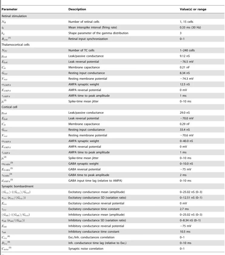
In Figure 3A the conductance variation ratio was fixed at 0.2 for both the excitatory and inhibitory components (an optimal value chosen in Fig. 3B). An exhaustive exploration of the conductance parameter space revealed the emergence of a ridge (dark red) within a narrow band, where the TE is highest for an ensemble of pairs of excitatory and inhibitory conductance amplitudes. This indicates that an adjustment in the balance between excitation and inhibition is required to optimize the information transfer.
On the left side of the narrow band (arrow ‘‘1’’ corresponding to the ‘‘quiet’’ regime domain shown in Fig. 3C; 0 bit/s), information transfer is inefficient due to the concomitant action of strong
Figure 2. Network topology affects the retinocortical transfer of sensory informationin computo.A. Transfer efficiency as a function of the thalamic population size. Each point represents the simulation of a modeled convergent circuit for three predefined TC AMPA synaptic weights in addition to a special case (dashed line) where the synaptic weight was adjusted to the thalamic population size on a per-simulation basis (see text for more details). The thickness of the curves represent the standard deviation across ten repetitions of the same retinal sensory simulation realized each time in the context of a different realization of the cortical synaptic bombardment. B. Transfer efficiency as a function of the TC AMPA synaptic weight for three predefined thalamic population size. C. Influence of the level of retinal input synchronization. The TE was measured for varying retinothalamic spike-time mean jitters and retinal synchronization levels (see text for more details). D. Transfer efficiency measured as a function of the thalamocortical spike-time mean jitter.
Figure 3. Background synaptic bombardment tunes the retinocortical signal transferin computo.A. Effect of the cortical input mean excitatory and inhibitory conductances on TE. Cortical input conductances are normalized relative to the rest conductance of the TC cells. For each trial, each model TC cell in the circuit received a unique realization of the synaptic noise conductances, obeying the same statistics across trials (this condition is referred as the uncorrelated condition in later figures). Arrows denote specific operating regimes, which are shown in C (arrows 1 to 3) and in Figure S3A (arrow 0). The conductance variation ratio was fixed to 0.2 for both the excitatory and inhibitory components, an optimal value denoted by the arrow in B. B. Similar to A for the SD of the conductances. The SD of the conductances were normalized relative to their respective means. The conductance amplitudes were set to 1.5 and 1.0 for the excitatory and inhibitory components of the synaptic noise, respectively. These optimal values are denoted by the arrow ‘‘2low’’ in A. C. Top. Membrane voltage traces for three operating regimes reflecting three distinct cortical
synaptic bombardment statistics. Each regime is shown by an arrow in A and B. The optimal regime was further separated into a low and a high conductance state. Bottom. Cortical spike-triggered averages relative to the number of thalamic spikes were calculated for each of the above cortical voltage traces. The number of thalamic spikes preceding each cortical spike was averaged and plotted as the black curve. Grayed areas indicate the SD of the thalamic spikes count across all cortical spikes. D. Numerical explorations as in A for a control circuit of normal biological size, and for an impaired circuit in which half of the thalamic cells were inactive. The TE difference was calculated for each point by substracting the TE obtained for the normal layer from the TE obtained for the impaired layer. White lines delineate the red ridge of optimal transfer found in the control condition, which is replicated in the two other graphs for comparison.
inhibition and weak excitation, resulting in an effective silencing of the TC cells.
The normalized total cortical input conductance (SGsynT=Grest; see Eq. 9) is a convenient way to characterize the relative strength of the cortical input action on the TC cells. In the band delineating optimal information transfer, the TE is highest for normalized total cortical input conductance ranging from ,2.5 to,4. Two optimal background conductance states connected by the ridge of optimal TE values can be qualitatively distinguished. The first state is a low conductance (LC) regime (denoted by the arrow ‘‘2low’’ in Fig. 3A; 95 bits/s for SGsynT=Grest~2:5) where the mean values of excitatory and inhibitory conductances are approximately comparable to the rest conductance. The second state is a high conductance (HC) regime (denoted by the arrow ‘‘2high’’ in Fig. 3A; 97 bits/s forSGsynT=Grest~4), characterized by a rest conductance that is approximately 50% smaller than the mean values of excitatory and inhibitory conductances. In the corresponding regimes of activity (LC and HC optimal regimes shown in Fig. 3C), the cortical spike-triggered average (STA) clearly indicates an increase of the thalamic synchrony a few milliseconds before the cortical spikes. No major differences were observed apart from slightly stronger voltage fluctuations in the relay cells for the HC state (TC cells membrane potentials SD after removal of spikes is 1.0 mV for LC and 1.4 mV for HC) and a slightly sharper peak for STA in the LC state. No significant STA was presented for the quiet regime since no cortical spikes were evoked.
On the right side of the narrow band (arrow ‘‘3’’, corresponding to the ‘‘saturated’’ regime domain shown in Fig. 3C; 31 bits/s), the inefficiency of the transfer is provoked conversely by a saturating level of excitation. The resulting spiking regime in the relay cells was sufficient to excite the cortical cell in a tonic mode and decorrelate its spiking from the timing of the retinal input, as shown in Figure 3C by the cortical spike-triggered average.
Next, we kept constant the excitatory and inhibitory conduc-tance amplitudes (SGexcT=Grest= 1.5 and SGinhT=Grest= 1.0 as found to be optimal in Figure 3A and corresponding to the LC state in Fig. 3C) and varied the conductance variation ratio (Fig. 3B). A ring shaped area of optimal transfer was found (arrow in Fig. 3B), flanked by areas where both either very low or very high fluctuations led to an inefficient transfer. Note that the amount of inhibitory fluctuations had limited importance com-pared to the amount of excitatory fluctuations as shown by the enlargement of the ring over the y-axis. One explanation resides in the fact that the inhibitory reversal potential is close to the actual resting potential of the model TC cells, effectively limiting the amplitude changes of the inhibitory synaptic bombardment fluctuations, and thus their effect on stochastic resonance (see below).
The mean firing rate of the TC cells occurring under optimal synaptic bombardment (35 Hz) was slightly higher than both the retinal and cortical firing rates (30 Hz) (Fig. 4C,Cnoise~0). The additional spikes responsible for the increased thalamic firing were caused by the CT input as expected from the high probability (0.7) to evoke a spike under optimal synaptic bombardment, even for retinothalamic AMPA events of small amplitude (Fig. S2B, gray curve). In contrast, in absence of contextual synaptic bombard-ment (denoted by the arrow ‘‘0’’ in Fig. 3A whereGsyn= 0; 1 bit/s; traces shown on Fig. S3A), thalamic spikes were solely evoked by the retinal inputs with a much lower probability (Fig. S2B, black curve) and TC cells relayed significantly fewer spikes than present in their retinal inputs. Depolarizing the thalamic cells with a positive constant current (AC, Eq. 13), as to mimic the effects of neuromodulation (see Discussion), shifted the optimal response
ridge seen in Figure 3A towards lowerGexcvalues, and increased the baseline TE observed in absence of synaptic bombardment (Fig. S3B; 50 bits/s forGsyn~0and a 0.6 nA constant current).
A common feature in the thalamocortical circuit is feedforward inhibition (FFI). FFI is defined here in a loose sense (not cell specific). It consists of a group of TC cells that influences excitatory cortical cells in layer 4 through direct connections and indirectly through local relay inhibitory neurons. In such FFI circuits, postsynaptic excitatory neurons are considered highly sensitive to the relative timing of action potentials among presynaptic TC neurons (reviewed in [32]). Therefore, we tested the impact of FFI in the current model circuit (see Methods). In the cortical cell, inhibitory GABAAdisynaptic events triggered by thalamocortical
inputs closely followed direct excitatory AMPA monosynaptic events. The FFI was controlled by the GABAA synaptic weight
and its time lag relative to the AMPA events. We numerically explored a range of GABAAsynaptic weights and time lags and
found that the sensory signal transfer efficiency was improved in the saturated regime by an average of more than 50% for GABAA
synaptic weights ranging from 3 to 5 nS and time lags up to 3 ms (Fig. S4B; note this range of synaptic weights corresponds to the combined inhibitory synaptic weight for the cortical cell in the 30 TC cells version of the model circuit). In contrast, TE in the optimal regimes was mostly unaffected for a large range of biologically realistic parameters, with only minor improvements characterized by a TE increased up to 3% and 5% for the LC state and the HC state regimes, respectively (Fig. S4A).
In the following sections, numerical simulations were performed using the optimal LC state of the synaptic bombardment. No FFI was implemented in the subsequent circuits.
Contextual synaptic bombardment adaptation to impaired topology
We investigated if the drop in transfer efficiency observed when changing the size of the thalamic population without readjusting the TC synaptic weight (Fig. 2B, solid curves) could be counter-balanced by a different tuning of the cortical input. This question explores a potentially important issue, since it aims at determining if pathological impairments of sensory afferent circuits associated with degenerative diseases such as age-related macular degener-ation, phantom limbs, tinnitus or strokes (see Discussion), could be compensated by corticofugal activity adaptation. We compared numerical explorations of the mean conductance amplitudes of the synaptic bombardment as was done in Figure 3A for an optimized network of realistic biological size (Fig. 3D, normal thalamic layer including 90 TC cells) and an impaired network where half of the TC cells did not receive any input (Fig. 3D, impaired thalamic layer including 45 active TC cells and 45 inactive TC cells). The TC synaptic weight of both normal and impaired thalamic layers are identical and set to the biological value which is optimized for a total of 90 TC cells.
synaptic transmission by boosting the responsiveness of the TC cells through cortical synaptic bombardment.
We speculate that this compensation could occur in early stages of macular degeneration, but it would not work in later stages when the thalamic layer is too massively impaired by a drastically
reduced number of retinal inputs. In our model, heavy compen-sation involved a large increase of the excitatory component of the CT input and drove the thalamocortical system into the saturated regime shown in Figure 3C, where the output of the thalamic population remained independent from the retinal afferent drive.
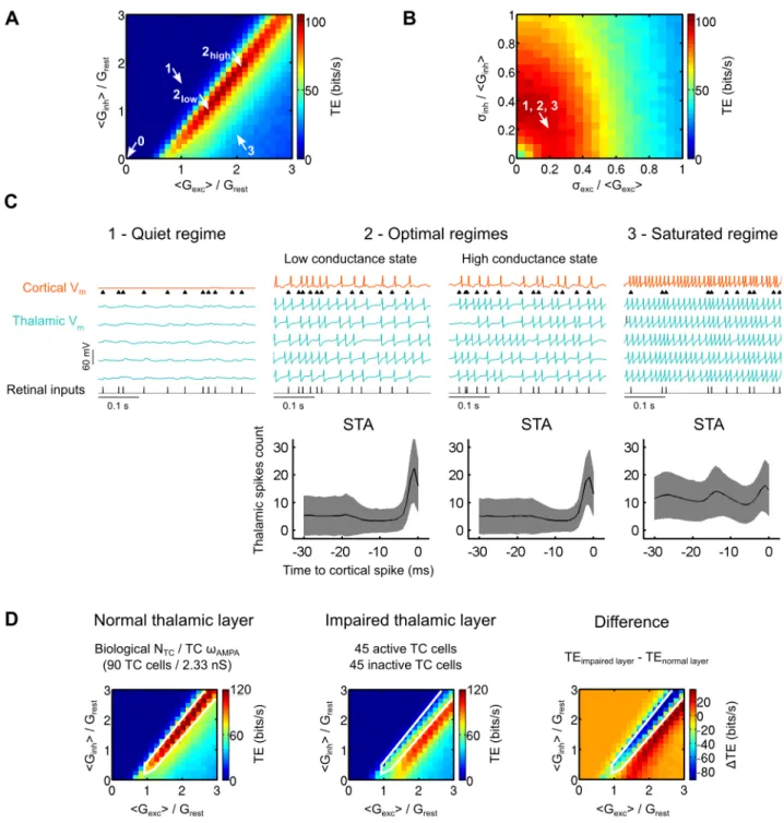
Figure 4. Impact of the CT inputs correlations on retinocortical information transfer efficiencyin computo.A. Illustration of the two synaptic bombardment correlation schemas used in this study. Colored cells receive an identical synaptic noise. Non-colored cells received an independent synaptic noise. Partially colored cells receive a partially correlated synaptic noise. See text for more details. B. Left. Mean pairwise spike correlations among the whole thalamic population as a function of the synaptic noise correlation strength,Cnoise. Right. Example distributions of the thalamic pairwise correlations forCnoise= 0.8 (indicated by the vertical dashed line in the left graph) for the homogeneous (upper) and heterogeneous (lower) schemas. C. Thalamic mean firing rate (6SD across the whole thalamic population) and cortical firing rate (6SD across non-overlapping windows of the cortical spike train). D. Thalamic coefficient of variation (6SD across the whole thalamic population) and cortical coefficient of variation (6SD across non-overlapping windows of the cortical spike train). E. Effect of the synaptic bombardment correlation strength on TE for both correlation schemas illustrated in A. F. Retinothalamic and thalamocortical partial sensory transfer efficiencies (TEpartial) for the homogeneous
Impact of the temporal coherence of the corticothalamic input across TC cells
Temporal correlations of corticothalamic inputs can be examined at two different scales in the thalamic layer, at the level of single cells or at the level of the cell population, with two very different outcomes for information transfer.
At the single cell level, the corticothalamic input directly excites TC cells and indirectly inhibits them via the NRT and local interneurons in the LGN [33]. The precise spatial and temporal organization of these inputs is not known. Inputs from layer 6 to TC and NRT cells may overlap if they originate from the same cortical columns [34], or not if they originate from different columns [13]. Therefore it can be hypothetized that different degrees of temporal correlation occurs in the target TC cells, between monosynaptic cortical feedback excitatory postsynaptic potentials (EPSPs) and disynaptic inhibitory postsynaptic poten-tials (IPSPs). To explore this question, we tested for a large range of correlation strengths (Cexc-inh) and correlation time lags (Dexc-inh)
between the excitatory and the inhibitory components of the synaptic bombardment (GexcandGinh, respectively; see Eq. 15 and 16). Positive correlation time lags caused the inhibition to lag behind the excitation. We observed only a very small decrease in the retinocortical transfer efficiency for high correlation level with no clear dependencies on the time lag (Fig. S5).
So far, the effects observed at the cell level can be explained by a classical gain control where the spike response probability of each individual TC cell is shaped by the characteristics of the noise bombardment [13,19,35].
Beyond this modulatory effect specific of each cell, the following simulations unravel another feature, critical in the control of information transfer, namely the temporal coherence of the synaptic bombardment across TC cells. To illustrate the functional impact of temporal coherence, two possible correlation schemas were explored at the population level, allowing a parametric exploration ranging from complete desynchronization (case examined so far in the previous parts) to full synchronization of the CT input across the whole thalamic cell population (Fig. 4A). In the ‘‘homogeneous’’ correlation case, the cortical projections were arbitrarily divided into two sets of additive noise sources, correlated and uncorrelated, whose relative influence could be titrated parametrically: i) a pool of ‘‘shared’’ CT axons was distributed jointly to all cells of the population and provided a common synaptic input, leading to cross-cell correlations, whereas ii) a pool of ‘‘independent’’ CT axons targeting different cells was distributed within the population, thus providing desynchronized synaptic drive. The differential recruitment of these two types of projections by the cortex can be seen as a simple way to impose different amounts of correlation across the thalamic cells.
We also explored a ‘‘heterogeneous’’ correlation case, where only one type of input could be integrated at once by the thalamic cells which received either shared or independent CT inputs from the cortex. In this latter case, gradual correlation levels are just implemented by spatial heterogeneity in the recruitment by CT shared axons, where a variable number of TC cells receive shared CT inputs while the remaining cells receive independent CT inputs. This spatially organized correlation schema is illustrated by islands of neighboring thalamic cells being densely contacted by common CT axons which would be either synchronously activated by the cortex or kept inactive.
Both the homogeneous (‘‘diffuse and shared’’) and heteroge-neous (‘‘spatially selective’’) correlation schemas are characterized by a correlation strength coefficient (Cnoise, see Eq. 17 and 18) ranging from 0 (no imposed correlation) to 1 (identical synaptic bombardment for every TC cells).
We gradually increased the correlation parameterCnoise while measuring the firing correlation of TC neurons pairs in model circuits (Eq. 20). Correlations in the cortical input provoked pairwise spike correlations in the thalamic layer (Fig. 4B, left). The two correlation schemas did not affect the population in a similar way. The homogeneous correlation schema induced an homoge-neous distribution of pairwise spike correlations across the population (Fig. 4B, upper right; distributions shown for
Cnoise~0:8 indicated by the vertical dashed line) while the heterogeneous correlation schema induced a bimodal distribution characterized by strong spike correlations only in a subset of TC cells (only receiving shared inputs) and no correlation other than the chance level for the remaining cells (only receiving indepen-dent inputs) (Fig. 4B, lower right).
Next, we compared the spiking activity of the cortical response with the average thalamic response. We measured both the firing rate and the spiking variability during fully synchronized (Cnoise~1) or uncorrelated (Cnoise~0) cortical bombardment in model circuits. Although the correlations introduced in the synaptic bombardment across cells did not affect the mean and standard deviation of the cortical input nor the average response of individual TC cells, it modulated both the firing rate (Fig. 4C) and the coefficient of variation of the cortical response (Fig. 4D). In the uncorrelated paradigm, the cortical firing rate remained lower than its thalamic input. In contrast, full correlation of the synaptic bombardment increased the firing rate of the cortical cell and equaled it to the firing rate of the TC cells. Similarly, the spiking variability depended upon the level of correlation of the synaptic bombardment. Note that variability in the cortical discharge was the largest during the uncorrelated paradigm.
We then explored the impact of the synaptic bombardment correlation on the efficiency of the global retinocortical informa-tion transfer for both correlainforma-tion schemas (Fig. 4E). We found that synaptic bombardment correlations injected at the thalamic level strongly decreased the TE of sensory signal transfer. The TE decrease was progressive resulting in a graded decoupling of the retinal stimulation and the cortical response. The starting and ending points were identical for both correlation schemas, only the rate of variation due to correlation increase were different in the two paradigms, being more linear for the homogeneous correla-tion schema than for the heterogeneous one. Full correlacorrela-tion of the synaptic bombardment (Cnoise~1) was still permissive for signal transfer albeit TE was 76% lower than that measured for uncorrelated bombardment (Cnoise~0).
We investigated further how the synaptic bombardment correlation across the thalamic population affected the transfer of sensory information in individual TC cells. We calculated the TE for partial information transfers between the retinal input and a TC cell response (retinothalamic TEpartial) and between a TC
cell input and the cortical response (thalamocortical TEpartial). The
synaptic bombardment correlation was varied using the homoge-neous schema ensuring symmetric variations in all TC cells. A single thalamic cell was thus arbitrarily chosen to calculate partial retinothalamic and thalamocortical TEs.
input and the cortical response. Increases in correlation levels of the CT synaptic bombardment degraded the retinocortical coupling while it improved the thalamocortical coupling. In contrast, the partial retinothalamic TE was unaffected by correlation changes in the CT inputs (Fig. 4F, circles). The latter finding obeys an invariance principle in the first order statistics seen by individual TC cells (note here that correlation changes across the TC cells do not affect the mean or the standard deviation of the synaptic bombardment conductances).
The simultaneous decrease of the global retinocortical TE and increase of the partial thalamocortical TE are both explained by a stochastic resonance effect between the synaptic bombardment noise and the response of every TC cells at the whole population level. The synaptic bombardment can sometime provoke spikes in TC cells which are decoupled from the retinal input if the fluctuations are depolarizing, and conversely prevent the gener-ation of TC spikes in response to retinal events when the fluctuations are hyperpolarizing. Therefore, in presence of highly synchronized corticothalamic noise, spikes provoked or prevented by the synaptic bombardment in TC cells are amplified simultaneously in the whole thalamic population, resulting in a more uniform response. High pairwise spike correlations among TC cells reveal this uniformity (Fig. 4B). The uniformity of the thalamic responses across TC cells further lead to increased spike transmission errors (Fig. 5A, spike transmission failures in the correlated condition) which is precisely what degrades the coupling (i.e. the global TE) between the retinal input and the cortical response. Another related consequence of the cortical input synchronization is the elevation of the thalamocortical synchrony, which boosts the thalamic population drive of the cortical cell. This effect is reflected by an increased partial TE value between any sampled TC cell and the cortical cell.
In summary, dissecting the analysis of information transfer properties at different levels of the circuit reveals that decorrelation in the synaptic bombardment of the corticothalamic input induces a stochastic facilitation process between the retinal input and the target cortical cell which only emerges at the whole thalamic population level (resulting from the collective action of all TC cells). This facilitation process optimizes the efficiency of the global retinocortical transfer of information when TC cells membrane potential fluctuations are decorrelated.
Impact of synaptic noise temporal correlations in reconstructed biological networks
Running biological exploration in parallel with simulation is a useful strategy for the refinement of the model parameters and allows checking their consistency in a biological situation. The explorations of the parameter space with the model suggested the use for BICNs of small fluctuation amplitudes for the synaptic bombardment. This strategy led to the important finding that small membrane fluctuations in individual biological cells (SD = 1– 1.4 mV in model cells and 0.9–3.5 mV in biological cells after removal of spikes; see Fig. 1C,D and Fig. 5A) —that may go unnoticed in in vivo recordings— have a strong effect on the transfer of information when considering the whole TC cell population synaptically converging to a same cortical cell. The use of BICNs (detailed below) also led to the observation that membrane properties of biological cells is an important element for information transfer, not only a the level of the single cell, but especially at the level of the circuit. Note that these experimental findings may not be fully captured by simulations due to inherent limitations of any model.
We examined more in-depth the impact of correlations in the synaptic bombardment by conducting information transfer analysis
on BICNs built from the recordings of 8 TC biological neurons (see Methods). BICNs were analogous to the model circuits tested in Figure 4. Pseudo-TC cells activities in BICNs replayed membrane potential traces recorded in biological TC neurons. The neurons were recorded in dLGN slices of rats and mice and stimulated with an input identical to the one used in model TC cells, using patch and sharp intracellular electrodes and the dynamic-clamp technique (see Methods).
First, we built 15 ‘‘small single-cell’’ BICNs (see Methods) each made of 10 pseudo-TC cells derived from the recordings of a single biological cell. We varied the correlation of the synaptic bombardment across the pseudo-TC cells as was previously done in the model circuits. The correlation parameter (Cnoise) ranged from 0 to 1 using the heterogeneous thalamocortical correlation schema. Illustration of recording sequences from a BICN is shown for both the uncorrelated (Cnoise~0) and correlated (Cnoise~1) conditions (Fig. 5A). Close examination of the voltage fluctuations in the uncorrelated condition revealed notable differences, reflecting variations in the injected synaptic noise conductances (Fig. 5A, insets). Variations in the voltage fluctuations were also present in the correlated condition, albeit much smaller, reflecting solely the trial-to-trial variability intrinsic to the biological TC cell. We found that, when compared to the uncorrelated condition, the correlated synaptic bombardment failed to elicit a number of cortical spikes in response to the retinal input (Fig. 5A, lower left bar graph). The retinocortical global transfer efficiency decreased with increasing levels of correlation in the thalamic layers (Fig. 5B), confirming the results obtainedin computoin previous sections. The average transfer efficiency drop in the 15 BICNs became highly significant for correlation strengths larger or equal to 0.33 (Fig. 5C; p = 0.00031 forCnoise= 0.33; p = 0.000015 forCnoise= 1; paired-sample t-test). Thesein vitroresults confirmed that an increase in the synaptic bombardment correlation led to a significant decrease of the retinocortical transfer efficiency.
Next, we constructed ‘‘large mixed-cell’’ BICNs (see Methods) with a number of pseudo-TC cells ranging from 0 to 130. We compared the TE for correlated (Cnoise~1) and partially decorrelated synaptic bombardments. Compared to the previous BICNs made of 10 pseudo-TC cells obtained from a single biological cell (Fig. 5A to 5C), the large mixed-cells BICNs (Fig. 5D, solid lines) were built from a collection of distinct biological cells which added cellular diversity and variability in the membrane potential fluctuations due to the differences in biological cell properties. We found that the TE was lower in the correlated condition for network sizes larger than,50 pseudo-TC cells, thus confirming the paradigm by which correlation in CT synaptic inputs decreases the efficiency of the sensory information transfer in the more realistic case of BICNs made of distinct biological TC cells.
individual small single-cell BICN composed of 10 pseudo-cells, with each sequence being duplicated a total of 13 times. We then averaged the TE for each thalamic layer size and each correlation condition of the above large single-cell BICNs (Fig. 5D, dashed lines). We found that TE averaged for the large single-cell BICNs was lower than TE obtained for the large mixed-cell BICNs, indicating that there was a positive contribution of the biological cellular diversity in the transfer of sensory information. For a size of 130 pseudo-cells, large mixed-cell BICNs TEs were 18% and 54% higher than the average TEs for large single-cell BICNs, in the correlated and partially decorrelated conditions, respectively.
In the light of these results obtained by combinedin vitroandin computo approaches, we propose that active decorrelation of background synaptic activity in the thalamic layer provides a powerful optimization mechanism —emerging from a population effect— controlling the efficiency of the retinocortical signal
transfer. In this framework, each TC cell is seen as a detector of the retinal stimulation and the brain could modulate the overall transfer efficiency via the CT feedback correlation by controlling the level of independency between the individual detectors, ranging from fully synchronized (lowest information rate) to desynchronized (highest information rate). In the next section, we further investigated the impact of cellular diversity on information transfer in model circuits.
Parametric study of cellular heterogeneity as a ‘‘decorrelation’’ source
In addition to the influence of synaptic inputs and the ongoing afferent activity, the putative diversity of intrinsic membrane properties encountered within a same cell class or across different cell classes due to the variety of their detailed morphology and the
Figure 5. Decorrelation of the corticothalamic synaptic noise boosts retinocortical signal transfer in BICNs.A. Top. Illustration of voltage traces for a small single-cell BICN (indicated by an arrow in B) receiving uncorrelated synaptic bombardment. Insets. Zoomed sections of the biological TC cells membrane voltage fluctuations. Bottom. Same BICN as above receiving a correlated synaptic bombardment. Numerous spike failures are observed compared to the uncorrelated synaptic bombardment. The lower left bar graph shows the mean (6SEM across all spikes) retinocortical spike transmission probability for both the uncorrelated and correlated conditions. B. Transfer efficiency as a function of the synaptic noise correlation strength in small single-cell BICNs (see Methods) normalized relative to the respective uncorrelated condition of each BICN (Cnoise= 0). Each curve represents a different BICN with varying synaptic bombardment correlation strength. The correlation was varied using the heterogeneous schema. Curves with similar colors represent BICNs built from the same biological TC neuron. C. Average TE drop for all small single-cell BICNs (6SEM across all BICNs) as a function of the synaptic bombardment correlation strength. D. TE measurements for large mixed-cell BICNs and average TE for large single-cell BICNs of varying size for both correlated and partially decorrelated conditions (see Methods).
distribution of their ionic channels may also contribute to the decorrelation of the cells activities. We therefore investigated in model circuits to which extent intrinsic cellular heterogeneity could affect the retinocortical global information transfer.
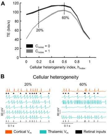
We introduced cellular heterogeneity in our convergent networks by randomizing key intrinsic parameters reflecting the number of channels, the morphology and the integration time constant of the TC neurons (see Methods). We defined a ‘‘cellular heterogeneity’’ index, ranging from 0 to 1, as the amount of variation of the randomized parameters, where a value of 0 meant there was no variation at all and a value of 1 meant that randomized parameters varied up to 100% around their respective original values (Eq. 4). This randomization was repeated for each TC cell. The synaptic bombardment parameters were kept identical to those used previously in model circuits made of non-randomized model TC cells. Note that as a consequence, the bombardment was no longer optimal after randomization of the TC cells intrinsic properties and, a priori, it would require a cell-per-cell adjustment to optimally adapt the bombardment to the new properties of each TC cell. To summarize, two types of decorrelation coexisted in these new simulations, respectively of extrinsic (synaptic bombardment) and intrinsic (biophysical cellular diversity) sources.
Starting with pools of identical TC cells (where the cellular heterogeneity index is 0 in Fig. 6A), in both the correlated and uncorrelated CT synaptic input condition, we found that moderate to high cellular heterogeneity was associated with an improved TE, up to a maximum of 60% of variation for all parameters, after which further cell variability led to degradation of transfer efficiency. Figure 6B illustrates the activities of cells for moderate (20%) and very high (60%) cellular heterogeneities. Comparing the two curves, it is important to note that cellular heterogeneity is very effective in rescuing the low information rate resulting from the correlated synaptic activity, as was previously observed in BICNs (Fig. 5D). Cellular heterogeneity has much less effects in presence of uncorrelated synaptic activity, especially for a moderate, presumably realistic, cell heterogeneity of around 20% (see Discussion).
In summary, these simulations show the diversity of possible mechanisms through which information transfer can be con-trolled, and the importance of decorrelated background activity in the gating of input from the sensory periphery to cortical areas.
Impact of coherent oscillations in the thalamic layer We finally considered an extreme mode of correlation, present in the brain in the form of widespread synchronized oscillations of various but specific frequencies, that are known to impair signal transfer during sleep [36,37], absence epilepsy [38], promote loss of consciousness [39] and show reduced magnitude during focal attention [40]. We investigated to which extent such oscillation-induced correlations imposed in the convergent structure of the thalamic network would affect signal transmission.
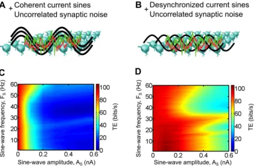
We induced oscillations in the thalamic layer of model circuits by injecting sine-wave currents of varying amplitude (AS) and frequency (fS) to every TC cells (Eq. 14) in addition to a cell-independent synaptic bombardment (no imposed correlation,
Cnoise~0). In a first case, the phases (wS) of the sine-wave currents were identical across all TC cells which resulted in coherent oscillations in the thalamic layer (Fig. 7A). In a second case, the oscillations were desynchronized by a homogeneous distribution of the sine-wave phases across the thalamic population (Fig. 7B).
We found that imposing coherent oscillations resulted in a large decrease of the TE for the full range of tested frequencies, as soon as the oscillation amplitude became large enough (Fig. 7C). In
contrast, the desynchronized oscillations were not as effective to decrease the TE. For particular oscillation frequencies, larger amplitudes, by at least three fold, were required to achieve a similar drop compared to the coherent oscillations (Fig. 7D). Moreover, the coherent oscillations achieved the same TE decrease for every tested frequencies while the desynchronized oscillations were more effective in dropping the transfer efficiency for the 30–60 Hz (gamma) frequency range. Changing the retinal input discharge frequency did not affect the shapes of the graphs (data not shown).
Consistent with recent reports showing task-dependent changes of the oscillatory synchrony in the alert animals (see Discussion), we propose that one important role of the cortical feedback is to modulate the spatial coherence of the thalamocortical oscillatory activities in order to regulate the efficiency of the retinocortical sensory transfer. Combined with a dynamic modulation of the first-order statistics of the CT input (classical single cell gain control), these mechanisms could be used by the brain to actively filter the information conveyed by the retinal ganglion cells to the cortical areas, reflecting both attentional processes and active stimulus filtering under the supervision of higher areas in the brain.
Discussion
In this paper we have quantified the impact of the corticofugal synaptic bombardment on information transfer at the scale of the whole thalamic population presynaptic to a cortical neuron, both in biological iteratively constructed networks and model circuits.
Cortically-induced fluctuations of the synaptic conductances were mimicked with a stochastic process and injected in the biological cells through dynamic-clamp. The main finding reported here is that the circuit simulating the convergence of thalamic neurons onto a common target cortical cell, constitutes a distributed array of input sources which are ideal targets for a top down control. Cortically-controlled stochastic facilitation in individual thalamic cells add up to form an emerging signal filtering property at the network level that promote accurate retinal spike transfer to cortex. We show that this property is critically controlled by the number of TC cells involved simultaneously in the convergence, the statistics of the cortically-driven synaptic bombardment, and the level of correlation imposed across membrane potential fluctuations of the TC cells.
Simulations limited to a realistic range of biophysical param-eters in synaptic weight and EPSP amplitude show that an optimal number of,90 TC cells was best adapted to favor the transfer of sensory information to the convergent circuit topology, which is characterized by weak TC synapses and a high degree of TC convergence. Cortically-induced thalamic voltage fluctuations could be adjusted to control the thalamic spike synchronization window thus sharpening the cortical spike-triggered average response and the efficiency of the sensory input transfer. Most importantly, we found that cortical input coherence was a key factor controlling the sensory signal information transfer efficiency to the target cortical cell. Simulation of coherence increase across TC cells by imposing additional correlated random fluctuations or coherent voltage oscillations in their membrane potential gradu-ally degraded the sensory signal transfer. In contrast, a relatively high amount of retinal afferent synchronization was critical to ensure efficient transfers.
Our approach calls however for some reservation: in its detailed implementation, the present BICN circuit does not implement in full the feedback between cortex and thalamus, since the simulated cortical input to the thalamic relay cells is not updated by the ongoing output activity of the thalamocortical stream. However,
our aim was to explore the functional impact of a parametrized cortical input signal whose statistical structure has the ‘‘color’’ of a ‘‘realistic’’ cortical feedback. One could still object that such added feedback could simulate as well a contextual noise at the thalamic level. Nevertheless, our working hypothesis posits that the ‘‘color’’ of this noise is dictated by the corticothalamic loop. There is indeed biological evidence supporting our theoretical framework: in contrast to the assumed distributed nature of the cortical feedback, it is well established that ongoing activity of intra-thalamic origin (as observedin vivo [41] and in vitro [42] in the deafferented slice) has a strong rhythmic dominance [43,44]. In order to test the impact of such oscillatory noise source, we imposed voltage oscillations in the TC cells and found that coherent oscillations have the property of reducing the sensory signal information transfer whereas desynchronized oscillations remain permissive.
We propose that the synaptic gating of sensory information in the thalamus may rely on transient and spatially-focalized modulations of the coherence level of the contextual cortical feedback.
Optimal size of the convergent circuit
The convergent synaptic organization of the thalamocortical circuit forms the structural kernel of our feedforward model. This rather simple topology of projecting relay neurons in the visual thalamocortical system [3] has not attracted the attention it deserves perhaps because of the yet unsolved technical challenge of identifying and record simultaneously all neurons belonging to the same convergent circuit. In this paper we reveal that this convergent topology might be essential for information transfer, first because, in a somewhat trivial way, it allows the recipient cortical cell to integrate sensory EPSPs evoked in all the relay cells simultaneously, and second, in a less obvious way, because the concomitant synaptic bombardment exerted by the descending corticothalamic feedback can result in stochastic facilitation of the feedforward input lines.
Figure 7. Impact of thalamocortical oscillations on the retinocortical information transfer efficiencyin computo.A and B. Sine-wave currents of varying amplitude and frequency were injected to every model TC cells in addition to retinal inputs and uncorrelated synaptic bombardment. The current oscillations were either coherent (same phase for every TC cells) or desynchronized (phase evenly distributed in the thalamic population). C and D. Transfer efficiency for both conditions shown in A and B, respectively.
We found the number of thalamocortical cell involved in the convergent network to be a key parameter controlling information transfer. An exhaustive exploration of the parameter space revealed that the efficiency of the transfer reached a peak for a population of,90 relay cells, with rather weak synapses (,1 mV EPSP), whose individual recruitment would never trigger per se a cortical spike. For larger population sizes, little or no gain was observed, indicating a saturation of the impact of the convergent afferent circuit.
These results are coherent with findings obtained in vivo. Thalamocortical inputs on layer 4 stellate neurons are thought to be among the strongest in the neocortex. However, recentin vivo data show that individual synapses are weak (EPSP,0,5–2 mV) and that a subnetwork of at least ,30 inputs needs to be synchronously active to drive the firing of a single cell in the visual cortex [31,45,46] and in the somatosensory cortex [47]. Our model is based on data from the visual thalamus and by exploration of the parameter space we found that statistically at least one third of the 90 cells are required to fire simultaneously in order to elicit a spike in the target cortical neuron. It should be noted that despite their different functional specialization, the topology of converging TC circuits to V1 and S1 seem to retain the similarity of having numerous and synchronous convergent inputs to layer 4 cortical cells (estimated to,85 in S1 [47]; and range between the extreme values of 15 to 125 in V1 [2,3]).
It is important to note that the optimal size of the convergent circuit we reported here was directly obtained from numerical explorations performed on the retino-thalamo-cortical model circuit. Put together, the above results led us to the suggestion that the value of,90 TC cells is of significance not only for the visual thalamic network but potentially for similar feedforward multi-layered networks as found in other sensory modalities. Nevertheless, although the model circuit parameters are extremely consistent and thoroughly constrained with biological data, it is likely that more exhaustive simulations are needed in order to estimate the actual optimal value of the population size adapted to other network topologies (with different synaptic convergence and divergence ratios), cellular properties and input statistics (which differ significantly across sensory modalities).
Synchrony detection and spike-timing in thalamocortical convergence
The activity of local groups of cells with neighboring receptive fields can be significantly correlated if the visual input itself has strong spatial and temporal correlations (for a review see [48]), as it is the case with natural scenes [49–51]. LGN cells with overlapping receptive fields of the same type (ON-center or OFF-center) often fire spikes that are synchronized within 1 msin vivo [1] and their precise correlation was found to be of considerable importance [52] in the coding of visual information. In our model circuit, synchronization of the LGN inputs to the cortical cell was directly controlled by the retinal spike synchronization parameter in a biologically realistic retinothalamic stage, where multiple retinal ganglion cells were connected through both convergent and divergent processes to the TC cells. In accordance with the works cited above, we found that a relative synchronization of the retinal afferents was critical to convey an efficient transfer to the cortical neuron (Fig. 2C).
Interestingly, the thalamocortical convergent circuit was adapt-ed to detect synchrony of the LGN cells. Successful propagation of retinal spikes to the cortical cell required the LGN spikes to fall within a,10 ms time window. This estimate corresponds to twice the peak half-width of the spike-triggered average for the optimal regimes in Figure 3C and is consistent with the ‘‘spiking
opportunity window’’ for thalamic spikes [53], thalamic synchro-nization tuning resulting from adaptation [54] and retinogenicu-late paired-spike transmission enhancements [55,56].
In contrast with the importance of the synchronization level of the retinal inputs, the precise timing of individual LGN action potentials within this spiking opportunity window was not a critical factor for an efficient information transfer to the cortical cell. We found that in the presence of background synaptic noise, information transfer remained relatively resistant to the deleterious effect of retinal and thalamic spike-time jitters. In a demanding test in which increasingly large delay jitters were randomly applied to the feedforward circuit (Fig. 2C,D), transfer efficiency decreased by less than 20% for delay jitters up to 3 ms in either the retinal or the thalamic inputs. This suggests that the deleterious effect of retinocortical propagation variability (estimated to 1 ms for the retinothalamic transmission [57] and to 0.4 ms for the thalamo-cortical transmission [58]) on signal transfer can be easily overcome by the gating effect of the corticothalamic feedback on the TC cell population.
Whether correlations among the retinal ganglion cells are strong enough to drive synchronously their thalamic targets remains a matter of debate. A possibility is to consider multiple arrays of small intermingled thalamocortical convergent networks, such as studied here, each capable of detecting and relay specific sets of synchronous retinal ganglion cells. In this framework, each set of synchronously active retinal ganglion cells could represent a distinct feature of the visual scene and convergent networks involved in the synchrony detection of the latter sets could propagate meaningful representations of the visual space to cortical layer 4. This intuitive proposition should be tested in larger scale computer simulations.
Feedforward inhibition in the cortex is another feature that could facilitate information transfer. Circuits with strong FFI can selectively gate synchronous over asynchronous inputs ([32]). This predicted that in our model —in which synchrony of thalamo-cortical inputs to cortex is paradoxically favored by uncorrelated corticothalamic noise— a strong cortical FFI could sharpen the synchrony of excitatory input and thus increase information transfer. We found that transfer efficiency in the quiet and optimal regimes was mostly unaffected for a large range of biologically realistic parameters. However, FFI could partially rescue infor-mation transfer in the saturated regime (Fig. S4).
The challenging task of modeling the activity of layer 6 corticothalamic neurons