MicroRNAs Induce Epigenetic
Reprogramming and Suppress Malignant
Phenotypes of Human Colon Cancer Cells
Hisataka Ogawa1,2, Xin Wu1, Koichi Kawamoto1,2, Naohiro Nishida1,2, Masamitsu Konno2, Jun Koseki3, Hidetoshi Matsui4, Kozou Noguchi1,2, Noriko Gotoh5, Tsuyoshi Yamamoto6, Kanjiro Miyata7, Nobuhiro Nishiyama7,8, Hiroaki Nagano1, Hirofumi Yamamoto1,
Satoshi Obika6, Kazunori Kataoka7, Yuichiro Doki1, Masaki Mori1*, Hideshi Ishii2,3*
1Department of Gastroenterological Surgery, Osaka University Graduate School of Medicine, Suita, Osaka, Japan,2Department of Frontier Science for Cancer and Chemotherapy, Osaka University Graduate School of Medicine, Suita, Osaka, Japan,3Department of Cancer Profiling Discovery, Osaka University Graduate School of Medicine, Suita, Osaka, Japan,4Faculty of Mathematics, Kyushu University, Fukuoka, Japan,
5Division of Cancer Cell Biology, Cancer Research Institute of Kanazawa University, Kakuma-machi, Kanazawa, Japan,6Department of Bioorganic Chemistry, Osaka University Graduate School of Pharmaceutical Science, Suita, Osaka, Japan,7Department of Materials Engineering, The University of Tokyo, Bunkyo-ku, Tokyo, Japan,8Tokyo Institute of Technology, Chemical Resources Laboratory, Yokohama, Midori-ku, Japan
*[email protected](HI);[email protected](MM)
Abstract
Although cancer is a genetic disease, epigenetic alterations are involved in its initiation and progression. Previous studies have shown that reprogramming of colon cancer cells using Oct3/4, Sox2, Klf4, and cMyc reduces cancer malignancy. Therefore, cancer reprogram-ming may be a useful treatment for chemo- or radiotherapy-resistant cancer cells. It was also reported that the introduction of endogenous small-sized, non-coding ribonucleotides such as microRNA (miR) 302s and miR-369-3p or -5p resulted in the induction of cellular re-programming. miRs are smaller than the genes of transcription factors, making them possi-bly suitable for use in clinical strategies. Therefore, we reprogrammed colon cancer cells using miR-302s and miR-369-3p or -5p. This resulted in inhibition of cell proliferation and in-vasion and the stimulation of the mesenchymal-to-epithelial transition phenotype in colon cancer cells. Importantly, the introduction of the ribonucleotides resulted in epigenetic repro-gramming of DNA demethylation and histone modification events. Furthermore,in vivo ad-ministration of the ribonucleotides in mice elicited the induction of cancer cell apoptosis, which involves the mitochondrial Bcl2 protein family. The present study shows that the intro-duction of miR-302s and miR-369s could induce cellular reprogramming and modulate ma-lignant phenotypes of human colorectal cancer, suggesting that the appropriate delivery of functional small-sized ribonucleotides may open a new avenue for therapy against human malignant tumors.
OPEN ACCESS
Citation:Ogawa H, Wu X, Kawamoto K, Nishida N, Konno M, Koseki J, et al. (2015) MicroRNAs Induce Epigenetic Reprogramming and Suppress Malignant Phenotypes of Human Colon Cancer Cells. PLoS ONE 10(5): e0127119. doi:10.1371/journal. pone.0127119
Academic Editor:Maria Cristina Vinci, Centro Cardiologico Monzino, ITALY
Received:January 19, 2015
Accepted:April 10, 2015
Published:May 13, 2015
Copyright:© 2015 Ogawa et al. This is an open access article distributed under the terms of the
Creative Commons Attribution License, which permits unrestricted use, distribution, and reproduction in any medium, provided the original author and source are credited.
Data Availability Statement:All relevant data are within the paper and its Supporting Information files.
Introduction
Every cancer cell is largely derived from stem or progenitor cells of normal somatic tissue via genetic and epigenetic alterations. These alterations inactivate growth-constraint tumor sup-pressor genes (TSGs) and activate growth-promoting oncogenes. Normal somatic cells are developed from a fertilized oocyte through an epigenetic program. Notably, the ectopic intro-duction of defined coding genes, OCT3/4, SOX2, KLF4, and c-MYC (OSKM), or OSK, which are exclusively expressed in embryonic stem cells (ESCs), induces full reprogramming of differ-entiated somatic cells back to pluripotent stem cells. We previously showed that the introduc-tion of OSKM in epithelial cancer cells of gastrointestinal organs modulates the malignant phenotype. Our findings suggested that reprogramming can suppress cancer invasion, drug re-sistance, and tumorigenicity through the re-activation of the tumor suppressor p16INK4A pathway by demethylation of the promoter sequence [1]. Moreover, a recent mouse study of transgenic OSK factors showed that epigenetic modifications are involved in tumor initiation and developmentin vivo. This study also demonstrated that, in combination with the
inactiva-tion of TSG signals such as mutant Apc, the modificainactiva-tions can orchestrate epigenetic alter-ations by the polycomb group complex to the core module of histone H3 at lysine-27 [2]. Taken together with previous findings [1–6], epigenetic alterations, regardless of the causes or results of the genetic changes, contribute to the occurrence and development of malignancies and may be attractive for the discovery of novel therapeutic targets against human cancer.
Therapeutic applications that augment endogenous signaling pathways should have mini-mal toxicityin vivo. In this regard, drug discovery from endogenous microRNAs (miRNAs,
miR), small-sized noncoding ribonucleotides (22 bp on average), are potential sources for in-novative therapeutic strategies [7,8]. Because synthesized mature miRs do not integrate into the genome, they should exert their functions without undesired genomic integration events and, if combined with a drug delivery system, miRs could exert these specific effects on thera-peutic targets [7,8].
Recent miR discovery studies have identified critical clusters, including miR-302s, which en-hance the reprogramming effect in collaboration with viral-mediated transcription factor transfer [9–12]. It has been shown that a set of three miRs (302s, 369s and miR-200c) selected from numerous miRs expressed exclusively in induced pluripotent stem cells (iPSCs)/ESCs, elicited reprogramming [13]; a significant role for miR-367 was also shown [12]. Reportedly, the introduction of miR-302s induced both cell cycle arrest by interfering with cy-clin-dependent kinases (Cdks) and global demethylation of DNA in liver cancer and melano-main vitro[4,5,14]. Here, we studied the effect of miR-302s and miR-369sin vitroandin vivo.
These results suggested that cancer reprogramming using miR-302s and miR-369s may be an effective treatment option for cancer therapy.
Materials and Methods
Cell culture
Colorectal cancer cell lines HT29, DLD-1, SW480, HCT116, Caco2, Colo201, RKO, and Lovo cells and teratocarcinoma cell line NTERA (NTera 2/cl.D1 [NT2/D1]) were obtained from RIKEN (Tsukuba, Japan). These cells were cultured in RPMI-1640 medium (Nakalai Tesque, Kyoto, Japan) with 10% FBS (Gibco Life Technologies, Tokyo, Japan) and 500μg/ml of penicil-lin-streptomycin.
Sports, Science, and Technology (http://www.mext. go.jp/english/; #23390199, #25112708, #25134711, #30253420, #26670604; M.M, K.M, H.I); a Grant-in-Aid from the Ministry of Health, Labor and Welfare (http://www.mhlw.go.jp/english/; #H23-003; M.M., H. I.); a grant from the National Institute of Biomedical Innovation (http://www.nibio.go.jp/english/index.html; #12-4; M.M., H.I.); and a grant from the Osaka University Drug Discovery Funds ( http://www.osaka-u.ac.jp/en/index.html; M.M., H.I.). Partial supports were received from Takeda Science and Medical Research Foundation (http://www.takeda-sci.or.jp/ index.html; M.M., H.I.), Princess Takamatsu Cancer Research Fund (http://www.ptcrf.or.jp/english; M.M., H.I.), Suzuken Memorial Foundation (http://www. suzukenzaidan.or.jp; M.K.), Yasuda Medical Foundation (http://www.yasuda-mf.or.jp; N.N.), Pancreas Research Foundation (http://www.jprf.or.jp/ shoreisho.html; K.K.), Nakatani Foundation (http:// www.nakatani-foundation.jp; H.I.), and Nakatomi Foundation of Japan (https://www.nakatomi.or.jp/en/ index.html; M.K.). The funders had no role in study design, data collection and analysis, decision to publish, or preparation of the manuscript.
Transfection
Specific miRs and negative control (NC) miR used inin vitroandin vivoanalysis were
pur-chased (Gene Design Inc., Osaka, Japan;S1 Table). Cells were transfected with specific miRs and NC miR using lipofection (LP) or carbonate apatite (CA). In LP, cells were transfected with miRs using Lipofectamine iMax (Invitrogen, Darmstadt, Germany) according to the manufacturer’s protocol.
Cell reprogramming
HT29 cells and DLD-1 cells were transfected with 10 nM of each miR using CA. Cells were in-cubated in RPMI-1640 with 10% FBS for 24 h and transfection was repeated every two days for a total of three times. After the third transfection, cells were seeded onto Matrigel-coated and mitomycin C-treated mouse embryonic fibroblasts (MEF). Cells were cultured in embryonal stem cell culture medium containing DMEM/F12 (Gibco Life Technologies, Tokyo, Japan), supplemented with 2 mM GlutaMAX, 20% knockout serum replacement (Gibco Life Technol-ogies), 0.1 mM nonessential amino acids (NEAA, Gibco Life TechnolTechnol-ogies), 10 ng/ml basic fi-broblast growth factor (bFGF, Wako, Tokyo, Japan), 55μM 2-mercaptoethanol (Gibco Life Technologies), 1% penicillin-streptomycin, and chemical inhibitors, including 0.5μM A83-01 (Stemgent, Cambridge, MA), 3μM CHIR99021 (Stemgent), and 0.5μM PD0325901 (Stem-gent), at 37°C in a 5% CO2incubator. Media was changed every two days and the cells were
maintained at 37°C in a 21% CO2incubator for an additional 21 days. During this period,
these cancer cells were monitored for the formation of ES-like colonies. These were picked for further analysis with Alkaline Phosphatase (AP) Live Stain (500×) (Invitrogen) using an all-in-one fluorescence microscope (BZ-9000; Keyence, Osaka, Japan) with digital photograph-ic capability for selection according to the manufacturer’s instructions. To study miRs transfec-tion efficiency, DLD-1 cells were transfected with BLOCK-iT Alexa Fluorescent Control (Invitrogen) with CA or LP. In brief, seeded DLD-1 cells in a 6-well plate were transfected with BLOCK-iT Alexa Fluorescent Control and photographed after transfection using a Keyence BZ-8000 microscope. The fluorescence intensity of transfected cells as observed using a FACS BD FACSAria III cell sorter.
Luciferase assay
The 30untranslated region (30-UTR) of CDK2 was amplified by RT-PCR using the primers 5’
-CTAGCTAGCTAGCCTTCTTGAAGCCCCCA-3' and 5’-CTAGCTAGCGAGCTACAAAC
TAAATTACA-3'. Primers were subcloned, ligated into the pmirGLO Dual-Luciferase miRNA Target Expression Vector (Promega) using NheI, and confirmed by direct sequencing. Lucifer-ase assays were conducted using 5 × 10³DLD-1 cells plated in a 96-well plate. Cells were trans-fected using Lipofectamine 3000 (Invitrogen) in OptiMEM reduced serum media (Gibco) with 200 ng of empty vector or Luciferase-CDK2 3’UTR vector and either NC miR or miR-302s (final concentration, 25 nmol/L). Luciferase activity was measured 24 h post-transfection using the Dual-Glo Luciferase Assay System (Promega) according to the manufacturer’s protocol. Relative luciferase level was calculated as (Sample Luc/Sample Renilla)/(Control Luc/Control Renilla), where Luc is raw firefly luciferase activity and Renilla is the internal transfection con-trol luciferase activity.
Cell proliferation assay
several concentrations of 5-FU for 72 h in a 96-well plate. Viable cells were evaluated using the Cell Counting Kit-8 (CCK-8) (Dojindo Molecular Technologies, Tokyo, Japan). To assess the influence of these indicated miRs on cell proliferation, HT29 cells and DLD-1 cells (5×10⁴ cells/well in a 12-well plate) were transfected with either miR-302s, miR-302s plus miR-369s, or NC miR, as described above. Cells were counted every day for three days, starting 24 h post-transfection.
Cell cycle analysis
For cell cycle analysis by flow cytometry, 5×10⁴DLD-1 cells were transfected with each miR in a 24-well plate, trypsinized after 72 h, washed with phosphate-buffered saline (PBS), and fixed in 70% ethanol on ice. After centrifugation, cells were stained with 50 mg/ml propidium iodide (PI) solution (Dojindo Molecular Technologies) and 0.1 mg/ml RNase A (Invitrogen) and ana-lyzed by flow cytometry using a FACS BD FACSAria III cell sorter. Each histogram was con-structed with data from at least 10,000 events and was used to calculate the percentage of the cell population in each phase.
Cell invasion assay
Transwell invasion assays were carried out in 24-well modified chambers pre-coated with Matrigel (BD BioCoat, BD Biosciences, Franklin Lakes, NJ). DLD-1 cells and SW480 cells were transfected with either 10 nM of miR-302s, miR-369s, miR-302s plus miR-369s and NC miR using CA. After 48 h, cells were trypsinized and re-suspended at a concentration of 10×104 cells/ml in serum-free medium, and 0.5 ml cell suspension was added to the top of each well. After 48 h incubation, cells migrating into the lower chamber, containing 10% FBS as a chemo-attractant, were fixed and stained with Wright-Giemsa stain (Diff-Quick; Sysmex, Kobe, Japan). Four random fields were counted in triplicate. Data are expressed as the median value with the standard error of the mean (s.e.m) of invaded cells in a relative ratio compared to NC miR-treated cancer cells.
Cell differentiation study
To determine the differentiation ability of the generated ES-like colony-forming cancer cellsin vitro, cells were cultured in floating cultivation to form embryoid bodies (EBs). After 4–5 days,
EBs were transferred to 0.1% gelatin-coated plates and cultured in RPMI-1640 with 10% FBS for a further 14 days.
RNA analysis
Total RNA was extracted using Trizol (Invitrogen, Tokyo, Japan). RNA quality was assessed with a NanoDrop ND-1000 spectrophotometer (NanoDrop Technologies, Wilmington, DE) at 260 and 280 nm (A260/280). Reverse transcription (RT) for miR was performedin vitrowith
the Taqman Reverse transcription microRNA Kit (Applied Biosystems, Tokyo, Japan). qPCR was performed with the universal Taqman PCR Master Mix (Applied Biosystems).
RT-PCR
RNU48 expression in vitro and RNU6B expression in vivo were used as an internal control to determine the relative expression of miR. RT for miR was performedin vitrowith miScript
Transcription System (Promega, Tokyo, Japan) and LightCycler FastStart Reaction Mix SYBR Green I in a Lightcycler (Roche, Tokyo, Japan). All results were normalized to a GAPDH control. RNA expression studies were independently repeated at least three times to confirm reproducibility.
Protein analysis
Protein levels were determined by immunoblotting. Antibodies against Bak (1:200, Santa Cruz, Santa Cruz, CA), Bid (1:200, Santa Cruz), Bcl-2 (1:1000, CST, Tokyo, Japan), Bcl-xl (1:1000, CST), Mcl-1 (1:1000, CST), Caspase8 (1:1000, CST), Caspase3 (1:1000, CST), CyclinD1 (1:200, Santa Cruz), CDK2 (1:200, Santa Cruz), CDK4 (1:200, Santa Cruz), E-cadherin (1:1000, CST), vimentin (1:1000, CST), Zeb1 (1:1000, CST), Phospho-Rb (Ser807/811) (1:1000, CST), and the internal control ACTB (1:2000, Sigma Aldrich, Tokyo, Japan) were used.
Immunoblotting
Cell pellets were lysed in ice cool radioimmunoprecipitation assay buffer with protease inhibi-tor. Protein concentrations were determined using the Bradford assay. Proteins were separated on 4–10% Mini-PROTEAN Precast Gels (BioRad, Tokyo, Japan). After overnight electropho-retic transfer onto PVDF membrane, membranes were blocked with Blocking One solution (Nacalai Tesque, Tokyo, Japan) for 1 h and incubated overnight at 4°C with the indicated pri-mary antibody. Finally, membranes were incubated with secondary anti-mouse or-rabbit whole IgG coupled to horseradish peroxidase (GE Healthcare Life Sciences, Tokyo, Japan) for 1 h at room temperature. Antibody binding to target proteins was visualized by chemilumines-cence using Amersham ECL Prime Western Blotting Detection Reagents (GE Healthcare Life Sciences).
Immunocytochemistry
Cells were washed twice with PBS and fixed in 4% paraformaldehyde for 15 min at room tem-perature. Cells were then washed twice with PBS and permeated with 0.1% TritonX-100 for 15 min at room temperature. Unspecific binding was blocked by incubating in goat or horse serum (Vectastain, Funakoshi, Tokyo, Japan) for 10 min at room temperature. Cells were incu-bated with primary antibodies overnight at 4°C. Primary antibodies used were polyclonal rab-bit anti-human Oct3/4 (1:400, MBL, Nagoya, Japan), polyclonal rabrab-bit anti-human Sox2 (1:400, MBL), monoclonal mouse human Nanog (1:2000, CST), monoclonal mouse anti-human Ki-67 Antigen (1:400, DAKO), and monoclonal rabbit anti-anti-human E-cadherin (1:200, CST). The next day, cells were washed twice with PBS and treated with secondary antibody for 30 min at room temperature. Secondary antibodies used were anti-rabbit IgG (H+L), F(ab’)2 fragment (Alexa Fluorr 488 Conjugate, 1:1000, CST) or anti-mouse IgG (H+L), F(ab’)2 frag-ment (Alexa Fluorr 555 Conjugate) (1:1000, CST). Nuclei were stained with ProLong Gold Antifade reagent with DAPI (Invitrogen). Images were captured with a Keyence BZ-8000 microscope.
DNA methylation analysis
the two samples were compared to determine the DNA methylation response to miR-302s transfection.
Histone methylation analysis
Histone extraction and measurement of global methylation levels of histone 3 lysine-4 (H3K4) were performed using the Global Histone H3-K4 Methylation kit according to the manufactur-er’s protocols (Abnova, Taipei, Taiwan).
Mouse study
Animal studies were conducted in strict accordance with the principles and procedures ap-proved by the Committee on the Ethics of Animal Experiments of Osaka University (approval number, 24-122-011). The tumorigenicity assay was performed using anin vivoxenograft
model. First, 5 × 10⁶cells suspended in a total volume of 200μL DMEM/Matrigel (1:1 (v/v) suspension) were injected into the flanks of 7-8-week-old female mice (BALB/cAJcl-nu/nu). When tumor volumes reached 100 mm3, as measured with calipers and calculated using the formula V = (ab2)/2, where a is the length and b is the width, tumors were harvested and cut into 3–4 mm3sections for serial transplantation. Sections were to confirm vasculaturization by immunohistochemistry using CD31, a vascular endothelial marker. Serially transplanted HT29 xenografts had the most abundant vasculature, was uniformly distributed over the whole tumor, and had the least amount of central necrosis. miRs/CA complexes were administered using a 30G needle via the tail vein. Tumor size and body weight were measured once every 3 days. Xenograft tumors and mouse blood were collected during sacrifice and preserved in 10% neutral-buffered formalin for histology and immunohistochemical studies or in RNAlater RNA Stabilization Reagent (Qiagen, Tokyo, Japan).
Carbonate apatite preparation
To prepare a CA transfection mixturein vitro, 2μg of each miR-302 (-a,-b,-c,-d), miR-369
(-3p, -5p) or NC miR was mixed with 4μL of 1 M CaCl2in 1 mL of serum-free bicarbonate
(44 mM)-buffered DMEM medium (pH 7.5) incubated at 37°C for 30 min, and used for trans-fection [15–17]. This method was mainly used forin vitroexperiments. Forin vivo
experi-ments, an inorganic solution (0.9 mM NaH2PO4, 1.8 mM CaCl2, pH 7.5) replaced the DMEM
solution. For one mouse, a mixture of 15μg of each miR was used for single injection. The solu-tion was centrifuged at 12,000 rpm for 3 min and the pellet dissolved in 200μl saline containing 0.5% albumin. Products in the solution were sonicated (38 kHz, 80 W) in a water bath for 10 min under 20°C to generate miR/CA complexes, which were injected intravenously within 5 min.
Patient samples
Clinical samples were collected during surgery from nine patients undergoing surgical resec-tion for colorectal cancer at Osaka University Hospital and its related hospitals in 2005. Pa-tients are summarized inS2 Table. All patients were in agreement with the use of resected specimens in this study according to the guidelines approved by the Institutional Research Board (approved protocol #213).
Immunohistochemical analysis
rehydrated in a graded ethanol series. Slides were heated in antigen retrieval buffer for 40 min, blocked with goat or horse serum for 20 min at room temperature, and incubated with mono-clonal mouse anti-human Ki67 antigen (1:400, DAKO), polymono-clonal rabbit anti-human Oct3/4 (1:100, MBL), polyclonal rabbit human Sox2 (1:200, MBL), or monoclonal mouse anti-human CK20 (DAKO) antibodies overnight at 4°C. The Vectastain ABC System (Vectastain, Funakoshi, Japan) was used to visualize antigen. Counter-staining was performed using hematoxylin.
Immunofluorescence analysis
Immunofluorescence analysis was performed on 3.5μm paraffin-embedded sections from xe-nografts to detect tumor vessels. Paraffin-embedded sections were de-paraffinized and treated as described for immunohistochemistry. Sections were blocked with goat serum for 20 min at room temperature and incubated with rat anti-mouse CD31 antibody (1:20, Dianova, Ham-burg, Germany) overnight at 4°C. The next day, the secondary antibody anti-rat IgG (H+L), F(ab’)2 Fragment (Alexa Fluorr555 Conjugate) (1:1000, CST) was applied. Sections were counter-stained with ProLong Gold Antifade Reagent with DAPI. Images were captured with a Keyence BZ-8000 microscope.
Alcian blue staining
Alcian blue staining was performed on 3.5μm paraffin-embedded sections from xenografts to detect mucus. In brief, the slides were de-pareffinized in Hemo-De (Farma), rehydrated in a graded ethanol series, immersed in 3% acetic acid for 3 min, and stained in alcian blue solution (1 g Alcian blue 8GX, 3 ml acetic acid, and 97 ml distilled water) for 20 min. Sections were then washed and the nuclei counterstained with Kernechtrot (TCI, Tokyo, Japan) for 5 min.
Apoptosis assay
The Annexin V-FITC Apoptosis Detection Kit (Biovision, Milpitas, CA) was used according to the manufacturer’s protocol forin vitroearly and late phase detection of apoptotic cells. Briefly,
to detect apoptosisin vitro, 20×104DLD-1 cells were transfected with miR-302s, miR-369s,
miR-302s plus miR-369s or NC miR using CA in a 6-well plate in triplicate 60 h before the assay. Cells were trypsinized and analyzed by flow cytometry using a FACS BD FACSAria III cell sorter. Three independent experiments were performed in triplicate.
To detect DNA breaks in apoptotic cellsin vitro, we used the DeadEnd Fluorometric
TUNEL System (Promega) according to the manufacturer’s protocol. In brief, cells were trans-fected with each miR using CA using the Nunc Lab-Tek II Chamber Slide System (Fisher Sci-entific, Tokyo, Japan). Slides were washed 48 h post-transfection and fixed in 4% formaldehyde in PBS for 25 min at 4°C. Slides were washed twice in PBS and permeabilized in 0.2% Triton X-100 in PBS for 5 min. Slides were then washed in PBS, equilibrated in Equilibration Buffer for 7 min at room temperature, and labeled with TdT reaction mix for 60 min at 37°C in a humidi-fied chamber to avoid light exposure. Slides were immersed in 2X SSC for 15 min to stop the reaction. Nuclei were counterstained using with ProLong Gold Antifade Reagent with DAPI. Apoptotic cells were detected by a Keyence BZ-8000 microscope.
To detect apoptotic cellsin vivo, a TUNEL assay was performed according to the
stop the reaction. Nuclei were counterstained using ProLong Gold Antifade Reagent with DAPI. Apoptotic cells were detected by a Keyence BZ-8000 microscope.
Results
Endogenous expression of miR-302s and miR-369s in primary tumors
and cancer cell lines of colon
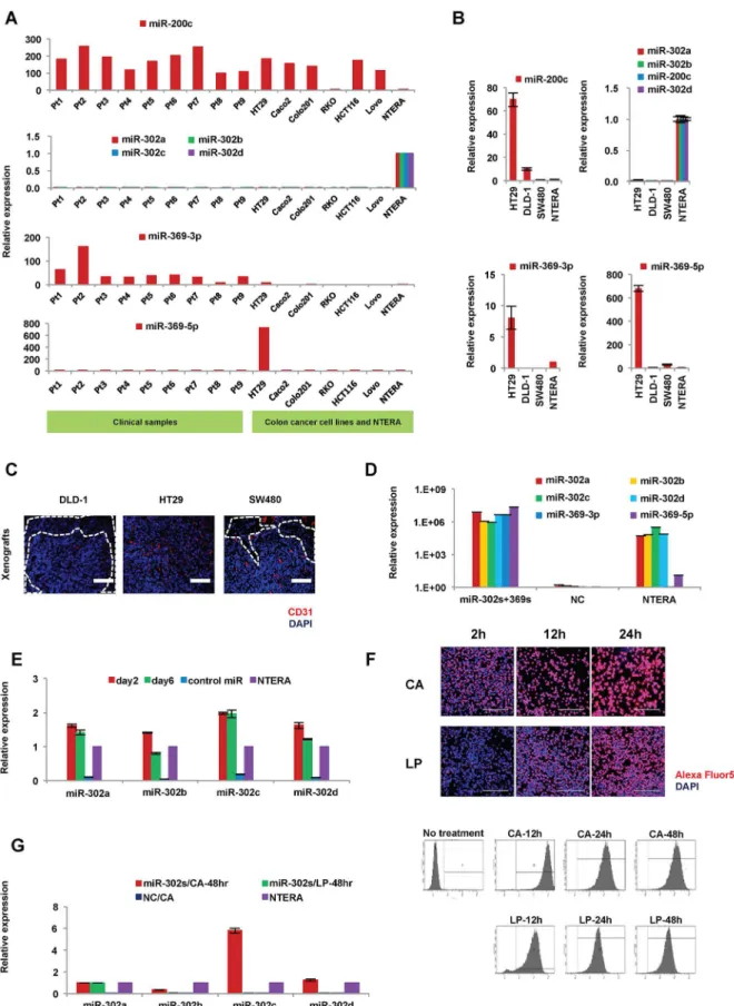
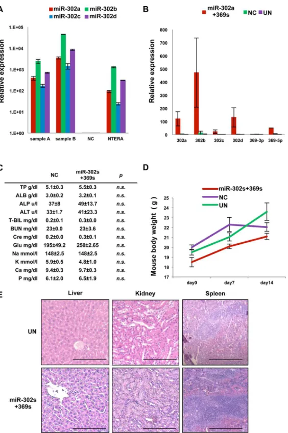
Previous studies indicated that miR-302a,-b,-c, and-d (miR-302s), miR369-3p, -5p (miR-369s) and miR-200c could elicit cellular reprogramming in normal somatic cells [13]. Here, we pal-need to reprogram cancer cells using these miRs. We started by investigating the endogenous expression of these miRs in colorectal cancer cell lines and clinical samples of colon cancer (Fig 1A and 1B). Expression of miR-302s in colon cancer was very low or undetectable com-pared to that in human teratoma NTERA cells, which reportedly express ESC-specific genes and are used as control cells to evaluate ESC-like gene expression in many studies [2,3,5,11]. In contrast, miR-200c expression was high in nine primary tumors examined and many cell lines, such as HT29. Expression levels of miR-302s and miR-369s were lower compared to that of miR-200c, suggesting that miR-200c expression is already high in many colon cancer cells and may be refractory to exogenous overexpression. Next, we transfected only miR-302s or miR-369s in colon cancer cells. To optimizein vivoexperiments, which mimic primary tumors,
we studied the immunofluorescent staining patterns of CD31, a vascular endothelial marker, using DAPI to counterstain nuclei, in xenografts of HT29, DLD-1 and SW480 cells (Fig 1C). HT29 xenografts had the most abundant vascularization, was uniformly distributed over the whole tumor, and the least amount of necrosis. Further cancer reprogramming analysis by miR-302s and miR-369s was performed mainly with HT29 cells and other supplementary cell lines.
Exogenous introduction of miR-302s and miR-369s elicits cellular
reprogramming
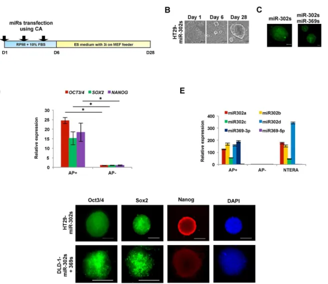
We transfected miR-302s plus miR-369s into colon cancer cell lines using CA to achieve miR expression levels similar to those of NTERA cells, which were used as a positive control (Fig 1D) [13]. We confirmed that miR-302s were effectively transfected (Fig 1E). Transfection efficiency using CA was higher than that of LP under our conditions (Fig 1F and 1G). There-fore, the CA method was used in all subsequent experiments.
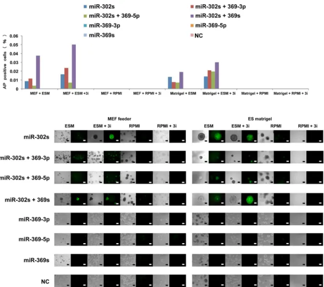
The present study indicates that AP imaging is a suitable method to analyzed not only so-matic cellular reprogramming but also cancer cell reprogramming [5]. The present study also shows that cells transfected with miR-302s plus miR-369s and cultured on MEF feeder cells in ES-certified medium containing 3i exhibit a marked increase in AP+ES-like colonies compared with those transfected with only miR-302s. In the absence of miRs or culturing in normal media, there is a decrease in the appearance of AP+colonies (Fig 3). The data suggest that miR-302s play an important role in reprogramming cancer cells in collaboration with miR-369s.
SW480 cells. HT29 xenografts exhibited many CD31-positive areas with little necrosis. Scale bars, 200μm. D) Relative expression of 302s and miR-369s in HT29 cells 24 h post-transfection of 30 nM miR-302s plus miR-miR-369s (n = 3). NC, negative control. E) Relative expression of miR-302s in HT29 cells at days 2 and 6 after transfection of miR-302s (n = 3). NC, negative control. F) To evaluate transfection efficiency, a fluorescently labeled dsRNA oligomer was transfected with carbonate apatite (CA) or lipofection (LP) into DLD-1 cells. Transfection efficiency was assessed by fluorescent microscopy and flow cytometry after a single transfection, as indicated. G) Relative expression of miR-302s in HT29 cells 48 h post-transfection by CA or LP (n = 3).
doi:10.1371/journal.pone.0127119.g001
Fig 2. Characteristics of colon cancer cells upon reprogramming induced by miR-302s or miR-302s plus miR-369s.A) Scheme of cancer reprogramming methods by transfection of miR-302s or miR-302s plus miR-369s. B) miR-302s-induced morphological changes in HT29 cells at days 1, 6, and 28. Reprogrammed HT29 have an embryonic stem cells-like appearance. Scale bars, 100μm. C) Cells stained using alkaline phosphatase (AP) live stain. Scale bars, 100μm. D) Relative expression ofOCT3/4,SOX2andNANOGcompared to AP−cells by qRT-PCR (n = 3). E) Relative expression of
miR-302s and miR-369s in AP+, AP−, and NTERA cells. Mean expression of each miR was compared to that in NTERA cells (n = 3). F) Immunofluorescence of
pluripotent stem cell markers Oct3/4, Sox2, and Nanog. Nuclei were stained with DAPI. Scale bars, 100μm (original magnification, ×200).
This may be facilitated by culturing in the presence of 3i on MEF feeder cells in ES-certified medium. We were therefore able to optimize the reprogramming conditions for cancer cells by utilizing synthesized compounds.
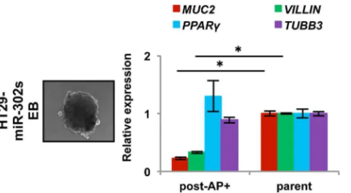
Reprogramming cell differentiation of colon cancer
Given that cellular reprogramming contributes cell pluripotency, we investigated whether miR-induced AP+cancer cells are pluripotent. We examined EB formation in floating cultures for 4–5 days, after which the EBs were cultured on 0.1% gelatin-coated dishes for an additional 14 days to induce differentiationin vitro. qRT-PCR showed that expression of endodermal
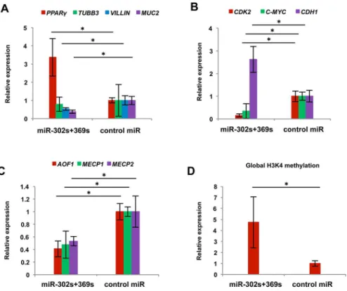
marker genes, such as VILLIN and MUC2, decreased, whereas expression of ectodermal mark-er gene TUBB3 and mesodmark-ermal markmark-er gene PPARY did not decrease. This suggests that re-programmed AP+cells have altered expression of differentiation markers in colon cancer cells, which are largely refractory to the induction of differentiation (Fig 4).
Fig 3. Cancer cell reprogramming conditions.Cells positive for the live stain alkaline phosphatase (AP) cells were counted as indicators of cancer reprogramming. HT29 cells were transfected with miRs as indicated and grown in culture under several conditions (culture medium, MEF feeder cells, and inhibitors). On day 21, AP staining was performed to evaluate reprogramming efficiency. Representative images are shown. Scale bars, 200μm.
Epigenetic reprogramming of colon cancer cells
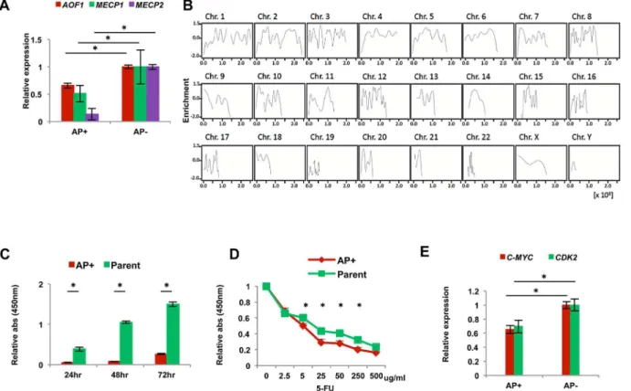
Previous reports showed that miR-302s lead to global DNA demethylation and upregulate NANOG, OCT3/4 and SOX2 via a signaling loop between miR-302s and regulatory enzymes involved in epigenetics, such as AOF1/AOF2, MECP1 and MECP2 [5,20,21]. Therefore, we compared the expression levels of those epigenetic enzyme genes between AP+and AP−
repro-grammed cancer cells. qRT-PCR showed that AP+cells expressed significantly lower levels of AOF1, MECP1, and MECP2 compared to AP−cells (Fig 5A). miR-302s control the level of
DNA demethylase, and we suspect that global demethylation is induced by miR-302s transfec-tion. Therefore, we performed immunoprecipitation using an anti-DNA methylation binding protein antibody with miR-302s-transfected DLD-1 colon cancer cells. Precipitated DNA was subjected to next-generation, high-throughput DNA sequencing. Data were consistent with the idea that miR-302 induces global DNA demethylation, on the DNA sequence level on each chromosome, compared to the mock experiment (Fig 5B). This result suggests that epigenetic reprogramming events occur on the genome-wide level in AP+reprogrammed cancer cells. Moreover, sequence level resolution indicated that several critical TSG loci were demethylated after miR-302s introduction, compared with mock control. This could play a role in the reacti-vation of the tumor suppression mechanism in malignant colon cancer cells (representative data shown inS1 Fig). Given that the reactivation of several TSGs supposedly involved in the inhibition of cancer cell growth and the sensitization of cancer cells to chemotherapeutic agents, we studied reprogrammed colon cancer cellsin vitro. The data indicate that
repro-grammed AP+cancer cells have reduced cell proliferation (Fig 5C) and a sensitized phenotype to exposure to 5-FU, chemotherapeutic agent, in culture (Fig 5D). This is consistent with previ-ous reports in other tumors [1,5,18]. The oncogene c-MYC is often involved in somatic cellular reprogramming and is an inevitable problem in tumorigenesis [22]. The present study indi-cates that the change of malignant phenotype might be due to decreased levels of c-MYC and CDK2 in AP+cells (Fig 5E), suggesting a potential therapeutic application.
miR-302s and miR-369s induce mesenchymal-to-epithelial transition
phenotypes
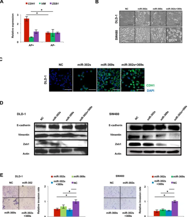
Somatic cellular reprogramming largely requires the mesenchymal-to-epithelial transition (MET), orchestrated by the suppression of pro-EMT, linked to aggressive cancer stem cell phe-notypes [23,24]. AP+cells showed an increase in CDH1 expression and a decrease in
Fig 4. Differentiation experiment with reprogrammed cancer cells.Embryoid body formation by HT29 cells induced by miR-302s. Scale bars, 100μm. qRT-PCR of the expression of differentiation markers, such as MUC2 and VILLIN (endodermal), PPARγ(mesodermal), and TUBB3 (ectodermal) in spontaneously differentiated AP+HT29 cells.
VIMENTIN expression compared with AP-cells (Fig 6A). Colon cancer cells transfected these miRs underwent a phenotype change from spindle-shaped to round-shaped in colon cancer cells (Fig 6B), consistent with an increase in CDH1 protein levels (Fig 6C and 6D). Interesting-ly, protein expression of Zeb1, a mesenchymal regulator, was also decreased in cells transfected with miR-369s (Fig 6D). An invasion assay using Matrigel-coated membranes demonstrated that transfection with miR-302s plus miR-369s resulted in a significant decrease in cancer cell invasion (Fig 6E). These results suggest that miR-302s plus miR-369s accelerate MET in colon cancer cells.
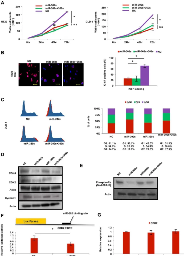
Cancer reprogramming inhibits the Cdk
–
Cyclin axis
We studied further the miR-induced effect on cell proliferation. Introduction of miR-302s and miR-302s plus miR-369s resulted in the inhibition of cell proliferation in colon cancer cells (Fig 7A). Immunocytochemistry showed that transfection of miR-302s resulted in the reduc-tion of Ki67-positive cells (Fig 7B). A cell cycle study of PI staining using flow cytometry showed that the percentage of cells in G1-phase was relatively increased compared with the NC (Fig 7C), suggesting the miR-302s inhibits entry into the S-phase of the cell cycle. Immuno-blot analysis also showed that CyclinD1, Cdk2 and Cdk4 proteins were decreased in miR-302s-transfected cells (Fig 7D), These results suggest that Cyclin/Cdk proteins are involved in
Fig 5. Phenotype of reprogrammed AP+and AP-cancer cells.A) Relative qRT-PCR expression of epigenetic regulators AOF1, MECP1 and MECP2 in
AP+cells compared with AP−cells (n = 3). B) Demethylation data was obtained from miR-302s-transfected DLD-1 cells by immunoprecipitation and
sequence analysis. Data indicate the fold of enrichment of methylation, given as a logarithmic ratio for each chromosome. Negative values indicate demethylation events upon miR-302s transfection compared to transfection with the NC. Each solid line is smoothly joined all over the chromosome. C) Cell proliferation assay with AP+and parent cells (n = 12). Data are shown in comparison with the parental cells at 24, 48 and 72 h. D) 5-fluorouracil sensitivity
assay of AP+and parent cells (n = 4). E) Relative qRT-PCR expression of the cell cycle-related genes CDK2 and C-MYC compared with AP−cells (n = 3).
Asterisk denotes a p-value in the Student’s t-test of less than 0.05 (mean±s.e.m.).
miR-dependent cell cycle inhibition. To address this hypothesis further, we studied the phos-phorylation of the Rb protein, a target of Cyclin/Cdk. Immunoblotting showed that miR-302s markedly inhibited Rb phosphorylation (Fig 7E). To determine whether miR-302s directly
Fig 6. miR-302s plus miR-369s induce the mesenchymal-to-epithelial transition phenotype.A) Relative qRT-PCR expression of MET-related genes CDH1, VIMENTIN (VIM) and ZEB1 compared with AP−cells (n = 3). B) Change in morphology of DLD-1 and SW480 cells after transfection of each miR.
Scale bars, 100μm. C) Immunofluorescence analysis of MET-related protein CDH1 in DLD-1 cells 48 h after each miR transfection. Nuclei were counterstained by DAPI. Scale bars, 100μm. D) Immunoblotting for MET-related proteins E-cadherin, Vimentin, and Zeb1. Actin was used as a loading control. Left, DLD-1 cells; right, SW480 cells. E) Matrigel invasion assay of DLD-1 cells and SW480 cells. Scale bars, 100μm. Relative invasion rates were determined by counting the number of cells that invaded through Matrigel compared to that in the mock control miR. Data are obtained from three independent experiments. Asterisk denotes a p-value in the Student t-test of less than 0.05 (mean±s.e.m.).
controls the 3’UTR of the CDK2 gene, we performed a luciferase reporter assay. The result showed that relative firefly luciferase activity of reporter plasmids containing the CDK2 3’
UTR binding site was significantly inhibited in colon cancer cells transfected with miR-302s (Fig 7F), indicating that miRs inhibits Cdk2 protein (Fig 7D), but not CDK2 mRNA (Fig 7G). These data suggest that miR-mediated reprogramming modulates the Cyclin–Cdk axis, a mechanism critical in cancer cell proliferation.
Induction of apoptosis in colon cancer cells
Since cell death was observed during the initial transfection period, we examined apoptosis using the fluorescent TUNEL assay. Data showed that transfection with the miRs induced the development of TUNEL-positive cells (A inS2 Fig). The results were confirmed by Annexin V staining, which demonstrated that miR-302s induced early and late apoptosis (B inS2 Fig). Im-munoblotting showed that expression of the apoptosis-related Bcl-2-family protein Mcl-1 was decreased in miR-302s-transfected colon cancer cells (C inS2 Fig), suggesting an involvement of the mitochondrial Bcl-2-family in miR-302s-induced apoptosis.
miR-mediated cancer reprogramming therapy
in vivo
The resultsin vitroshow that miR-302s and miR-369s play a role as tumor suppressor miRs
and influence cancer reprogramming, acceleration of MET, inhibition of the Cyclin/Cdk axis, suppression of cell growth, and apoptosis. Therefore, we also analyzed the effect of these miRs
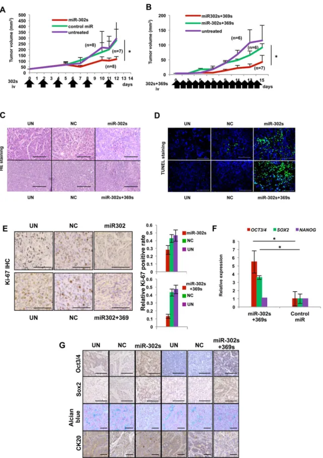
in vivo. Because miRs are unstablein vivo, miR-302s was administered by injection into the tail
vein six times. Systemic administration of miR-302s resulted in the inhibition of tumor growth compared with the control miR and a mock experiment as controls (Fig 8A). We then studied the combination of miR-302s and miR-369s by injecting these miRs into the tail vain 14 times. Administration of miR-302s and miR-369s also resulted in the inhibition of tumor growth compared with the control miR and experimental control groups (Fig 8B). Those two experi-ments demonstrated that significant tumor growth inhibition occurs when mice are with miR-302s alone or miR-miR-302s and miR-369s compared with controls, without apparent adverse ef-fects. Histological examination of these tumor tissues showed a focal involvement of cell debris with denatured nuclei in tumors (Fig 8C). TUNEL staining confirmed DNA fragmentation, a characteristic of apoptosis (Fig 8D). The number of Ki67-positive cells was significantly de-creased in miRs-treated tumors compared with NC miR tumors (Fig 8E). In terms of tumor differentiation, it was revealed that miR-302s plus miR-369s-treated tumors were strongly pos-itive for Oct3/4 and Sox2 (Fig 8F and 8G). Alcian blue staining for goblet-like cell differentia-tion showed a decrease in mucus producdifferentia-tion in tumors treated with 302s alone or miR-302s plus miR-369ss (Fig 8G). CK20, a differentiation marker for colon adenocarcinoma, was also repressed in tumors treated with miR-302s plus miR-369ss (Fig 8G).
transfected with miR-302s, miR-302s plus miR-369s, or negative control (NC) miR. Positively stained-cells were counted from three random fields in three independent experiments. Scale bars, 100μm (original magnification, ×200). C) Cell cycle analysis of DLD-1 cells transfected with miR-302s, miR-302s plus miR-369s, or NC miR. Cells were stained by propidium iodide. Left, representative image; right, median cell populations (%) in three cell cycle phases (G1, S and G2) from three independent experiments. D) Immunoblotting of CDK4, CDK2, and CyclinD1 in DLD-1 cells transfected with 302s, 369s, miR-302s plus miR-369s, or NC miR. Actin was used as a loading control. E) Immunoblotting with anti-phosphorylated Rb (Ser807/811) antibody in DLD-1 cells transfected with miR-302s, miR-369s, miR-302s plus miR-369s, or NC miR. Actin was used as a loading control. F) Schematic of CDK2-3’-UTR-containing reporter constructs. A luciferase reporter assay with DLD-1 cells transfected with a luciferase-CDK2 3’UTR vector and miR-302s was performed. NC, negative control. Data were normalized by empty vector co-transfection and were obtained from three independent experiments. G) qRT-PCR of CDK2 expression in DLD-1 cells 48 post-transfection with miR-302s, miR-369s, or NC miR (n = 3). Relative ratios are shown. Asterisk denotes a p-value in the Student t-test of less than 0.05 (mean±s.e.m).
Fig 8. Anti-tumor effect by systemic administration of miR-302s plus miR-302sin vivo.A) Tumor-bearing mice were treated with systemic
qRT-PCR indicated that expression of MUC2 and VILLIN, enterocyte-like cell differentia-tion markers associated with axial microfilaments and conserved in adenocarcinoma [25], de-creased, whereas expression of PPARY increased (Fig 9A). These results showed miR-302s plus miR-369s modulate the colonic differentiation state and could elicit a shift toward another line-agein vivo. Moreover, we confirmed the decrease of CDK2 and c-MYC and increase of CDH1
expression at the mRNA levelin vivo, consistent with the resultsin vitro(Fig 9B). We next
studied the alteration of epigenetic regulators in tumors treated with miR-302s plus miR-369ss. Results showed thatAOF1,MECP1 and MECP2expression levels were lower in miR-302s plus
miR-369ss-treated tumors (Fig 9C). Therefore, we studied global H3K4 methylation. The re-sults indicated that H3K4 methylation was increased in miR-302s plus miR-369s-treated tu-mors (Fig 9D) and suggested that miR-302s plus miR-369s may decrease AOF1, MECP1 and MECP2 expression, leading to global epigenetic modification [20].
We performed qRT-PCR to investigate whether systemic administration of miRs was dis-tributed into the deep areas of tumors. The data indicated that systemic administration of miRs led to excellent distribution in xenografts (Fig 10A and 10B). Although systemic administra-tions of miR-302s plus miR-369s were repeated for 14 consecutive days, we did not detect any noticeable adverse effects in laboratory blood tests, body weight or histology of the liver, kidney and spleen (Fig 10C, 10D and 10E).
Discussion
Epigenetic events are critical in dictating cellular differentiation and reprogramming of somatic and cancer cells [1,2]. The present study showed that the combination of 302s plus miR-369s could modulate biological behaviors of colorectal cancer cells. A state-of-the-art chroma-tin immunoprecipitation sequencing experiment demonstrated that global epigenetic alter-ations such as DNA demethylation occur in miR-302s-transfected colorectal cancer cells. This may be beneficial in reactivating tumor suppressor genes, usually inactivated during the estab-lishment of cancer through a complex series of epigenetic events [1]. We also found that ad-ministration of miRs resulted in methylation of H3K4, an active marker of chromatin that is often inactivated by abnormal epigenetic control by the p16/INK4A tumor suppressor. Report-edly, H3K4 plays a critical role in the cell cycle control of cancer stem cells, acting to slow down cycling [1]. Given the present study showed that a combination of 302s and miR-369s induced epigenetic modulations of histones and DNA, there may be a justification for the use of these miRs as novel therapeutic agents.
Here, we used miR-302s and miR-369s, based on expression analysis, to boost endogenously low expression with exogenously high expression. In this regard, we did not use miR-200c in this study, since the additional expression of miR-200c in endogenously miR-200c-expressing cancer cells did not indicate an apparent anti-cancer effect. One of the common features shared by these three miRs, which can induce cellular reprogramming in somatic cells [11], is the
of CA-equipped miR-302s plus miR-369s, NC miR, or CA only daily, with a total of 14 injections, similar to (A). C) Representative hematoxylin and eosin-stained images of tumor sections from the indicated experimental groups. Areas with denaturated nuclei were predominantly observed in xenografts treated with miR-302s/CA complexes and miR-302s plus miR-369s/CA complexes. Scale bars, 100μm (magnification ×200). D) Confirmation of intratumoral apoptosis by TUNEL staining in tumor sections obtained from xenografts treated with CA-mediated miR-302s, miR-302s plus miR-369s, NC, or CA only. Scale bars, 100μm (magnification ×200). E) Immunohistochemistry analysis of the proliferation marker Ki-67 in xenografts treated with miR-302s, miR-302s plus miR-369s, NC miR or the mock control. Representative images are shown. Scale bars, 100μm (magnification ×200). Ki-67-positive cells were counted in three random fields from different three xenografts in each group. F) Relative expression of pluripotency-related markers OCT3/4, SOX2 and NANOG in the indicated experimental groups. Data are shown in comparison with the NC miR group. (n = 3). G) Representative immunohistochemical images of tumor sections from the indicated experiment groups. Oct3/4 and Sox2 were predominantly observed in xenografts treated with 302s and 302s plus miR-369s. Alcian blue staining showed the decrease of mucin production in these miR-treated-groups. CK20 expression was also decreased in xenografts treated with miR-302s plus miR-369s. Scale bars, 100μm (magnification ×200).
ability to inhibit the epithelial–to–mesenchymal transition (EMT). Reportedly, the miR-200 family is recognized as a master regulator of the epithelial phenotype by targeting ZEB1 and ZEB2, two important transcriptional repressors of cell adherence (E-cadherin) and polarity (CRB3 and LGL2) genes [22]. The miR-200 family has also been shown to inhibit the EMT, stimulating the epithelial phenotype and cancer cell migration by direct targeting of the E-cad-herin transcriptional repressors ZEB1 and ZEB2 [23]. The miR-302s target several important molecules, such as the receptor for TGFβ(α στρονγ ινδυχερ οϕEMT), histone H3K4 demethy-lase [24], and Cdk2/4/6 [12]. The miR-369s alone inhibit the EMT and stimulate the epithelial phenotype. The present study indicated that miR-369s inhibit the EMT in collaboration with miR-302s, which is compatible with a database search that indicates that miR-369s target ZEB1 (http://www.microrna.org/microrna/home.do). We hypothesize that miR-369s exert part of their function of EMT inhibition in collaboration with other miRs. Thus, similarly to iPS in-duction stimulated by the epithelial program [25], inhibition of the mesenchymal phenotype by these miRs may be beneficial in eradicating the metastatic potential of cancer. Eventually, the introduction of miR-302s should result in the induction of reprogramming [5] and the sup-pression of the malignant potential of tumors [12].
Considering cellular reprogramming,‘the c-Myc issue’may be important. It has been re-ported that c-Myc is involved in reprogramming events and inevitably activated during the course of cellular reprogramming through virus-mediated reprogramming, which can lead to
Fig 9. Gene expression of reprogrammed cancer cellsin vivo.A) Relative expression of the
differentiation-related genes PPARγ, TUBB3, VILLIN and MUC2 in the indicated experimental groups. Data are shown in comparison with the negative control (NC) miR group. (n = 3). B, C) Relative expression of CDK2, C-MYC and CDH1 (B) and epigenetic regulators AOF1, MECP1 and MECP2 (C) in the indicated experimental groups. Data are shown in comparison with the NC miR group. (n = 3). D) Relative global histone 3 lysine-4 methylation status of xenografts. Data are shown in comparison with the NC miR group. (n = 3). Asterisk denotes a p-value in the Student t-test<0.05 (mean±s.e.m.).
carcinogenesis [22,26]. The present study indicates that a combination of 302s and miR-369s results in the suppression of endogenous c-Myc and inactivates tumors. Thus, miR-based reprogramming may be beneficial, given that a combination of miR-302s and miR-367 could carry out reprogramming without apparent c-Myc alterations [8,9,11,20,21]. The present study shows that introduction of the miRs resulted in sensitization to 5-FU, induction of the G0/ G1-phases of the cell cycle, and induced apoptosis via the mitochondrial Bcl-2 family. Anin vivostudy indicated that miRs modulated the differentiation process, and may direct other
dif-ferentiation lineages and inactivate malignant potential, as detected by difdif-ferentiation markers CK20 and Alcian blue. Thus, we suspect that reprogramming therapy may overcome resistance against conventional chemotherapy by epigenetic modifications, although further studies are undoubtedly necessary. In the application of nucleotide medicinein vivo, a proper drug
deliv-ery system may facilitate targeting efficiency against cancer stem cells [27,28]. Chemical modi-fications, such as bridged nucleotides, might be beneficial in increasing the anti-cancer effects and decreasing off-target effects [27,28]. Since small RNAs are unlikely to be incorporated into DNA strands in the nucleus, a reprogramming strategy may be worth considering as a novel future treatment strategy [27,28].
Supporting Information
S1 Fig. DNA methylation of around tumor suppressor gene (± 10 Kb).Each solid lines are fitted by Gaussian function. The intensities and the deviation of function correspond to fold enrichment and the detected sequence range, respectively.
(TIF)
S2 Fig. Apoptotic induction.A, Fluorescent TUNEL staining was performed to detect apopto-tic HT29 cells transfected with miR302, miR302 plus miR-369s, or negative control (NC) miR. Apoptotic cells are indicated by an arrow. B, Propidium iodide and Annexin V-FITC staining was performed in DLD-1 cells 60 h post-transfection with miR302, miR-369s, miR302 plus miR-369s, or NC miR. Apoptotic cells were measured by flow cytometry. Early (Annexin-posi-tive only) and late (both Annexin and PI-posi(Annexin-posi-tive) apoptotic cells were detected. Three inde-pendent experiments were performed. C, Immunoblotting of the apoptosis-related proteins Bak, Bid, Bcl-xl, Bcl2, Mcl1, Caspase-8, and Caspase-3 in HT29 and DLD-1 cells transfected with miR302, miR-369s, miR302 plus miR-369s, or NC miR. Actin was used as a loading con-trol. Asterisk denotes a p-value in the Student t-test of<0.05 (mean ± s.e.m.).
(TIF)
S1 Table. Sequence of mature miRNAs.
(TIFF)
S2 Table. Patients characteristics of clinical samples.Stage was according to TNM classifica-tion (UICC 7th) Abbreviaclassifica-tions; CEA.carcinoembryonic antigen, CA19-9.carbohydrate antigen 19–9, RS. Rectosigmoid, Ra. Upper rectum, Rb.Lower rectum A. Ascending, D. Descending, S. Sigmoid, wel. well differentiated adenocarcinoma, mod. moderately differentiated adenocarci-noma.
(TIFF)
Acknowledgments
funding sources played no role in data collection and analysis, decision to publish, or prepara-tion of the manuscript.
Author Contributions
Conceived and designed the experiments: HI MM K. Kataoka MK. Performed the experiments: HO KN N. Nishida MK NG KM TY N. Nishiyama HN HY SO K. Kawamoto YD. Analyzed the data: JK HM. Contributed reagents/materials/analysis tools: JK HM XW. Wrote the paper: HO HI MM MK XW.
References
1. Miyoshi N, Ishii H, Nagai K, Hoshino H, Mimori K, Tanaka F, et al. Defined factors induce reprogram-ming of gastrointestinal cancer cells. Proc Natl Acad Sci USA 2010; 107: 40–45. doi:10.1073/pnas. 0912407107PMID:20018687
2. Ohnishi K, Semi K, Yamamoto T, Shimizu M, Tanaka A, Mitsunaga K, et al. Premature termination of reprogramming in vivo leads to cancer development through altered epigenetic regulation. Cell 2014; 156: 663–677. doi:10.1016/j.cell.2014.01.005PMID:24529372
3. Zhang X, Cruz FD, Terry M, Remotti F, Matushansky I. Terminal differentiation and loss of tumorigenici-ty of human cancers via pluripotency-based reprogramming. Oncogene 2013; 32: 2249–2260, 2260. e2241-2221. doi:10.1038/onc.2012.237PMID:22777357
4. Lin SL, Chang DC, Chang-Lin S, Lin CH, Wu DT, Chen DT, et al. Mir-302 reprograms human skin can-cer cells into a pluripotent ES-cell-like state. RNA 2008; 14: 2115–2124. doi:10.1261/rna.1162708
PMID:18755840
5. Koga C, Kobayashi S, Nagano H, Tomimaru Y, Hama N, Wada H, et al. Reprogramming Using micro-RNA-302 Improves Drug Sensitivity in Hepatocellular Carcinoma Cells. Ann Surg Oncol 2014; 21: Suppl 4:S591–600. doi:10.1245/s10434-014-3705-7PMID:24740829
6. Tsuno S, Wang X, Shomori K, Hasegawa J, Miura N. Hsa-miR-520d induces hepatoma cells to form normal liver tissues via a stemness-mediated process. Sci Rep 2014; 4: 3852. doi:10.1038/srep03852
PMID:24458129
7. Kong YW, Ferland-McCollough D, Jackson TJ, Bushell M. microRNAs in cancer management. Lancet Oncol 2012; 13: e249–258. doi:10.1016/S1470-2045(12)70073-6PMID:22652233
8. Lanford RE, Hildebrandt-Eriksen ES, Petri A, Persson R, Lindow M, Munk ME, et al. Therapeutic silenc-ing of microRNA-122 in primates with chronic hepatitis C virus infection. Science 2010; 327: 198–201. doi:10.1126/science.1178178PMID:19965718
9. Hu S, Wilson KD, Ghosh Z, Han L, Wang Y, Lan F, et al. MicroRNA-302 increases reprogramming effi-ciency via repression of NR2F2. Stem Cells 2013; 31: 259–268. doi:10.1002/stem.1278PMID:
23136034
10. Lipchina I, Studer L, Betel D. The expanding role of miR-302-367 in pluripotency and reprogramming. Cell Cycle 2012; 11: 1517–1523. doi:10.4161/cc.19846PMID:22436490
11. Liao B, Bao X, Liu L, Feng S, Zovoilis A, Liu W, et al. MicroRNA cluster 302–367 enhances somatic cell reprogramming by accelerating a mesenchymal-to-epithelial transition. J Biol Chem 2011; 286: 17359–
17364. doi:10.1074/jbc.C111.235960PMID:21454525
12. Anokye-Danso F, Trivedi CM, Juhr D, Gupta M, Cui Z, Tian Y, et al. Highly efficient miRNA-mediated re-programming of mouse and human somatic cells to pluripotency. Cell Stem Cell 2011; 8: 376–388. doi:
10.1016/j.stem.2011.03.001PMID:21474102
13. Miyoshi N, Ishii H, Nagano H, Haraguchi N, Dewi DL, Kano Y, et al. Reprogramming of mouse and human cells to pluripotency using mature microRNAs. Cell Stem Cell 2011; 8: 633–638. doi:10.1016/j. stem.2011.05.001PMID:21620789
14. Lin SL, Chang DC, Ying SY, Leu D, Wu DT. MicroRNA miR-302 inhibits the tumorigenecity of human pluripotent stem cells by coordinate suppression of the CDK2 and CDK4/6 cell cycle pathways. Cancer Res 2010; 70: 9473–9482. doi:10.1158/0008-5472.CAN-10-2746PMID:21062975
15. Takeyama H, Yamamoto H, Yamashita S, Wu X, Takahashi H, Nishimura J, et al. Decreased miR-340 Expression in Bone Marrow Is Associated with Liver Metastasis of Colorectal Cancer. Mol Cancer Ther 2014; 13: 976–985. doi:10.1158/1535-7163.MCT-13-0571PMID:24448820
17. Hossain S, Yamamoto H, Chowdhury EH, Wu X, Hirose H, Haque A, et al. Fabrication and intracellular delivery of doxorubicin/carbonate apatite nanocomposites: effect on growth retardation of established colon tumor. PLoS One 2013; 8: e60428. doi:10.1371/journal.pone.0060428PMID:23613726 18. Miyazaki S, Yamamoto H, Miyoshi N, Wu X, Ogawa H, Uemura M, et al. A Cancer Reprogramming
Method Using MicroRNAs as a Novel Therapeutic Approach against Colon Cancer: Research for Re-programming of Cancer Cells by MicroRNAs. Ann Surg Oncol. 2014 Nov 11. [Epub ahead of print]
19. Singh U, Quintanilla RH, Grecian S, Gee KR, Rao MS, Lakshmipathy U. Novel live alkaline phospha-tase substrate for identification of pluripotent stem cells. Stem Cell Rev 2012; 8: 1021–1029. doi:10. 1007/s12015-012-9359-6PMID:22426885
20. Lin SL, Chang DC, Lin CH, Ying SY, Leu D, Wu DT. Regulation of somatic cell reprogramming through inducible mir-302 expression. Nucleic Acids Res 2011; 39: 1054–1065. doi:10.1093/nar/gkq850PMID:
20870751
21. Wang J, Hevi S, Kurash JK, Lei H, Gay F, Bajko J, et al. The lysine demethylase LSD1 (KDM1) is re-quired for maintenance of global DNA methylation. Nat Genet 2009; 41: 125–129. doi:10.1038/ng.268
PMID:19098913
22. Okita K, Ichisaka T, Yamanaka S. Generation of germline-competent induced pluripotent stem cells. Nature 2007; 448: 313–317. PMID:17554338
23. Findlay VJ, Wang C, Watson DK, Camp ER. Epithelial-to-mesenchymal transition and the cancer stem cell phenotype: insights from cancer biology with therapeutic implications for colorectal cancer. Cancer Gene Ther 2014; 21: 181–187. doi:10.1038/cgt.2014.15PMID:24787239
24. Mani SA, Guo W, Liao MJ, Eaton EN, Ayyanan A, Zhou AY, et al. The epithelial-mesenchymal transi-tion generates cells with properties of stem cells. Cell 2008; 133: 704–715. doi:10.1016/j.cell.2008.03. 027PMID:18485877
25. Kennedy MT, Jordan RC, Berean KW, Perez-Ordonez B. Expression pattern of CK7, CK20, CDX-2, and villin in intestinal-type sinonasal adenocarcinoma. J Clin Pathol 2004; 57: 932–937. PMID:
15333652
26. Nakagawa M, Koyanagi M, Tanabe K, Takahashi K, Ichisaka T, Aoi T, et al. Generation of induced plu-ripotent stem cells without Myc from mouse and human fibroblasts. Nat Biotechnol 2008; 26: 101–106. PMID:18059259
27. Bader AG, Brown D, Winkler M. The promise of microRNA replacement therapy. Cancer Res 2010; 70: 7027–7030. doi:10.1158/0008-5472.CAN-10-2010PMID:20807816