Article
J. Braz. Chem. Soc., Vol. 22, No. 6, 1125-1129, 2011. Printed in Brazil - ©2011 Sociedade Brasileira de Química
0103 - 5053 $6.00+0.00
A
*e-mail: [email protected]
Citrinin Derivatives from the Soil Filamentous Fungus
Penicillium
sp. H9318
Guangmin Yao,a,d Fred Musoke Sebisubi,b,e Lok Yung Christopher Voo,c Coy Choke Ho,c Ghee Teng Tanb and Leng Chee Chang*,a,b
aDepartment of Chemistry and Biochemistry, College of Science and Engineering,
University of Minnesota Duluth, 1039 University Drive, Duluth, MN 55812, USA
bDepartment of Pharmaceutical Sciences, College of Pharmacy, University of Hawaii Hilo,
34 Rainbow Drive, Hilo, HI 96720, USA
cBiotechnology Program, School of Science and Technology, University Malaysia Sabah,
88999 Kota Kinabalu, Sabah, Malaysia
dHubei Key Laboratory of Natural Medicinal Chemistry and Resources Evaluation, School of
Pharmacy, Tongji Medical College, Huazhong University of Science and Technology, 13 Hangkong Road, Wuhan 430030, P. R. China
eDivision of Pharmaceutical Services, Ministry of Health,
PO Box 7272 Kampala, Uganda
A investigação do extrato orgânico produzido na fermentação microbiológica de Penicillium
sp. H9318 conduziu ao isolamento de um novo alcaloide isoquinolínico, o composto (5S)-3,4,5,7-tetrametil-5,8-di-hidróxi-6(5H)-isoquinolinona (1), juntamente com quatro outros conhecidos compostos derivados da citrinina (2-5). Uma atividade inibitória signiicativa no ensaio da inibição da formação dos halos (HFI), foi exibida pela citrinina (2), semelhante àquela observada pelo Streptomyces 85E, enquanto os compostos 1, 3, 4 e 5 não mostraram atividade inibitória com respeito ao ensaio HFI quando testados com 20 µg/disco. Em relação ao ensaio de citotoxicidade, a citrinina (2) demonstrou uma atividade inibitória mais fraca quando comparada as linhagens de células cancerosas MCF-7 (IC50 71,93 µmol L−1), LNCaP (IC
50 77,92 µmol L
−1), LU-1 (147,85 µmol L−1) e KB (IC
50 65,93 µmol L
−1).
Investigation of a microbial fermentation organic extract of Penicillium sp. H9318 led to the isolation of a new isoquinolinone alkaloid, (5S)-3,4,5,7-tetramethyl-5,8-dihydroxyl-6(5H )-isoquinolinone (1), along with four known citrinin derivatives (2-5). Citrinin (2) exhibited signiicant inhibitory activity against Streptomyces 85E in the hyphae formation inhibition (HFI) assay, while compounds 1, 3-5 were not active when tested at 20 µg/disk in the HFI assay. Citrinin (2) further demonstrated a weak inhibitory activity against MCF-7 (IC50 71.93 µmol L
−1), LNCaP (IC50 77.92 µmol L−1), LU-1 (147.85 µmol L−1) and KB (IC
50 65.93 µmol L
−1) cell lines, respectively, in the cytotoxicity assay.
Keywords: Streptomyces 85E, isoquinolinone alkaloid, citrinin, Penicillium sp. H9318, kinase inhibitor
Introduction
The phosphorylation of proteins on serine/threonine and tyrosine residues by protein kinases is one of the major regulatory mechanisms in biological processes including apoptosis, cell proliferation, cell differentiation,
and metabolism.Deregulated phosphorylation associated with these pathways can result from genetic alterations acquired early in tumorigenesis, and are often the cause of cancer. In this regard, protein kinases have emerged as promising inhibitory targets in cancer treatment.1,2 Aerial
During the course of our search for novel protein kinase inhibitors from natural resources,5-10 an
acetone-soluble extract of the fungus Penicillium sp. H9318 showed inhibitory activity towards both mammalian protein phosphatase 1 (PP1) and protein phosphatase 2A (PP2A) in an in vitro phosphatase assay.5 The
n-BuOH-soluble extract exhibited inhibitory activity in the hyphae formation inhibition (HFI) assay with Streptomyces 85E giving a 20 mm clear zone of inhibition at 80 µg/disk. These preliminary results encouraged us to investigate the fungus Penicillium sp. H9318 for the discovery of novel protein kinase inhibitors. A bioassay-directed fractionation of the organic extract has resulted in the isolation of a novel isoquinolinone alkaloid 3,4,5,7-tetramethyl-5,8-dihydroxyl-6(5H)-isoquinolinone, namely, isoquinocitrinin A (1), and four known compounds, citrinin (2), penicitrone A (3), penicitrinols A (4) and B (5). Herein, we report the isolation, structure elucidation, and biological activities of compounds 1-5.
Results and Discussion
The fungus Penicillium sp. H9318 was isolated from a soil sample collected at a heath forest in Maliau Basin, Sabah, Malaysia. The CHCl3-soluble partition (26.6 g) of the n-BuOH extract of fermented Penicillium sp. H9318 signiicantly inhibited the growth of hyphae formation in Streptomyces 85E at a concentration of 80 µg/disk. A bioassay-guided isolation of the active fractions by repeated Sephadex LH-20 and silica gel MPLC afforded compounds 1-5. The known compounds
2-5 were identiied as citrinin (2),11-13 penicitrone A (3)
(also known as dicitrinin A),14 penicitrinol A (4) 15 and
penicitrinol B (5),16 respectively, by spectroscopic analysis
and comparison with literature data.
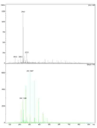
Compound 1 was obtained as a colorless oil, with the molecular formula C13H15NO3 determined from the 1H and
13C NMR data (Table 1) and HRESIMS (m/z 234.11248
[M+H]+, calc. 234.11247), indicating seven degrees of
unsaturation. The UV absorption bands at λmax 249, 269, 293, 307, 330 nm indicated the presence of an extended conjugated chromophore. The 1H NMR spectrum of
1 (Table 1) displayed signals of an aromatic proton at
dH 8.85 (1H, s, H-1), three aromatic methyls at dH 2.66
(3H, s, H-10), 2.57 (3H, s, H-9), 1.89 (3H, s, H-12); and a tertiary methyl at dH 1.62 (3H, s, H-11). The
13C NMR
and DEPT spectrum exhibited 13 carbon signals, including one carbonyl (dC 188.5), one sp
2 methine (d
C 143.9), six sp 2
quaternary carbons (dC 178.8, 161.1, 152.4, 131.9, 126.0
and 108.5), one oxygen-bearing sp3 quaternary carbon
(dC 73.6), and four methyls (dC 29.3, 22.9, 16.8 and 8.3).
One carbonyl and four double bonds from the 13C NMR
spectra accounted for ive degrees of unsaturation, thus the remaining two degrees of unsaturation requires the presence of two rings in 1.
Comparison of its 1H and 13C NMR data (Table 1) with
those of citrinin (2) showed that these compounds differ in the presence of C-3 and C-4 sp2 quaternary carbons (d
C 161.1
and 131.9) in 1 rather than the C-3 and C-4 sp3 methines
(dC 82.0 and 33.4) in 2. The
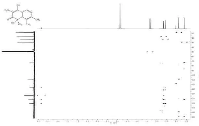
1H and 13C assignments of 1 were
conirmed by analysis of the HMBC spectrum (Figure 1). Long-range correlations were observed between the signal of H-1 (dH 8.85) and C-3 (dC 161.1); between CH3-9 (dH 2.57)
and C-3 (dC 161.1), C-4 (dC 131.9); as well as between CH3-10
(dH 2.66) with C-3 (dC 161.1), C-4 (dC 131.9) and C-4a
(dC 152.4). Another major difference in the
13C NMR spectrum
of compounds 1 and 2 was that C-12 was changed from a carbonyl group (dC 174.0) in 2 into a methyl group (dC 8.3)
in 1. Also, HMBC correlations of 1 from CH3-12 (dH 1.89, s)
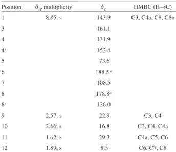
Table 1. NMR spectroscopic data for compounds 1 in CD3OD (500 MHz)
Position dH,multiplicity dC HMBC (H→C)
1 8.85, s 143.9 C3, C4a, C8, C8a
3 161.1
4 131.9
4a 152.4
5 73.6
6 188.5a
7 108.5
8 178.8a
8a 126.0
9 2.57, s 22.9 C3, C4
10 2.66, s 16.8 C3, C4, C4a
11 1.62, s 29.3 C4a, C5, C6
12 1.89, s 8.3 C6, C7, C8
to C-6 (dC 188.5), C-7 (dC 108.5), and C-8 (dC 178.8) revealed
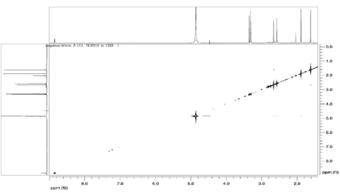
that the methyl C-12 was located at C-7. Furthermore, the cross peaks of the methyl CH3-11 (dH 1.62, s) with C-4a (dC 152.4),
C-5 (dC 73.6) and C-6 (dC 188.5), along with a NOESY
correlation observed between CH3-11 and CH3-10 conirmed that the methyl C-11 was connected to an oxygen bearing sp3
quaternary carbon at C-5. In consideration of the molecular formula and the 13C NMR chemical shift, C-5 (d
C 73.6) and
C-8 (dC 178.8) were substituted by hydroxyl groups, and a N
atom must be assigned to position 2. In spite of our utmost efforts, the absolute coniguration at C-5 of compound 1 has yet to be ascertained. Therefore, compound 1 was elucidated as 3,4,5,7-tetramethyl-5,8-dihydroxyl-6(5H)-isoquinolinone and named isoquinocitrinin A.
Several similar novel isoquinoline alkaloids have been reported from the genus of Penicillium,17 Aspergillus,18
Streptomyces19,20 and Chaetomium.21 Most isoquinoline
alkaloids discovered from plants exhibit complicated structures, which were mainly synthesized from tyrosine, whereas most isoquinolines from lichens, fungi, and sponges show simple skeletons and are probably biosynthesized via mixed pathways.21
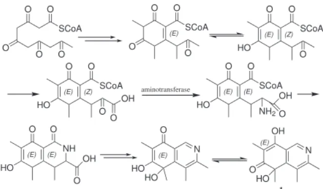
Compound 1 possesses a carbon skeleton similar to citrinin (2), and it is possibly biosynthesized via mixed routes. The proposed biosynthesis of compound 1 proceeds via an enzyme-bonded poly-β-ketone chain22 and the amino
acid formation is accomplished by an aminotransferase (Scheme 1).
Citrinin (2) was irst isolated from P. citrium in 1931 and it has been isolated from ten or more species of Penicillium and Aspergillus. Compounds 3-5 are known citrinin dimer derivatives. Until now, more than ten citrinin dimers have
been discovered from natural resources.14,16 This is the
irst report of an isoquinolinone alkaloid from the genus Penicillium possessing a carbon skeleton similar to that of citrinin.
Compounds 1-5 were evaluated for their inhibitory activities against Streptomyces 85E in the hyphae formation inhibition assay, according to an established protocol.4 Compound 2 exhibited signiicant inhibitory
activity and gave a 21 mm clear zone of inhibition (ZOI) at 80 µg/disk, 17 mm clear ZOI at 40 µg/disk, 12 mm ZOI at 20 µg/disk, and a 10 mm bald ZOI at 2.5 µg/disk. All other isolates were inactive. It is hypothesized that compounds which inhibit hyphae formation in Streptomyces 85E may block the proliferation of cancer. In the human breast cancer MCF-7 cells, proliferation can be blocked in numerous ways, including serum deprivation, pharmacological inhibition of speciic kinase and steroid hormone pathways.23,24 Therefore, compound
2 was further evaluated for its cytotoxicity activity using several cancer cell lines. Compound 2 demonstrated a weak activity with the MCF-7 (IC50 71.93 µmol L−1 or
18 µg mL−1), LNCaP (IC
50 77.92 µmol L
−1 or 19.5 µg mL−1),
and LU-1 (147.85 µmol L−1 or 37.0 µg mL−1), KB cells
(IC50 65.93 µmol L−1 or 16.5 µg mL−1), respectively.
Citrinin (2), a well-known mycotoxin, is both nephrotoxic13 and carcinogenic.25 It was reported to show
moderate cytotoxicity against the mouse NS-1 cell line with LD99 values of 25 µg mL−1,15 and antifungal activities
against Candida albicans, Cryptococcus neoformans, Aspergillus fumigatus, and A. niger at a concentration of 100 µg/disk.14 It was documented too that penicitrone
A (also known as dicitrinin A) (3) showed moderate cytotoxicity against the mouse NS-1 cell line with LD99 values of 6.3 µg mL−1, 15 and displayed scavenging activity
with IC50 values of 55.3 mmol L−1,16 while penicitrinol B(5)
did not show cytotoxicity against P388, A-549, BEL-7402, and HL-60 cells (IC50> 50 µmol L−1).16
Experimental
General procedures
Optical rotations were measured using a JASCO P-1010 automatic polarimeter. UV spectra were measured on a HP 8453 UV-Visible spectrophotometer. FT-IR spectra were acquired on a Perkin-Elmer Spectrum BX spectrometer using AgCl ilm. Mass spectra and high-resolution mass spectra were taken with a BioTOF II ESI mass spectrometer. 1D and 2D NMR spectra were recorded in MeOH-d4 and DMSO-d6 on a Varian INOVA Unity (500 MHz) spectrometer. The chemical shift
Scheme 1. Proposed biogenesis pathway of compound 1.
O SCoA O O O O O SCoA O O O (E) O SCoA O (E) HO O (Z) N O (E) HO HO O SCoA O (E) HO NH2 (E) OH O aminotransferase O SCoA O (E) HO O (Z) OH O O O (E) HO (E) OH O NH N (E) OH O HO 1
Figure 1. Key HMBC correlations (H→C) for compound 1.
(d) values are given in ppm with the residual CD3OD signals (3.31 ppm for 1H and 49.15 ppm for 13C) as
internal standard, and the coupling constants (J) are in Hz. Chromatographic fractions and pure compounds were monitored by TLC, detected by absorption of UV light at 254 nm and a color reaction by spraying with a solution of 10%, v/v, sulfuric acid/EtOH followed by 5 min heating at 120 °C. MPLC (medium pressure column chromatography) was carried out on a Büchi pump system and a Büchi column packed with Merck silica gel 60 and/or reversed-phase C18.
Fungal material
The fungal strain H9318 was isolated from the soil sample (MB 75) collected under an unidentiied tree in a “Kerangas” or a heath forest at about 3000 feet (900 meters) above sea level located within the rim perimeter (south side) of Maliau Basin, Sabah,26 Malaysia in May 2001. The
morphological characteristics of the Penicillium structure of H9318 as observed through light microscopic are circular conidia, the Ampulliforum philiades are shorter than the metula, 3 to 4 metulas were observed to branch from a single smooth stipe (of which some metulas were non-uniform in length). These characteristics points most likely to the subgenus Furcatum.27 The strain is maintained in the
Laboratory Collection at the University of Malaysia Sabah. The isolation medium used was dichloran rose bengal chloramphenicol (DRBC) medium with NaCl (2.5%) at pH 5.6. Fungi were puriied, and the producing strain was prepared on potato dextrose agar slants containing NaCl 10%, m/v, and stored at 4 °C. Conidia of single colonies of microfungi were kept in anhydrous silica gel particles at 4 °C. It was then ready to inoculate to a seed medium.
Fermentation
The composition of the seed and production medium (in g L−1) was as follows: yeast extract (10), peptone (10),
sucrose (10), KH2PO4 (1), and MgSO4•7H2O (0.3). The seed medium was prepared with distilled water, and the pH was adjusted to 5.5 prior to sterilization. The medium was dispensed at 50 mL per 250 mL in Belco bafled shaker lasks. A single colony of Penicillium from the agar plate was used as inoculum into each lask of seed media, cultured at 30 °C at 250 rpm for two days. An aliquot (1.5%) strain H9318 from the seed medium was inoculated into the production medium (similar composition) and were incubated at 30 °C at 220 rpm for seven days and were then harvested.
Extraction and isolation
The microfungi fermentation culture (90 L) was centrifuged, and the supernatant was iltered and partitioned with n-BuOH. Both the n-BuOH-soluble and aqueous layers were tested against the hyphae formation inhibition (HFI) assay. The organic extracts were concentrated, suspended in H2O (1:1, v/v) and subsequently partitioned with hexane, CHCl3, and EtOAc (3 × 500 mL each). The CHCl3-soluble partition (26.6 g) of n-BuOH extract of fermented Penicillium sp. H9318 signiicantly inhibited the growth of hyphae formation in Streptomyces 85E at a concentration of 80 µg/disk, and was subjected to bioassay-guided fractionation. This extract was chromatographed using silica gel CC, eluting with a gradient of CH2Cl2 -MeOH, to afford ten fractions (H1-H10). Fraction H2 (5.3 g) was further separated on Sephadex LH-20 eluting with CHCl3-MeOH (1:1, v/v) to give one major fraction, which was crystallized from CH2Cl2 to afford the yellow crystalline compound 2 (2 g). Fraction H4 (100 mg, CH2Cl2-MeOH (50:1, v/v)) was chromatographed on silica gel and then Sephadex LH-20 (CHCl3-MeOH (1:1, v/v)), to afford compounds 3 (10.5 mg), 4 (5.6 mg), and 5
(7.0 mg). Fraction H6 (80 mg, CH2Cl2-MeOH (30:1, v/v)) was separated further on Sephadex LH-20 (CHCl3-MeOH (1:1, v/v)) and then puriied by reversed-phase C18 MPLC, eluting with 50% MeOH to give compound 1 (8.5 mg).
Isoquinocitrinin A (1): colorless oil; [α]D23−5.2 (c 0.42,
MeOH); λmax/nm (MeOH) (logε): 207 (4.15), 229 (sh, 3.78), 249 (sh, 3.66), 269 (3.54), 293 (3.58), 307 (3.59), 330 (sh, 3.53); IR (AgCl, ilm) νmax/cm−1 3350, 2928, 1600, 1508,
1383, 1237, 1124, 1071, 910; 1H and 13C NMR see Table 1;
LRESIMS m/z 234 (M+); LRESIMS m/z 232 (M−); HRESIMS [M+H]+ m/z Found: 234.11248. Calc. for
C13H16NO3: 234.11247.
Hyphae formation inhibition assay
The inhibition assay observed with hyphae formation in Streptomyces 85E was performed on puriied isolates as described previously.4 The mycelia fragments of
MeOH were employed as positive and negative controls, respectively. An inhibition zone of greater than 9 mm is considered active. Subfractions were tested at 80 µg/disk on 7 mm ilter disks. Active compounds were tested at lower concentrations (20, 10, 5, 2.5 µg/disk). The assays were performed in duplicate.
Cytotoxicity assay
The standard protocol for the assessment of cellular toxicity measures the ability of cultured cells to proliferate in the presence of test samples, and subsequently quantitates total protein content with sulforhodamine B dye as a measure of the percentage of surviving cells.28
The cytotoxic potential of citrinin was determined against the KB (human oral epidermoid carcinoma), LNCaP (androgen-sensitive human prostate adenocarcinoma), MCF-7 (human breast adenocarcinoma), and LU-1 (human lung carcinoma) cell lines. Cells were seeded in 96-well plates (2-5 × 10⁻⁴ cells mL−1), and six two-fold serial
dilutions of samples in 10% DMSO (10 µL) were added to each well. The plates were incubated for 72 h at 37 °C, after which cell viability was determined with sulforhodamine B staining. IC50 values were determined as the concentration of sample required to inhibit cell growth by 50% relative to a control treated with 0.5% DMSO, and represent the average of triplicate values obtained from two independent experiments.28
Supplementary Information
HRESIMS, 1H and 13C NMR spectra, HMQC, HMBC
and NOESY spectra for compound 1 are available free of charge at http://jbcs.sbq.org.br as a PDF ile.
Acknowledgments
We thank J. Davies for providing the strain of Streptomyces 85E; and B. Ostrowski (UMN-TC NMR Facility) for assistance with Varian Unity Inova 500 MHz NMR measurements. Financial support from the American Chemical Society-Institutional Research Support, and the Research Council Seed grant, UH Hilo (L. C. C). This work was also supported by a grant in UMS (to H.C.C).
References
1. Persidis, A.; Nat. Biotechnol. 1998, 16, 1082. 2. Cohen, P.; Nat. Rev. Drug Discovery2002, 1, 309.
3. Hong, S. K.; Matsumoto, A.; Horinouchi, S.; Beppu, T.; Mol. Genet. Genomics 1993, 236, 347.
4. Waters, B. D.; Saxena, G.; Wanggui, Y.; Kau, D.; Wrigley, S.; Stokes, R.; Davies, J.; J. Antibiot. 2002, 55, 407.
5. Ong, S. M.; Voo, L. Y. C.; Lai, N.S.; Stark, M. J. R.; Ho, C. C.; J. Appl. Microbiol.2007, 102, 680.
6. Xiang, W.;Chang, L. C.;Planta Med.2006, 72, 735. 7. Shao, N.; Yao, G. M; Chang, L. C.; J. Nat. Prod. 2007, 70, 869. 8. Yao, G. M; Vidor, N. B.; Foss, A. P.; Chang, L. C.; J. Nat. Prod.
2007, 70,901.
9. Yao, G. M; Chang, L. C.; Org. Lett. 2007, 9, 3037.
10. Yao, G. M; Kondratyuk, T. P.; Tan, G. T.; Pezzuto, J. M.; Chang, L. C.; J. Nat. Prod. 2009, 72, 319.
11. Marinho, A. M. R.; Rodrigues-Filho, E.; Moitinho, M. L.; Santos, L. S.; J. Braz. Chem. Soc. 2005, 16, 280.
12. Barber, J.; Cornford, J.L.; Howard, T. D; Sharples, D.; J. Chem. Soc., Perkin Trans. I 1987, 1, 2743.
13. Krogh, P.; Hasselager, E; Friis,P.;Acta Pathol. Microbiol. Scand., Sect. B: Microbiol. Immunol. 1970, 78, 401.
14. Clark, B. R.; Capon, R. J.; Lacey, E.; Tennant, S.; Gill, J. H.;
Org. Biomol. Chem. 2006, 4, 1520.
15. Wakana, D.; Hosoe, T.; Itabashi, T.; Okada, K.; Campos Takaki, G. M.; Yaguchi, T.; Fukushima, K.; Kawai, K.; J. Nat. Med.
2006, 60, 279.
16. Lu, Z.-Y.; Lin, Z.-J.;Wang, W.-L.; Du, L.; Zhu, T.-J.; Fang, Y.-C.; Gu, Q.-Q.; Zhu,W.-M.; J. Nat. Prod. 2008,71, 543. 17. Trisuwan, K.; Rukachaisirikul, V.; Sukpondma, Y.; Phongpaichit,
S.; Preedanon, S.; Sakayaroj, J.; Tetrahedron2010, 66, 4484. 18. Kohno, J.; Hiramatsu, H.; Nishio, M.; Sakurai, M.; Okuda, T.;
Komatsubara, S.; Tetrahedron1999, 55, 11247.
19. Hawas, U. W.; Shaaban, M.; Shaaban, K. A.; Speitling, M.; Maier, A.; Kelter, G.; Fiebig, H. H.; Meiners, M.; Helmke, E.; Laatsch, H.; J. Nat. Prod.2009, 72, 2120.
20. Solecka, J.; Sitkowski, J.; Bocian, W.; Bednarek, E.; Kawecki, R.; Kozerski, L.; J. Antibiot. 2009, 62, 581.
21. Li, G.-Y.; Li, B.-G.; Yang, T.; Liu, G.-Y.; Zhang, G.-L.; Org. Lett. 2006, 8, 3613.
22. Rodig, O. R.; Ellis, L. C.; Glover, I.T.; Biochemistry 1966, 5, 2458. 23. Osborne,C.K.; Hobbs, K.;Clark, G. M.; Cancer Res. 1985,
45, 584.
24. Lobenhofer, E. K.; Huper, G.; Iglehart, J. D.; Marks, J. R.; Cell Growth Differ. 2000, 11,99.
25. Ueno, Y.;Kubota, K.; Cancer Res. 1976, 36, 445.
26. Voo, C. L.; Lai, N. S.; Kibat, C.; Puah, S. H.; Hew, C. S.; Lee, K-H.; Ho, C. C.; J. Trop. Biol. Conserv.2007,3, 11.
27. Pitt, J. I.; The Genus Penicillium andits Teleomorphic States Eupenicillium and Talaromyces, Academic Press: London, 1979. 28. Skehan, P.; Storeng, R.; Scudiero, D.; Monks, A.; McMahon, J.; Vistica, D.; Warren, J.T.; Bokesch, H.; Kenney, S.; Boyd, M. R.; J. Natl. Cancer Inst. 1990,82,1107.
Supplementary Information
S
I
J. Braz. Chem. Soc., Vol. 22, No. 6, S1-S4, 2011. Printed in Brazil - ©2011 Sociedade Brasileira de Química 0103 - 5053 $6.00+0.00
*e-mail: [email protected]
Citrinin Derivatives from the Soil Filamentous Fungus
Penicillium
sp. H9318
Guangmin Yao,a,d Fred Musoke Sebisubi,b,e Lok Yung Christopher Voo,c Coy Choke Ho,c Ghee Teng Tanb and Leng Chee Chang*,a,b
aDepartment of Chemistry and Biochemistry, College of Science and Engineering,
University of Minnesota Duluth, 1039 University Drive, Duluth, MN 55812, USA
bDepartment of Pharmaceutical Sciences, College of Pharmacy, University of Hawaii Hilo,
34 Rainbow Drive, Hilo, HI 96720, USA
cBiotechnology Program, School of Science and Technology, University Malaysia Sabah,
88999 Kota Kinabalu, Sabah, Malaysia
dHubei Key Laboratory of Natural Medicinal Chemistry and Resources Evaluation, School of
Pharmacy, Tongji Medical College, Huazhong University of Science and Technology, 13 Hangkong Road, Wuhan 430030, P. R. China
eDivision of Pharmaceutical Services, Ministry of Health,
PO Box 7272 Kampala, Uganda
Figure S2.1H NMR spectrum of isoquinocitrinin A (1, CD
3OD, 500 MHz).
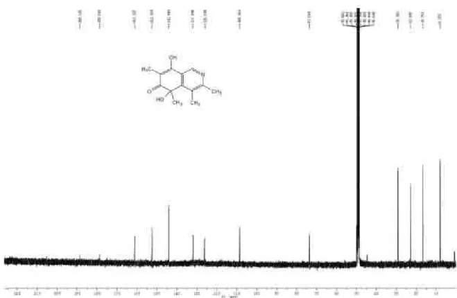
Figure S3.13C NMR spectrum of isoquinocitrinin A (1, CD
Figure S5. HMBC of isoquinocitrinin A (1, CD3OD).
Figure S6. NOESY spectrum of isoquinocitrinin A (1, CD