Jebmh.com
Original Article
J. Evid. Based Med. Healthc., pISSN- 2349-2562, eISSN- 2349-2570/ Vol. 3/Issue 26/Mar. 31, 2016 Page 1182
STUDY OF HEAD INJURIES WITH REFERENCE TO EXTRADURAL HAEMORRHAGE
S. Krishna Prasad1, P. Brahmaji Master2
1Professor, Department of Forensic Medicine & Toxicology, S. V. S. Medical College, Mahaboobnagar.
2Associate Professor, Department of Forensic Medicine & Toxicology, Government Medical College, Ananthapuramu.
ABSTRACT
OBJECTIVES
An autopsy study of extradural haemorrhage in head injuries was done. Extradural haemorrhage is a common manifestation in trauma in vehicular accidents, head injuries due to blunt force, falls, being hit by a moving object, etc.
METHODS
Data was collected from the police, relatives, and photographic evidences from the scene, post-mortem findings. 78 cases of deaths due to extradural haemorrhage in head injuries were studied.
RESULTS
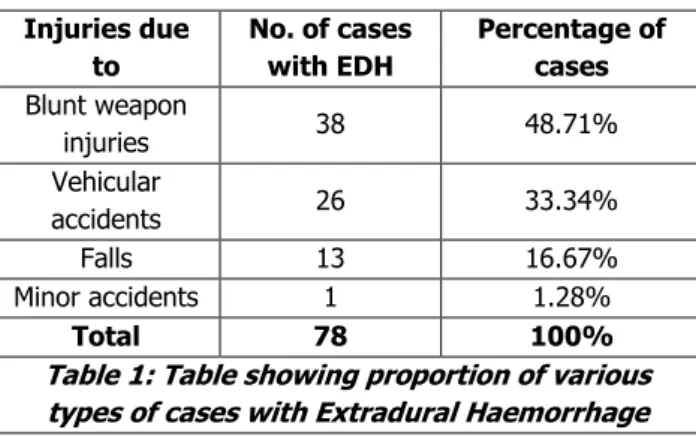
The presence of EDH in injuries with blunt weapons, vehicular accidents, falls, minor accidents were 48.71%, 33.33%, 16.67%, 0.6%.
CONCLUSION
Extradural haemorrhages are very common in blunt weapon injuries followed by vehicular accidents, falls and minor accidents.
KEYWORDS
Extradural haemorrhage, Blunt Injuries, Vehicular accidents, Falls, Minor accidents.
HOW TO CITE THIS ARTICLE: Prasad SK, Master PB. Study of head injuries with reference to extradural haemorrhage.
J. Evid. Based Med. Healthc. 2016; 3(26), 1182-1184. DOI: 10.18410/jebmh/2016/271
INTRODUCTION: Any trauma to head or face that has the potential for damaging the brain can have devastating consequences. Normally the brain is protected within the bony skull, but it is not well restrained within this compartment and injuries to the brain result from differences between the motion of the solid skull and the
relatively ‘fluid’ brain. The clinical significance of any space
occupying lesion within the cranial cavity is the effect that the raised intracranial pressure caused has on brain structure and function. Bleeding compresses the brain and, if it continues for sufficient time, and in sufficient quantity, can raise the intracranial pressure. As pressure increases, blood flow to the brain decreases and, if the pressure reaches the point where it equals or exceeds arterial blood pressure, the blood flow to the brain will cease. The dura is strong and bluish connective tissue membrane and is firmly attached to the skull. Extradural haemorrhage is caused almost exclusively due to trauma. Extradural haemorrhage is a common manifestation in head injuries due to blunt trauma due to falls, blow with blunt weapon, traffic injuries, crushing of the head, etc., At the moment of impact, the skull moves relative to the dura beneath it, and the dura is stripped from the bone. This produces an empty extradural space at the site of trauma.
A blood vessel may be injured at the same time and results into haemorrhage depending upon the site of blood vessel.1
METHODS: The present study has been carried out in the Department of Forensic Medicine, Government Medical College, Ananthapuramu on 78 cases containing cases of falls, vehicular accidents, blunt injuries to head during the period 2013-2015. Data was collected from the police, relatives, and photographic evidences from the scene, post-mortem findings.
Inclusion Criteria: Head injury cases due to blunt force.
Exclusion Criteria: Head injury cases due to sharp force.
RESULTS:
Injuries due to
No. of cases with EDH
Percentage of cases Blunt weapon
injuries 38 48.71%
Vehicular
accidents 26 33.34%
Falls 13 16.67%
Minor accidents 1 1.28%
Total 78 100%
Table 1: Table showing proportion of various types of cases with Extradural Haemorrhage
Submission 19-02-2016, Peer Review 06-03-2016, Acceptance 14-03-2016, Published 31-03-2016. Corresponding Author:
Dr. S. Krishna Prasad,
Professor, Department of Forensic Medicine & Toxicology, S. V. S. Medical College, Mahaboobnagar.
Jebmh.com
Original Article
J. Evid. Based Med. Healthc., pISSN- 2349-2562, eISSN- 2349-2570/ Vol. 3/Issue 26/Mar. 31, 2016 Page 1183
Fig. 1: Autopsy of skull showing extradural haemorrhage
Fig. 2: Pie-chart showing proportion of cases with extradural haemorrhages due to various reasons
Extradural haemorrhages are seen in the form of clots. These are usually present in the temporoparietal area, or in the frontotemporal area or parieto-occipital area. Occasionally, these are also seen in frontal or posterior fossa. Of all the 78 cases, 13 were due to falls with 16.67%, 26 were due to vehicular accidents with 33.34%, 38 were due to blunt injuries with 48.71%, 1 was a minor accident with 1.28%.
DISCUSSION: Accumulation of blood and blood clot
between the inner aspect of the skull and the outermost layer of brain membranes (dura matter) is known as extradural haemorrhage.2 It is the least common of all the
three brain membrane haemorrhages.3 EDH is most
commonly due to tear of middle meningeal artery and an associated fracture is present.4 Pure extradural
haemorrhage only is less common and it is most commonly with subdural haemorrhage.5 In a typical case, there is a
history of head injury which starts the bleeding, and will usually cause temporary unconsciousness followed by normal consciousness and the time interval between them is known as Lucid Interval.6 Bleeding from damaged
perforating veins, dural sinuses, in which case the development of symptoms will be slower.7
Study conducted by Graham et al7 showed that this type
of haemorrhage is common in 5-10% of fatal head injuries. Study conducted by Koc et al8 showed significant percentage
of EDH (20-30%) can also occur in frontal and posterior cranial fossa and is more common in children in these
regions. Separate studies conducted by Phonprasert et al9
and Servedai et al10 showed that imaging studies have
confirmed that about 50% cases are associated with subdural haemorrhage and/or contusional brain injury. Studies conducted by Viljoen and Wessels11 showed that
acute and chronic EDH can be differentiated by histopathological examination. Henderson et al12 said in their
report that it can also occur spontaneously. A study conducted by Mustafa and Subramaniam,13 Matsumoto et
al14 showed that the extradural haemorrhages occurdue
vascular malformations. A study conducted by Rodriguez et al15 showed that EDH occurs also with anticoagulant
therapy.
CONCLUSION: A study on extradural haemorrhage in head injuries is conducted on 78 cases. Of them the presence of EDH in falls, vehicular accidents, injuries with blunt weapons, minor accidents were 17.9%, 33.33%, 48.71%, 0.6%. Extradural haemorrhages are very common in blunt weapon injuries followed by vehicular accidents, falls and minor accidents.
BIBLIOGRAPHY:
1. Narayan Reddy KS. The essentials of forensic medicine and toxicology. 1999;18th ed:208.
2. Knight. Lawyers guide to forensic medicine. Cavendish Publishing Limited 1998;2nd edn:110.
Jebmh.com
Original Article
J. Evid. Based Med. Healthc., pISSN- 2349-2562, eISSN- 2349-2570/ Vol. 3/Issue 26/Mar. 31, 2016 Page 1184 4.
http://radiopaedia.org/articles/extradural-haemorrhage.
5. Helen L Whitwell. Forensic neuropathology, Hodder Arnold publications. CRC press London 2005;2nd
edn:62.
6. Jason Payne-James, Cliona McGovern, Richard Jones, et al. Simpson forensic medicine: Irish version, Taylor and Francis group CRC Press 2014;13th edn:116.
7. Graham DI, Gennarelli TA, McIntosh TK. Trauma. In Graham DI and Lantos Pl, eds. Greenfield’s Neuropathology, London: Arnold, Chapter 14, 2002;7th
edn.
8. Koc RK, Pasaoglu A, Menku A, et al. Extradural haematoma of the posterior cranial fossa. Neurosurg Rev 1998;21(1):52-57.
9. Phonprasert C, Suwanwela C, Hongsaprabhas C, et al. Extradural haematoma: analysis of 138 cases. J Trauma 1980;20(8):679-683.
10.Servedai F, Nanni A, Nasi MT, et al. Evolving brain lesions in the first 12 hours after head injury: analysis
of 37 comatose patients. Neurosurgery
1995;37(5):899-906.
11.Viljoen JJ, Wessels LS. Subacute and chronic extradural haematomas. S Afr J Surg 1990 Dec;28(4):133-137. 12.Henderson RT, Pittock S, Piepgras DG, et al. Acute
spontaneous spinal epidural haematoma. Arch Neurol 2001;58(7):1145-1146.
13.Mustafa M, Subramanian N. Spontaneous extradural haematoma causing spinal cord compression. Int Orthopaedics 1996;20(6):383-384.
14.Matsumoto K, Akagi K, Abekura M, et al. Vertex epidural haematoma associated with traumatic arteriovenous fistula of the middle meningeal artery:a case report. Surg Neurol 2001;55(5):302-304. 15.Rodriguez Y, Baena R, Gaetani P, et al. Spinal epidural