Physical Interactions and Functional
Coordination between the Core Subunits of
Set1/Mll Complexes and the Reprogramming
Factors
Zhenhua Yang, Jonathan Augustin, Jing Hu, Hao Jiang*
Department of Biochemistry and Molecular Genetics and the UAB Stem Cell Institute, University of Alabama at Birmingham School of Medicine, Birmingham, Alabama, United States of America
Abstract
Differentiated cells can be reprogrammed to the pluripotent state by overexpression of defined factors, and this process is profoundly influenced by epigenetic mechanisms includ-ing dynamic histone modifications. Changes in H3K4 methylation have been shown to be the predominant activating response in the early stage of cellular reprogramming. Mecha-nisms underlying such epigenetic priming, however, are not well understood. Here we show that the expression of the reprogramming factors (Yamanaka factors, Oct4, Sox2, Klf4 and Myc), especially Myc, directly promotes the expression of certain core subunits of the Set1/ Mll family of H3K4 methyltransferase complexes. A dynamic recruitment of the Set1/Mll complexes largely, though not sufficiently in its own, explains the dynamics of the H3K4 methylation during cellular reprogramming. We then demonstrate that the core subunits of the Set1/Mll complexes physically interact with mainly Sox2 and Myc among the Yamanaka factors. We further show that Sox2 directly binds the Ash2l subunit in the Set1/Mll com-plexes and this binding is mediated by the HMG domain of Sox2. Functionally, we show that the Set1/Mll complex core subunits are required for efficient cellular reprogramming. We also show that Dpy30, one of the core subunits in the complexes, is required for the efficient target binding of the reprogramming factors. Interestingly, such requirement is not neces-sarily dependent on locus-specific H3K4 methylation. Our work provides a better under-standing of how the reprogramming factors physically interact and functionally coordinate with a key group of epigenetic modulators to mediate transitions of the chromatin state involved in cellular reprogramming.
Introduction
The seminal discovery that differentiated cells can be reprogrammed to induced pluripotent stem cells (iPS cells) by four transcription factors Oct4, Sox2, Klf4, and c-Myc (O, S, K, M, the
OPEN ACCESS
Citation:Yang Z, Augustin J, Hu J, Jiang H (2015) Physical Interactions and Functional Coordination between the Core Subunits of Set1/Mll Complexes and the Reprogramming Factors. PLoS ONE 10(12): e0145336. doi:10.1371/journal.pone.0145336
Editor:Wei Xu, University of Wisconsin - Madison, UNITED STATES
Received:October 6, 2015
Accepted:December 2, 2015
Published:December 21, 2015
Copyright:© 2015 Yang et al. This is an open access article distributed under the terms of the Creative Commons Attribution License, which permits unrestricted use, distribution, and reproduction in any medium, provided the original author and source are credited.
Data Availability Statement:All relevant data are within the paper.
Funding:This work was supported by the University of Alabama at Birmingham Start-up fund. The funders had no role in study design, data collection and analysis, decision to publish, or preparation of the manuscript.
Yamanaka factors) [1] represents a major conceptual breakthrough in our understanding of the fundamental mechanisms controlling cell identity, and has a huge potential to revolutionize regenerative medicine. However, a number of issues, including inefficient and incomplete reprogramming and tumorigenic risks, need to be resolved before fully realizing the potential of iPS cells [2]. Directed by key transcription factors, cellular reprogramming is accompanied by extensive remodeling of epigenetic marks, and mounting evidence supports the profound influence of epigenetic regulators on reprogramming [3–11]. Moreover, chemicals acting on epigenetic modifications have been shown to be able to functionally replace some of the origi-nal transcription factors in reprogramming or enhance the reprogramming efficiency [12–14]. These findings underscore the importance of a deep comprehension of the epigenetic mecha-nisms for improved reprogramming.
At the very early stage of reprogramming upon OSKM expression, H3K4 methylation was found to be a predominant activating response globally—it is established de novo or signifi-cantly enhanced at large subsets of pluripotency-related or developmental gene promoters pre-ceding the loading of the general transcription machinery [15,16]. These results suggest that the reprogramming factors, rather than RNA polymerase II, directly or indirectly promote the dynamic changes of the histone mark, and thereby initiate a concerted change in the target chromatin environment which may epigenetically prime the subsequent transcription change. Some other pluripotency-associated genes gain promoter H3K4 methylation at the late or final stage of reprogramming [15,17,18]. However, the functional significance of the locus-specific H3K4 methylation in reprogramming is less clear. Moreover, it remains incompletely under-stood how the reprogramming factors elicit the alteration of locus-specific H3K4 methylation, which can be potentially affected by many factors including the local enzyme concentration through regulated recruitment, the enzymatic activities, and the chromatin and histone status.
In mammals, the most notable H3K4 methyltransferases are the Set1/Mll family complexes [19–21]. Apart from some specialized subunits, these complexes contain either Set1a, Set1b, Mll1, Mll2, Mll3, or Mll4 as the catalytic subunit and Wdr5, Rbbp5, Ash2l, and Dpy30 as inte-gral core subunits that are also important for the efficient methylation activity of the complexes [22–27]. Several of these subunits have been linked to either the maintenance or the execution of pluripotency. Wdr5, Ash2l, and Set1a are important for maintenance of an undifferentiated state of mouse embryonic stem (ES) cells [28–30], and Wdr5 and Set1a are also essential for cellular reprogramming to pluripotency [28,29]. However, Rbbp5 or Dpy30 knockdown (KD) in mouse ES cells does not significantly affect their self-renewal, but prevents their efficient dif-ferentiation [27]. Other than the presence of Wdr5 in several complexes different from H3K4 methyltransferases [31–34], reasons for the discrepant effects among these subunits remain unclear. Although depletion of these subunits all affect bulk H3K4 methylation, it is difficult to functionally attribute the cellular effects to the locus-specific methylation.
Mechanisms that control the recruitment of histone modifiers remain a central and largely open question in epigenetics. Several types of mechanisms may be involved in the genomic recruitment of H3K4 methyltransferases [19,35]. Preexisting H3K4 methylation is likely estab-lished by mechanisms intrinsic to DNA sequence including CpG islands which interact with the CXXC domain in some of the H3K4 methyltransferase complex components such as CFP1 [36,37]. Transcription factors can recruit histone modifiers to genomic sites for histone modi-fications and transcription [26,38]. It has not been systematically examined whether the Yamanaka factors, all of which are transcription factors, physically recruit the methylation enzymes to their genomic targets in cellular reprogramming.
cellular reprogramming through physical interactions and functional coordination with the reprograming factors. Our results provide a better understanding of the epigenetic priming and regulation involved in cellular reprogramming toward pluripotency.
Materials and Methods
Lentiviruses, cell culture, reprogramming, and gene knockdown
The lentiviral constructs for FUW-M2rtTA (plasmid #20342) [39], TetO-FUW-OSKM (plas-mid #20321) [40], TetO-FUW-Oct4 (plasmid #20323) [41], TetO-FUW-Sox2 (plasmid #20326) [41], TetO-FUW-Klf4 (plasmid #20322) [41], and TetO-FUW-Myc (plasmid #20324) [41] were all from Addgene. FLAG-HA-tagged individual mouse O, S, K and M-expressing lentiviral constructs were made based on TetO-FUW-OSKM. Lentiviruses for Rbbp5 and Dpy30 shRNAs and viral particle production were described [27].
HEK293T cells were obtained from American Type Culture Collection (ATCC, CRL-11268), and cultured in DMEM [Gibco] with 10% fetal bovine serum [FBS] [Gibco]. Oct4-GFP mouse embryonic fibroblasts (MEFs) harbor a GFP reporter gene downstream of exon 5 of the endogenousOct4locus [41], and were purchased from Stemgent (Cat# 08–0028, San Diego, CA) at passage 2. They were cultured in MEF culture medium (L-glutamine-containing DMEM with 10% FBS, 0.1 mM non-essential amino acids, and 55μMβ-mercaptoethanol, all
from Invitrogen), and passage was kept at minimum. For stable KD, MEFs were infected with lentiviruses expressing control, Rbbp5, or Dpy30 shRNA, followed by puromycin (2μg/ml)
selection for 2–3 days starting from 2 days after infection. For reprogramming, MEFs were infected with FUW-M2rtTA and TetO-FUW-OSKM lentiviruses, and were cultured on the next day on 0.1% gelatin-coated tissue-culture plates in the reprogramming medium (knockout DMEM [Gibco], 15% ES-certified FBS [Gibco], 2mM L-glutamine, 0.1mM non-essential amino acids, and 0.1mMβ-mercaptoethanol, recombinant leukemia inhibitory factor [LIF] [27], 50μg/ml vitamin C [42], and 2μg/ml doxycycline). P493-6 cells [43] were a kind gift from
Alanna Ruddell (Fred Hutchinson Cancer Research Center, Seattle) with the permission of Dirk Eick (Helmholtz Center Munich, Germany), and were cultured in RPMI 1640 medium with 10% FBS.
Protein-protein interaction by co-immunoprecipitation (co-IP)
For co-IP in MEFs, MEFs were infected with indicated lentivirus expressing individual repro-gramming factors, followed by induction by doxycycline at 1μg/ml for 48 hours. For co-IP in
293 cells, FLAG-HA-tagged mouse O, S, K and M cDNAs were cloned into pcDNA5/FRT/TO (Invitrogen). Cell lines stably expressing these factors were generated by co-transfection of these individual constructs with pOG44 (Invitrogen) into Flp-In T-REx-293 cells (Invitrogen), followed by hygromycin B selection. Selected clones were induced by doxycycline at 1μg/ml for
Protein purification and
in vitro
binding assays
His-tagged mouseSox2was cloned into pET28a (Novagen), and induced by Isopropylβ -D-1-thiogalactopyranoside in BL21 STAR DE3E.colicells (Invitrogen). Cell pellets were lysed in the lysis buffer containing 800mM NaCl, 50mM Tris [pH7.5], 10% Glycerol, 0.5% NP40, 1X EDTA-free protease inhibitor cocktail (Roche), 50mM Imidazole, 0.5mg/ml Lysozyme, and 5mMβ-mercaptoethanol. Cell lysate was sonicated and cleared by centrifugation, and then incubated with Ni-NTA resin (GE healthcare). The resin was extensively washed by the lysis buffer and then by the binding buffer (50 mM Tris pH 7.4, 300 mM NaCl, 10% Glycerol, 0.1% NP40, 30mM Imidazole), and was checked for bound protein by SDS-PAGE and coomassie blue staining. Sf9 insect cells (Invitrogen) were infected with baculoviruses expressing F-Ash2l and F-Wdr5 [27] for 72 hours. Cells were lysed in 500mM NaCl, 50mM Tris [pH7.5], 10% Glycerol, 0.1% NP40, protease inhibitor cocktail (Roche), and cleared by centrifugation. The supernatant was incubated with anti-FLAG M2 resin (Sigma) at 4°C for 6 hours and exten-sively washed with BC500 and 0.5% NP40, followed by BC100 and 0.1% NP40. Bound proteins were eluted with 0.4mg/ml FLAG peptide (Sigma) in BC100 and 0.1%NP40.
For in vitro binding assays, 4μg His-Sox2 on resin was pre-incubated with 100μl binding
buffer (above) containing 1% bovine serum albumin (BSA), and further incubated at 4°C over-night after addition of 400ng purified F-Ash2l or F-Wdr5. Resin was extensively washed with the binding buffer (no BSA) and was checked for bound proteins by SDS-PAGE and western blotting.
Western blotting, RNA extraction, chromatin immunoprecipitation
(ChIP), and quantitative PCR (qPCR)
Antibodies were described before [27] or are as follows: anti-HA [12CA5] (Roche,
11583816001, for western); anti-HA (Abcam, ab9110, for ChIP); anti-Myc (Santa Cruz Bio-technology, sc-764x) anti-Oct4 (Santa Cruz BioBio-technology, sc-8628x). RNA extraction and ChIP assays were done as described [27]. qPCR was performed with SYBR Advantage qPCR Premix (Clontech) on a ViiA7 Real-Time PCR System (Applied Biosystems). Primers used are listed inTable 1.
Results
Reprogramming factors promote expression of core subunits of the
Set1/Mll complexes
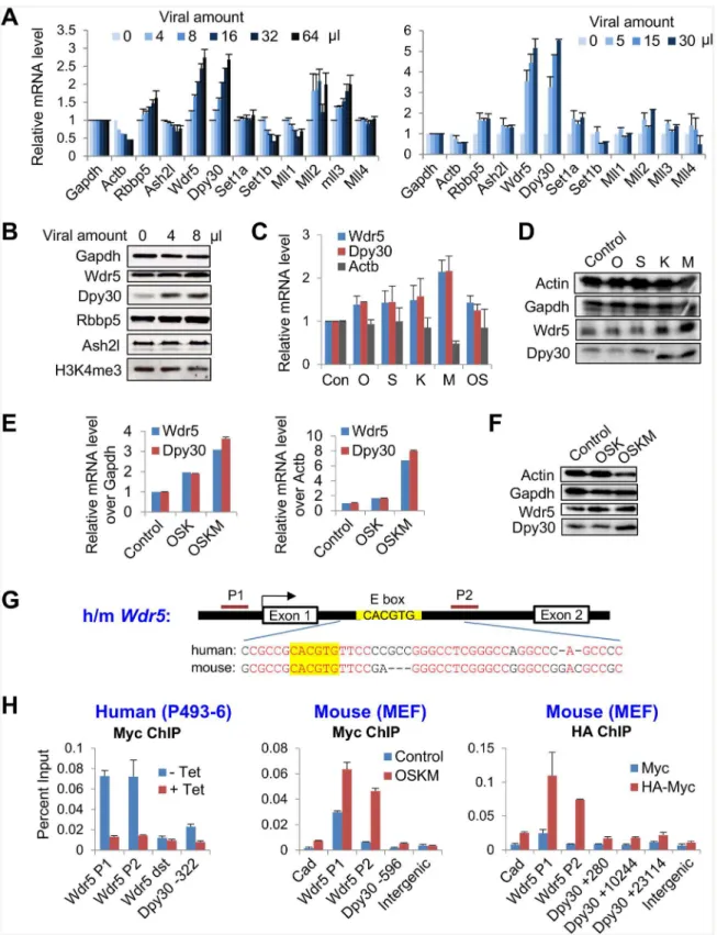
To address the initial effects on the machineries responsible for epigenetic priming upon the ectopic expression of the reprogramming factors OSKM, we determined the expression of all of the major integral subunits in the Set1/Mll complexes including the catalytic and core sub-units upon expression of increasing doses of OSKM in mouse embryonic fibroblasts (MEFs). The mRNA levels of several subunits, especially Wdr5 and Dpy30, were markedly up-regulated by OSKM in a dose-dependent manner (Fig 1A). We confirmed such up-regulation at the pro-tein level (Fig 1B). Global H3K4 trimethylation (H3K4me3), however, was not enhanced at the initial stage following OSKM expression (Fig 1B), probably due to the modest effect on other subunits (especially the catalytic subunits) of the complexes, which are also required for meth-ylation. These results also suggest that locus-specific dynamics of histone methylation, rather than the change of the global methylation, set the stage for changes of transcriptional programs in the initial stage of reprogramming. This notion is supported by a recent report [17].
We next sought to identify the specific factors among OSKM that promote the expression of
Wdr5andDpy30expression (Fig 1C) at the RNA level. Myc had the strongest effect (Fig 1C), although the fold increase of Myc expression over uninfected MEFs is typically lower than those of the other reprogramming factors (which are usually expressed at low or undetectable levels in uninfected MEFs). The results were confirmed at the protein level (Fig 1D). As Oct4 and Sox2 are known to often act together on target gene transcription [44,45], we co-infected MEFs with viruses expressing Oct4 and Sox2. Such co-infection, however, did not increase the effect onDpy30andWdr5expression (Fig 1C). We cannot exclude the possibility that cells may have only taken up virus encoding one factor, but this possibility should be low, given that
Table 1. Primers for qPCR.
App. Species gene Forward start Forward primer (5’-3’) Backward start Reverse primer (5’-3’)
RT mouse Gapdh CATCTTCTTGTGCAGTGCCAG GGCAACAATCTCCACTTTGCC
RT mouse Actb CCTTCAACACCCCAGCCATGTACG GGCACAGTGTGGGTGACCCCGTC
RT mouse Rbbp5 TGGACAGAACTACCCAGAGGA CCATCGTTACAGCCAACAGC
RT mouse Ash2l TGACACCTTTGGAATAGACACG AAGGCACTAAGGCACATTTCTT
RT mouse Wdr5 GTCTCAGCCGTTCATTTCAACC CGAAGGACACTGGAGGATTGT
RT mouse Dpy30 ACCCTCACTCTGAGTACGGG GGACTGTAGATCCACCTTCTGT
RT mouse Set1a TGCTGTCCCTCGTAGACTGG GGCTCTTTCCGTTTTACCTTGA
RT mouse Set1b GTGAAGTCCGGTGAGCACAA CAGGAGGCGATTCGGTCTTTG
RT mouse Mll1 GCAGATTGTAAGACGGCGAG GAGAGGGGGTGTTCCTTCCTT
RT mouse Mll2 GATGAGAATGGCTCGTTGTGG TCTATCTTGTCACACTTCCGGTA
RT mouse Mll3 TCAGTGCCATTCAAGGGTCC ACCCATGAGTGGATGGTGAGA
RT mouse Mll4 GAGGACTCGCTCATGTCCCT GCGGAGATAGGTGTGGCTC
RT mouse Oct4 ACATCGCCAATCAGCTTGG AGAACCATACTCGAACCACATCC
RT mouse Sox2 ACAGATGCAACCGATGCACC TGGAGTTGTACTGCAGGGCG
RT mouse Klf4 GCACACCTGCGAACTCACAC CCGTCCCAGTCACAGTGGTAA
RT mouse Myc CCACCAGCAGCGACTCTGA TGCCTCTTCTCCACAGACACC
RT IRES AACAGACCTTGCATTCCTTTGGCG TAAGGCCGGTGTGCGTTTGTCTAT
RT GFP TCTTGTAGTTGCCGTCGTCCTTGA TGACCCTGAAGTTCATCTGCACCA
RT mouse Nanog CCTCCAGCAGATGCAAGAACTC CTTCAACCACTGGTTTTTCTGCC
ChIP human Wdr5 P1 TSS -266 AGGGCACACTAGCACTTTCCTGTA TSS -187 TTGACTGGTGCAGGCCCTTCTAAT
ChIP human Wdr5 P2 TSS +391 ACTGGGTCCTCTTTCCCGCA TSS +492 ACTGAGTGGCTTGGTGGGTCT
ChIP human Wdr5 dst Chr 9: 134,211,630 AGCTCCATGCCACTGCCTTACTTA Chr 9: 134,211,824 TGCCTTCTTTCTCCACAGCAGCTA
ChIP human Dpy30 TSS -332 TGAGGAAGTTCAGTTGGTCGAGGA TSS -231 ACCAAGTGACCAGCCCAGTTAGAA
ChIP mouse Cad TSS -388 GCCATGTCGCAGCCAAGAAGATTT TSS -307 CAATGGCCGCTTCAGCCTTAAACA
ChIP mouse Wdr5 P1 TSS -254 ACCAAGAGGTTTCCCAACAGTCCT TSS -92 ACTGGTGTTCGGTAACTGCAGACT
ChIP mouse Wdr5 P2 TSS +373 CACTGGGAGTGGAATTCTCTG TSS +486 CTAGGACACAAATGAGGGTTGT
ChIP mouse Dpy30 TSS -596 TCACAAGCTTGAACCATCACCCAG TSS -494 TCTGCCTCCCAAGTGCTGGAATTA
ChIP mouse Dpy30 TSS +280 ACTGTGAACCCAGAGGTTGTGCTA TSS +359 AGGTGTATGAGAAGGGTTGAGCCA
ChIP mouse Dpy30 TSS +10244 TCTTGGGCAGTAACTGTAGCAGCA AAGTGCTTGCCAGTACTCAGCTCT
ChIP mouse Dpy30 TSS +23114 CCGTGCTTCCCACAAAGCAAATGT CATGCCACCAGCAACATTAGCACA
ChIP mouse Intergenic Chr8: 72,806,101 AAGGGGCCTCTGCTTAAAAA Chr8: 72,806,240 AGAGCTCCATGGCAGGTAGA
ChIP mouse Fgf4 TSS +681 ACCGGTAGACAGGAGATGAG TSS +769 ACTCTAAGCCTCTTGGGATCT
ChIP mouse Irf6 Ref. (15) GAGGGAGGACAGACACCTGA GCCGTCCCAAAACTACTTGA
ChIP mouse Cdh1 Ref. (15) CCGAGCTCAGTGTTTGCTC CAGGACCCTCCACATACCTG
ChIP mouse Sall4 Ref. (61) AACCTGCATTCTCCTACAGACC TTTCTTTAATGCCTGCATTTTG
ChIP mouse Oct4-A Ref. (61) GACGGCAGATGCATAACAAA AGGAAGGGCTAGGACGAGAG
ChIP mouse Oct4-B Ref. (61) TGTGAACTTGGCGGCTTC CCTCCACTCTGTCATGCTCA
ChIP mouse Postn Ref. (15) TATGCTCTGCTGCTGCTGTT AACAAGCCAGGGACTTACCC
Fig 1. The reprogramming factors promote the expression of the core subunits of Set1/Mll complexes.(A) MEFs were infected with increasing dose of FUW-M2rtTA and TetO-FUW-OSKM viruses in two independent infection experiments, and treated with doxycycline for 2 days. The mRNA levels of Set1/ Mll complex subunits was determined by RT-qPCR and normalized byGapdh. Average±range of values from duplicate assays are plotted. (B) MEFs were
co-infection of MEFs with our high titer viral mix separately encoding O, S, K, and M effi-ciently reprogrammed the MEFs to pluripotency (data not shown). Therefore, we conclude that although all four factors contribute to the initial up-regulation of the core subunits of H3K4 methyltransferase complexes, Myc plays a major role in such regulation.
As Myc is not essential but stimulatory for induction of iPS cells [46,47], we sought to com-pare the effects of the reprogramming factors with and without Myc on the expression of the Set1/Mll complex subunits. Our results showed that, while OSK modestly enhanced both the mRNA and the protein levels of Wdr5 and Dpy30, OSKM strongly increased the expression of these core subunits (Fig 1E and 1F).
To determine if Myc directly regulatesWdr5andDpy30expression, we performed ChIP assays for Myc in human P493-6 cells and in MEFs that ectopically express Myc either by OSKM virus or HA-tagged Myc virus. P493-6 is an immortalized B cell line that expresses a tet-racycline-repressibleMyctransgene but negligible endogenousMyc[43]. We found that Myc bound to both human and mouseWdr5gene at the promoter and a region in intron 1 close to a canonical E box (5’-CACGTG-3’) surrounded by extraordinarily conserved intronic sequences (Fig 1G and 1H). Similarly, Myc also binds to the promoter region ofDpy30gene (Fig 1H). Moreover, after a more thorough examination, we found 6 canonical E boxs at +5451, +10088, +14600, +15385, +19350, +23063 bp in the mouseDpy30gene body (about 24.5 kb in total size), while no E box was found in the 20kb region downstream the gene. Our ChIP assay showed that, in addition to the binding at TSS, HA-Myc also bound to regions near at least two E boxes in theDpy30gene body at +10088 and +23063 bp (Fig 1H, right panel). These results show extensive albeit relatively weak binding of Myc toDpy30gene. Thus, Myc directly promotes the expression of two core subunits of the H3K4 methyltransferase com-plexes with a potential functional implication.
Dynamic recruitment of Dpy30 during reprogramming
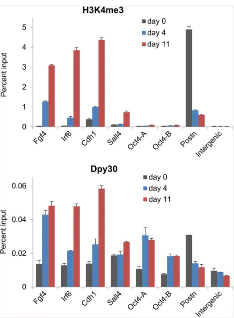
The direct promotion of the expression of the Set1/Mll complex core subunits by the repro-gramming factors suggests that these subunits may be functionally involved in reprorepro-gramming the epigenetic landscape back to that of pluripotency. To understand the molecular mecha-nisms underlying the dynamics of H3K4 methylation during cellular reprogramming, we sought to determine the binding dynamics of the enzymatic complexes in this process. Consis-tent with previous reports [15,17,18], we found that H3K4me3 was markedly enhanced at promoters of pluripotency-associated genes includingFgf4,Irf4,Cdh1, andSall4, but reduced atPostn, a somatic determinant gene, on days 4 and 11 during reprogramming (Fig 2, top). We then found that Dpy30 was increasingly recruited toFgf4,Irf4,Cdh1, andSall4promoters,
TetO-FUW-Oct4 and TetO-FUW-Sox2 viruses (OS), in addition to FUW-M2rtTA. After induction with doxycycline for 2 days, the mRNA (C) and protein (D) levels of indicated Set1/Mll complex subunits were determined by RT-qPCR and normalized byGapdh(C) and western blotting (D). Average±SD from 3
independent infections are plotted in (C). (E and F) MEFs were infected with viral mixes that expressed individual Oct4, Sox2, Klf4 (OSK) or Oct4, Sox2, Klf4, and c-Myc (OSKM), in addition to FUW-M2rtTA, and MEFs infected with only FUW-M2rtTA virus was used as control. After induction with doxycycline for 2 days, the mRNA levels (E) of indicated core subunits were determined by RT-qPCR and normalized byGapdh(E, left) orActb(E, right). Average±range of
values from 2 independent infections are plotted. The indicated proteins were determined by western blotting (F). (G) Structure of human and mouseWdr5
genes showing the highly conserved intronic sequences flanking the canonic E box. Identical residuals between human and mouse are in red. (H) Left: P493-6 cells were cultured in the absence (-Tet, Myc on) or presence of (+Tet, Myc off) tetracycline, and used for Myc ChIP followed by qPCR on indicated loci. Middle: MEFs were infected with only FUW-M2rtTA virus (control) or with FUW-M2rtTA and TetO-FUW-OSKM viruses (OSKM), induced with doxycycline, and used for Myc ChIP followed by qPCR on indicated loci. Right: MEFs were infected with FUW-M2rtTA and TetO-FUW-Myc virus (Myc) or FUW-M2rtTA and TetO-FUW-HA-Myc viruses (HA-Myc), induced with doxycycline, and used for HA ChIP followed by qPCR on indicated loci. Wdr5 dst, aWdr5
downstream region.Cadis a previously established Myc target [63] and used as a positive control. Note that there are E boxes at +10088 and +23063 bp in theDpy30gene body. Average±SD from triplicate assays are plotted, except for Myc ChIP in MEFs, for which Average±range of values from duplicate
assays are plotted. The differences between blue and red bars in all three panels are statistically significant (P<0.05 in 2-tailed Student’st-test) for all loci except for the“Wdr5 dst”and the“Intergenic”sites.
while leavingPostnin this process (Fig 2, bottom). The changes of local H3K4me3 and Dpy30 binding are in general correlated for most of these loci, but not in a very quantitative manner for all loci (Fig 2). Consistent with findings thatOct4promoter does not significantly gain H3K4 methylation until in the final stage [15,17], our results here also show little H3K4me3 at theOct4promoter on days 4 or 11 during reprogramming (Fig 2, top). Interestingly, Dpy30 binding significantly increased at theOct4promoter (comparable to that atIrf6andCdh1) from day 0 to 4 during reprogramming, without eliciting much increase of local H3K4me3 (Fig 2). These results suggest that the reprogramming factors mediate the changes in local histone modifications primarily through affecting the recruitment of the enzymes to those loci, while other mechanisms are also involved, possibly in a gene-dependent manner.
Fig 2. Dynamic recruitment of Dpy30 during cellular reprogramming.MEFs were infected with FUW-M2rtTA and TetO-FUW-OSKM viruses and cultured under the reprogramming condition. At days 0, 4 and 11 during reprogramming, cells were used for H3K4me3 and Dpy30 ChIP followed by qPCR on indicated loci. Oct4-A and Oct4-B are two different primers at theOct4promoter [61]. Average±range of values from
duplicate assays are plotted, and results are representative of 3 independent biological repeats.
Interaction of the Set1/Mll complex core subunits with the
reprogramming factors
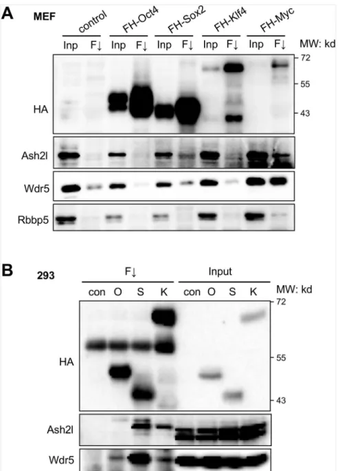
The dynamic recruitment of Dpy30 following OSKM expression suggests that the Set1/Mll complexes may be recruited to relevant chromatin targets by their physical interaction with the reprogramming factors. We thus characterized such interactions between the core subunits of the Set1/Mll complexes with each of OSKM. We found that, Sox2 and Myc, when individually expressed in MEFs, bind to endogenous Ash2l and Wdr5 (Fig 3A). Possible binding of Oct4 and Klf4 with these subunits was difficult to detect in infected MEFs due to the background sig-nal in control MEFs (Fig 3A). We then performed co-IP in 293 cells expressing individual Oct4, Sox2 or Klf4, and found that they all bound to endogenous Ash2l and Wdr5, yet with varying affinity (Fig 3B). Among OSK, Oct4 was found to be the weakest in binding to the core subunits including Wdr5 and Ash2l, while Sox2 was the strongest in binding to these two sub-units of Set1/Mll complexes (Fig 3B).
The HMG domain of Sox2 mediates its direct binding to Ash2l of Set1/Mll
complexes
We next determined which domain of Sox2 mediates its binding to Set1/Mll complex core sub-units by examining the binding of various truncations of Sox2 with endogenous Ash2l and Wdr5 (Fig 4A). While Sox2 (1–123) containing the HMG domain was sufficient to bind to Ash2l and Wdr5, the Sox2 mutant missing the HMG domain (ΔHMG) failed to bind to these proteins (Fig 4B), indicating that the HMG domain mediates the binding of Sox2 to Ash2l and Wdr5 (and thus to the Set1/Mll complexes).
We then asked if Sox2 directly binds to the Set1/Mll complex core subunits. We recombi-nantly expressed His-tagged Sox2 in bacteria, FLAG-tagged Ash2l and Wdr5 in baculovirus-infected insect cells, and purified these proteins by affinity chromatography (Fig 4C). Our in vitro binding assays using these purified proteins showed that the Sox2 directly binds to Ash2l, but not to Wdr5, under a stringent binding condition (Fig 4D).
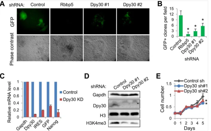
Set1/Mll complex core subunits are required for efficient reprogramming
Having established the physical interactions between the reprogramming factors with the Set1/ Mll complex core subunits, we asked whether these subunits are functionally important in helping fulfill the mission of the reprogramming factors. Through the doxycycline-inducible expression of OSKM in a single polycistronic viral vector [40], we could achieve efficient repro-gramming of primary Oct4-GFP MEFs, in whichGFPis placed downstream of the endogenous
Dpy30 is required for efficient recruitment of Oct4 to its genomic targets
As reprogramming is driven by the binding of the reprogramming factors to their chromatin target sites and the following execution of the transcriptional programs, we hypothesized that the Set1/Mll complex activity is required for the efficient binding of the reprogramming factors to the chromatin targets. Given the key role of Oct4 in a successful reprogramming to
Fig 3. Interaction of the Set1/Mll complex core subunits with the reprogramming factors.(A) MEFs were infected with only FUW-M2rtTA virus (control), or with FUW-M2rtTA and virus expressing the indicated FLAG-HA-tagged individual reprograming factor. After induction with doxycycline for 2 days, co-immunoprecipitation was performed by anti-FLAG M2 resin. Inp, input (4%); F#, anti-FLAG immunoprecipitation. (B) 293 cells were stably transfected with empty vector (con) or plasmid expressing the indicated FLAG-HA-tagged individual reprograming factor. Co-immunoprecipitation was performed by anti-FLAG M2 resin. Input (2.5%) was loaded.
Fig 4. The HMG domain of Sox2 mediates its direct binding to Ash2l of Set1/Mll complexes.(A) Diagram for truncation mutants of FLAG-HA-tagged Sox2 showing their domain structures and whether the protein binds to Ash2l as summarized from (B). (B) 293T cells were transfected with indicated FLAG-HA-tagged Sox2 mutants, and co-IP was performed by anti-FLAG M2 resin. Wdr5 blotting for the right panel is not shown due to a fortuitous cross-reactivity withΔHMG. TAD: transcriptional activation domain. Inp, input (2.5%); F#, anti-FLAG immunoprecipitation. (C) Purified proteins were examined by SDS-PAGE along with the molecular weight ladder on the left, and visualized by coomassie blue staining. (D) In vitro binding assay for Ni bead-bound His-Sox2 with FLAG-Ash2l and FLAG-Wdr5, and examined by western blotting using anti-FLAG antibody. Ni-beads that was not used for His-His-Sox2 purification was used as the mock control in the binding assay. Input (20%) was loaded.
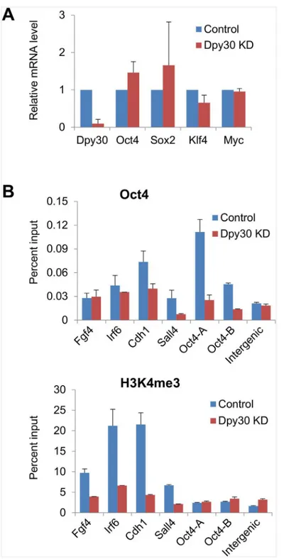
pluripotency, we focused on the genomic binding of Oct4. We performed OSKM-mediated reprogramming on the control and stable Dpy30 KD MEFs with titrated OSKM viral amounts so that the reprogramming factors were expressed at the comparable levels in the Dpy30 KD versus control cells (Fig 6A). In support of our hypothesis, Dpy30 KD markedly reduced the recruitment of Oct4 to several pluripotency gene promoters including the endogenousOct4
promoter 7 days after OSKM induction (Fig 6B, top). In an attempt to study molecular mecha-nisms underlying the regulation of Oct4 recruitment by Dpy30, we examined the effect of Dpy30 KD on H3K4 methylation at relevant gene promoters. We found that Dpy30 KD greatly reduced H3K4me3 at the promoters ofFgf4,Irf6,Cdh1, andSall4(Fig 6B, bottom). Minimal H3K4me3, however, was at theOct4promoter at this stage of reprogramming, and conse-quently, Dpy30 KD had little effect on H3K4me3 at these promoters (Fig 6B, bottom). These results provide a mechanistic basis for the importance of Dpy30 (and potentially other core subunits of the Set1/Mll complexes) in cellular reprogramming.
Discussion
Cellular reprogramming is essentially an epigenetic process, and an efficient reprogramming probably requires a close coordination between the initiating transcription factors and the responsive epigenetic factors. Our work here outlines a model (Fig 7) for such coordination
Fig 5. The Set1/Mll complex core subunits are required for efficient reprogramming to pluripotency.(A) Oct4-GFP MEFs stably depleted of Rbbp5 or Dpy30 were used in reprogramming for 16 days, and imaged under microscope. (B) GFP positive clones per field, as averaged±SD from 10 random fields, were quantified by counting under microscope. Results are representative of over 5 biological repeats.*P<0.05 in 2-tailed Student’st-test between control and each knockdown. (C) The relative mRNA levels of indicated genes were determined by RT-qPCR after reprogramming, and normalized byGapdh. Dpy30 shRNA#1 was used for KD. Results are representative of over 5 biological repeats. (D) Western blotting for MEFs after stable knockdown. (E) Growth curves of the control and Dpy30 KD MEFs as measured by described method [27]. Results are representative of two biological repeats, and average±SD from triplicate measurements are plotted.*P<0.05 in 2-tailed Student’st-test between control and each knockdown.
Fig 6. Dpy30 is required for efficient recruitment of Oct4 to its genomic targets.MEFs infected with control shRNA or Dpy30 shRNA#1 (KD) were used in reprogramming for 7 days. (A) Expression ofDpy30
andOSKMwas determined by RT-qPCR and normalized byGapdh. Average±SD from 3 independent
infection and reprogramming assays are plotted. (B) ChIP assays for Oct4 and H3K4me3 were performed followed by qPCR on indicated gene promoters or an intergenic site. Average±range of values from duplicate assays are plotted, and results are representative of 3 independent infection and reprogramming assays.
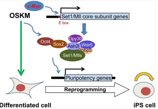
between OSKM and the core subunits of Set1/Mll complexes, the major H3K4 methyltrans-ferases in mammalian cells. OSKM, especially Myc, stimulates the expression of the core sub-units, most prominently Dpy30 and Wdr5. Sox2 also directly interacts with Ash2l and likely guides the localization of the Set1/Mll complexes to the promoters of pluripotency genes. These complexes remodels the local chromatin to help the further recruitment of reprogram-ming factors including Oct4. These coordinated actions of the transcription factors and chro-matin modifiers eventually lead to the change of the cell fate to pluripotency.
Our findings that OSKM can bind to the Set1/Mll complex core subunits combined with the increased recruitment of these subunits to genomic sites following OSKM induction sug-gest a direct role of the reprogramming factors in recruiting the H3K4 methyltransferases to epigenetically prime the transcription programs for reprogramming. The increase of Dpy30 binding and H3K4me3 at the pluripotency-associated genes is unlikely a mere result of passive spread of overall increased levels of Dpy30 and histone methylation, because (i) the reduced Dpy30 binding and H3K4me3 atPostn(Fig 2) following OSKM expression suggests a regulated and locus-specific dynamics of methyltransferase binding, and (ii) the increase of the Dpy30 recruitment to several promoters (e.g.Irf6,Cdh1, andSall4) occurs at the later stage of the reprogramming (Fig 2), while the overall upregulation of Dpy30 occurs shortly after OSKM expression. The reduced Dpy30 binding and H3K4me3 atPostnand presumably other loci that get silenced during reprogramming cannot be explained by a direct recruitment of Set1/ Mll core subunits by OSKM to those sites, and is most likely an indirect result initiated by the reprogramming factors. The recruitment of Dpy30 (and presumably the H3K4 methyltransfer-ase complexes) at theOct4promoter is insufficient to enhance H3K4me3 at the same site in
Fig 7. A model for the role of the core subunits of Set1/Mll complexes in cellular reprogramming.This model depicts two different levels of mechanisms by which the reprogramming factors coordinate with the core subunits of Set1/Mll complexes to facilitate cellular reprogramming of differentiated cells to pluripotency. SeeDiscussionfor details.
the intermediate stage of reprogramming. Our results suggest that, while regulated recruitment of the epigenetic machinery is the major determinant, multiple mechanisms are responsible for the dynamic epigenetic regulation involved in reprogramming.
Our findings are consistent with previous reports on the direct interaction of Myc with Ash2l [49] and that of Oct4 with Wdr5 [28]. They are also consistent with the broad associa-tion of promoters gaining H3K4me2 and targets for OSKM, predominantly Oct4 and Sox2 [15]. Since Sox2 binds to the core subunits of Set1/Mll complexes much more strongly than Oct4, it is likely that Sox2 may assist its close partner Oct4 to bring the H3K4 methyltrans-ferases to their common targets. The interaction of the Set1/Mll complexes with key cell fate-regulatory transcription factors appears to be highly conserved in evolution, as a Wdr5 ortho-log inC.eleganswas recently shown to interact with Sox2 and CEH-6 (OCT) [50].
Although Myc regulates a large number of expressed genes in the genome [51–54], it is unlikely that its regulation of all of the targets is critical. The phylogenetic conservation of the Myc binding site within intron 1 ofWdr5is extraordinary for an intronic sequence, suggesting that this regulation is likely to be physiologically important. Our results may partially explain the previously finding thatMyc-null mouse embryos have greatly reduced bulk level of H3K4me3 [55]. The up-regulation ofWdr5andDpy30upon OSKM (especially Myc) expres-sion is not sufficient to enhance global H3K4me3, but it is likely to facilitate alteration of gene-specific methylation, or prepare for the much elevated methylation at the later stage of repro-gramming. As the Myc regulatory network is pervasively involved in the regulation of cell growth, metabolism, and tumorigenesis [56], Set1/Mll complexes are unlikely to be specifically involved in regulating maintenance or acquisition of pluripotency. In this sense, depletion of many subunits in the Set1/Mll complexes commonly affects cell proliferation in a cell context-dependent manner [28,29,57]. It is possible that the altered cell proliferation or other potential cellular effects upon Dpy30 KD may affect the reprogramming efficiency. However, the effect of cell proliferation on reprogramming is not definitive or unidirectional since both positive and negative effects of cell proliferation on reprogramming have been reported [58,59]. Fur-thermore, dense colonies still form in Rbbp5 or Dpy30 KD cells during reprogramming, except that these colonies show no or little GFP signals (Fig 5A). Finally, the impaired target binding of Oct4 upon Dpy30 KD suggests an important role of Dpy30 for the molecular activity of the reprogramming factors through regulating the chromatin accessibility.
complementary mechanism by which Dpy30 facilitates the recruitment of a reprogramming factor. We note that this alternative mechanism does not necessarily exclude the involvement of the H3K4 methylation activity of the enzymatic complexes that may affect global chromatin environment and thus secondarily affect the local genomic accessibility (i.e., indirect effect for a particular locus).
Based on this and our previous work [27], we conclude that certain core subunits of the Set1/Mll complexes including Rbbp5 and Dpy30 are important for the plasticity of cell identity in the two-way transitions between the pluripotent and differentiated states. Loss of Rbbp5 or Dpy30 results in a rigid transcriptional program and a fixed cell identity. Our findings have added functional significance and mechanistic basis for the epigenetic priming of genes for reprogramming, and further demonstrate a profound impact of the epigenetic machinery on the plasticity of cell fate. This conclusion is also in line with a recent report that demonstrates an important role of the H3K4 methylation activity in ensuring robust transdifferentiation of hindgut cells into motor neurons inC.elegans[50].
Acknowledgments
We thank Rudolf Jaenisch for making the reprogramming factor lentiviral vectors available through Addgene, and Dirk Eick and Alanna Ruddell for providing the P493-6 cells.
Author Contributions
Conceived and designed the experiments: HJ. Performed the experiments: ZY JA JH. Analyzed the data: ZY JA HJ. Wrote the paper: HJ.
References
1. Takahashi K, Yamanaka S. Induction of pluripotent stem cells from mouse embryonic and adult fibro-blast cultures by defined factors. Cell. 2006; 126(4):663–76. Epub 2006/08/15. doi:10.1016/j.cell.2006. 07.024PMID:16904174.
2. Hanna JH, Saha K, Jaenisch R. Pluripotency and cellular reprogramming: facts, hypotheses, unre-solved issues. Cell. 2010; 143(4):508–25. Epub 2010/11/16. doi:10.1016/j.cell.2010.10.008PMID:
21074044; PubMed Central PMCID: PMC3032267.
3. Doege CA, Inoue K, Yamashita T, Rhee DB, Travis S, Fujita R, et al. Early-stage epigenetic modifica-tion during somatic cell reprogramming by Parp1 and Tet2. Nature. 2012; 488(7413):652–5. Epub 2012/08/21. doi:10.1038/nature11333PMID:22902501.
4. Mansour AA, Gafni O, Weinberger L, Zviran A, Ayyash M, Rais Y, et al. The H3K27 demethylase Utx regulates somatic and germ cell epigenetic reprogramming. Nature. 2012; 488(7411):409–13. Epub 2012/07/18. doi:10.1038/nature11272PMID:22801502.
5. Zhao W, Li Q, Ayers S, Gu Y, Shi Z, Zhu Q, et al. Jmjd3 inhibits reprogramming by upregulating expres-sion of INK4a/Arf and targeting PHF20 for ubiquitination. Cell. 2013; 152(5):1037–50. Epub 2013/03/ 05. doi:10.1016/j.cell.2013.02.006PMID:23452852.
6. Barrero MJ, Boue S, Izpisua Belmonte JC. Epigenetic mechanisms that regulate cell identity. Cell stem cell. 2010; 7(5):565–70. Epub 2010/11/03. doi:10.1016/j.stem.2010.10.009PMID:21040898. 7. Bhutani N, Brady JJ, Damian M, Sacco A, Corbel SY, Blau HM. Reprogramming towards pluripotency
requires AID-dependent DNA demethylation. Nature. 2010; 463(7284):1042–7. Epub 2009/12/23. doi:
10.1038/nature08752PMID:20027182; PubMed Central PMCID: PMC2906123.
8. Singhal N, Graumann J, Wu G, Arauzo-Bravo MJ, Han DW, Greber B, et al. Chromatin-Remodeling Components of the BAF Complex Facilitate Reprogramming. Cell. 2010; 141(6):943–55. Epub 2010/ 06/17. doi:10.1016/j.cell.2010.04.037PMID:20550931.
9. Gaspar-Maia A, Alajem A, Polesso F, Sridharan R, Mason MJ, Heidersbach A, et al. Chd1 regulates open chromatin and pluripotency of embryonic stem cells. Nature. 2009; 460(7257):863–8. Epub 2009/ 07/10. doi:10.1038/nature08212PMID:19587682.
11. Papp B, Plath K. Epigenetics of reprogramming to induced pluripotency. Cell. 2013; 152(6):1324–43. Epub 2013/03/19. doi:10.1016/j.cell.2013.02.043PMID:23498940; PubMed Central PMCID: PMCPmc3602907.
12. Huangfu D, Maehr R, Guo W, Eijkelenboom A, Snitow M, Chen AE, et al. Induction of pluripotent stem cells by defined factors is greatly improved by small-molecule compounds. Nature biotechnology. 2008; 26(7):795–7. Epub 2008/06/24. doi:10.1038/nbt1418PMID:18568017.
13. Huangfu D, Osafune K, Maehr R, Guo W, Eijkelenboom A, Chen S, et al. Induction of pluripotent stem cells from primary human fibroblasts with only Oct4 and Sox2. Nature biotechnology. 2008; 26 (11):1269–75. Epub 2008/10/14. doi:10.1038/nbt.1502PMID:18849973.
14. Mikkelsen TS, Hanna J, Zhang X, Ku M, Wernig M, Schorderet P, et al. Dissecting direct reprogram-ming through integrative genomic analysis. Nature. 2008; 454(7200):49–55. Epub 2008/05/30. doi:10. 1038/nature07056PMID:18509334; PubMed Central PMCID: PMC2754827.
15. Koche RP, Smith ZD, Adli M, Gu H, Ku M, Gnirke A, et al. Reprogramming factor expression initiates widespread targeted chromatin remodeling. Cell stem cell. 2011; 8(1):96–105. Epub 2011/01/08. doi:
10.1016/j.stem.2010.12.001PMID:21211784; PubMed Central PMCID: PMC3220622.
16. Sindhu C, Samavarchi-Tehrani P, Meissner A. Transcription factor-mediated epigenetic reprogram-ming. The Journal of biological chemistry. 2012; 287(37):30922–31. Epub 2012/09/07. doi:10.1074/ jbc.R111.319046PMID:22952239; PubMed Central PMCID: PMCPmc3438925.
17. Hussein SMI, Puri MC, Tonge PD, Benevento M, Corso AJ, Clancy JL, et al. Genome-wide characteri-zation of the routes to pluripotency. Nature. 2014; 516(7530):198–206. doi:10.1038/nature14046 http://www.nature.com/nature/journal/v516/n7530/abs/nature14046.html#supplementary-information. PMID:25503233
18. Polo JM, Anderssen E, Walsh RM, Schwarz BA, Nefzger CM, Lim SM, et al. A molecular roadmap of reprogramming somatic cells into iPS cells. Cell. 2012; 151(7):1617–32. Epub 2012/12/25. doi:10. 1016/j.cell.2012.11.039PMID:23260147; PubMed Central PMCID: PMC3608203.
19. Ruthenburg AJ, Allis CD, Wysocka J. Methylation of lysine 4 on histone H3: intricacy of writing and reading a single epigenetic mark. Mol Cell. 2007; 25(1):15–30. Epub 2007/01/16. S1097-2765(06) 00872-0 [pii] doi:10.1016/j.molcel.2006.12.014PMID:17218268.
20. Sims RJ 3rd, Nishioka K, Reinberg D. Histone lysine methylation: a signature for chromatin function. Trends Genet. 2003; 19(11):629–39. PMID:14585615.
21. Shilatifard A. The COMPASS family of histone H3K4 methylases: mechanisms of regulation in develop-ment and disease pathogenesis. Annu Rev Biochem. 2012; 81:65–95. Epub 2012/06/06. doi:10.1146/ annurev-biochem-051710-134100PMID:22663077.
22. Dou Y, Milne TA, Tackett AJ, Smith ER, Fukuda A, Wysocka J, et al. Physical association and coordi-nate function of the H3 K4 methyltransferase MLL1 and the H4 K16 acetyltransferase MOF. Cell. 2005; 121(6):873–85. Epub 2005/06/18. S0092-8674(05)00449-6 [pii] doi:10.1016/j.cell.2005.04.031PMID:
15960975.
23. Dou Y, Milne TA, Ruthenburg AJ, Lee S, Lee JW, Verdine GL, et al. Regulation of MLL1 H3K4 methyl-transferase activity by its core components. Nat Struct Mol Biol. 2006; 13(8):713–9. Epub 2006/08/01. nsmb1128 [pii] doi:10.1038/nsmb1128PMID:16878130.
24. Milne TA, Briggs SD, Brock HW, Martin ME, Gibbs D, Allis CD, et al. MLL targets SET domain methyl-transferase activity to Hox gene promoters. Mol Cell. 2002; 10(5):1107–17. Epub 2002/11/28. S1097276502007414 [pii]. PMID:12453418.
25. Wysocka J, Swigut T, Milne TA, Dou Y, Zhang X, Burlingame AL, et al. WDR5 associates with histone H3 methylated at K4 and is essential for H3 K4 methylation and vertebrate development. Cell. 2005; 121(6):859–72. Epub 2005/06/18. S0092-8674(05)00355-7 [pii] doi:10.1016/j.cell.2005.03.036PMID:
15960974.
26. Demers C, Chaturvedi CP, Ranish JA, Juban G, Lai P, Morle F, et al. Activator-mediated recruitment of the MLL2 methyltransferase complex to the beta-globin locus. Molecular cell. 2007; 27(4):573–84. Epub 2007/08/21. doi:10.1016/j.molcel.2007.06.022PMID:17707229; PubMed Central PMCID: PMC2034342.
27. Jiang H, Shukla A, Wang X, Chen WY, Bernstein BE, Roeder RG. Role for Dpy-30 in ES Cell-Fate Specification by Regulation of H3K4 Methylation within Bivalent Domains. Cell. 2011; 144(4):513–25. Epub 2011/02/22. S0092-8674(11)00059-6 [pii] doi:10.1016/j.cell.2011.01.020PMID:21335234. 28. Ang YS, Tsai SY, Lee DF, Monk J, Su J, Ratnakumar K, et al. Wdr5 mediates self-renewal and
repro-gramming via the embryonic stem cell core transcriptional network. Cell. 2011; 145(2):183–97. Epub 2011/04/12. doi:10.1016/j.cell.2011.03.003PMID:21477851; PubMed Central PMCID: PMC3097468. 29. Bledau AS, Schmidt K, Neumann K, Hill U, Ciotta G, Gupta A, et al. The H3K4 methyltransferase
Development (Cambridge, England). 2014; 141(5):1022–35. Epub 2014/02/20. doi:10.1242/dev. 098152PMID:24550110.
30. Wan M, Liang J, Xiong Y, Shi F, Zhang Y, Lu W, et al. The trithorax group protein Ash2l is essential for pluripotency and maintaining open chromatin in embryonic stem cells. The Journal of biological chemis-try. 2013; 288(7):5039–48. Epub 2012/12/15. doi:10.1074/jbc.M112.424515PMID:23239880; PubMed Central PMCID: PMC3576105.
31. Cai Y, Jin J, Swanson SK, Cole MD, Choi SH, Florens L, et al. Subunit composition and substrate spec-ificity of a MOF-containing histone acetyltransferase distinct from the male-specific lethal (MSL) com-plex. The Journal of biological chemistry. 2010; 285(7):4268–72. Epub 2009/12/19. doi:10.1074/jbc. C109.087981PMID:20018852; PubMed Central PMCID: PMC2836030.
32. Wang YL, Faiola F, Xu M, Pan S, Martinez E. Human ATAC Is a GCN5/PCAF-containing acetylase complex with a novel NC2-like histone fold module that interacts with the TATA-binding protein. The Journal of biological chemistry. 2008; 283(49):33808–15. Epub 2008/10/08. doi:10.1074/jbc. M806936200PMID:18838386; PubMed Central PMCID: PMC2590711.
33. Suganuma T, Gutierrez JL, Li B, Florens L, Swanson SK, Washburn MP, et al. ATAC is a double his-tone acetyltransferase complex that stimulates nucleosome sliding. Nature structural & molecular biol-ogy. 2008; 15(4):364–72. Epub 2008/03/11. doi:10.1038/nsmb.1397PMID:18327268.
34. Li X, Wu L, Corsa CA, Kunkel S, Dou Y. Two mammalian MOF complexes regulate transcription activa-tion by distinct mechanisms. Molecular cell. 2009; 36(2):290–301. Epub 2009/10/27. doi:10.1016/j. molcel.2009.07.031PMID:19854137; PubMed Central PMCID: PMC2768600.
35. Smith E, Shilatifard A. The chromatin signaling pathway: diverse mechanisms of recruitment of histone-modifying enzymes and varied biological outcomes. Mol Cell. 2010; 40(5):689–701. Epub 2010/12/15. doi:10.1016/j.molcel.2010.11.031PMID:21145479; PubMed Central PMCID: PMC3037032. 36. Thomson JP, Skene PJ, Selfridge J, Clouaire T, Guy J, Webb S, et al. CpG islands influence chromatin
structure via the CpG-binding protein Cfp1. Nature. 2010; 464(7291):1082–6. Epub 2010/04/16. nature08924 [pii] doi:10.1038/nature08924PMID:20393567.
37. Lee JH, Skalnik DG. CpG-binding protein (CXXC finger protein 1) is a component of the mammalian Set1 histone H3-Lys4 methyltransferase complex, the analogue of the yeast Set1/COMPASS complex. J Biol Chem. 2005; 280(50):41725–31. PMID:16253997.
38. Rampalli S, Li L, Mak E, Ge K, Brand M, Tapscott SJ, et al. p38 MAPK signaling regulates recruitment of Ash2L-containing methyltransferase complexes to specific genes during differentiation. Nature struc-tural & molecular biology. 2007; 14(12):1150–6. Epub 2007/11/21. doi:10.1038/nsmb1316PMID:
18026121.
39. Hockemeyer D, Soldner F, Cook EG, Gao Q, Mitalipova M, Jaenisch R. A drug-inducible system for direct reprogramming of human somatic cells to pluripotency. Cell stem cell. 2008; 3(3):346–53. Epub 2008/09/13. doi:10.1016/j.stem.2008.08.014PMID:18786421.
40. Carey BW, Markoulaki S, Hanna J, Saha K, Gao Q, Mitalipova M, et al. Reprogramming of murine and human somatic cells using a single polycistronic vector. Proceedings of the National Academy of Sci-ences of the United States of America. 2009; 106(1):157–62. Epub 2008/12/26. doi:10.1073/pnas. 0811426106PMID:19109433; PubMed Central PMCID: PMC2629226.
41. Brambrink T, Foreman R, Welstead GG, Lengner CJ, Wernig M, Suh H, et al. Sequential expression of pluripotency markers during direct reprogramming of mouse somatic cells. Cell stem cell. 2008; 2 (2):151–9. Epub 2008/03/29. doi:10.1016/j.stem.2008.01.004PMID:18371436; PubMed Central PMCID: PMC2276627.
42. Esteban MA, Wang T, Qin B, Yang J, Qin D, Cai J, et al. Vitamin C enhances the generation of mouse and human induced pluripotent stem cells. Cell stem cell. 2010; 6(1):71–9. Epub 2009/12/29. doi:10. 1016/j.stem.2009.12.001PMID:20036631.
43. Pajic A, Spitkovsky D, Christoph B, Kempkes B, Schuhmacher M, Staege MS, et al. Cell cycle activa-tion by c-myc in a burkitt lymphoma model cell line. Internaactiva-tional journal of cancer Journal internaactiva-tional du cancer. 2000; 87(6):787–93. Epub 2000/08/24. PMID:10956386.
44. Ambrosetti DC, Basilico C, Dailey L. Synergistic activation of the fibroblast growth factor 4 enhancer by Sox2 and Oct-3 depends on protein-protein interactions facilitated by a specific spatial arrangement of factor binding sites. Molecular and cellular biology. 1997; 17(11):6321–9. Epub 1997/10/29. PMID:
9343393; PubMed Central PMCID: PMCPmc232483.
46. Nakagawa M, Koyanagi M, Tanabe K, Takahashi K, Ichisaka T, Aoi T, et al. Generation of induced plu-ripotent stem cells without Myc from mouse and human fibroblasts. Nature biotechnology. 2008; 26 (1):101–6. Epub 2007/12/07. doi:10.1038/nbt1374PMID:18059259.
47. Wernig M, Meissner A, Cassady JP, Jaenisch R. c-Myc is dispensable for direct reprogramming of mouse fibroblasts. Cell stem cell. 2008; 2(1):10–2. Epub 2008/03/29. doi:10.1016/j.stem.2007.12.001
PMID:18371415.
48. Lengner CJ, Camargo FD, Hochedlinger K, Welstead GG, Zaidi S, Gokhale S, et al. Oct4 expression is not required for mouse somatic stem cell self-renewal. Cell stem cell. 2007; 1(4):403–15. Epub 2007/ 12/27. doi:10.1016/j.stem.2007.07.020PMID:18159219; PubMed Central PMCID: PMC2151746. 49. Ullius A, Luscher-Firzlaff J, Costa IG, Walsemann G, Forst AH, Gusmao EG, et al. The interaction of
MYC with the trithorax protein ASH2L promotes gene transcription by regulating H3K27 modification. Nucleic acids research. 2014. Epub 2014/05/02. doi:10.1093/nar/gku312PMID:24782528. 50. Zuryn S, Ahier A, Portoso M, White ER, Morin MC, Margueron R, et al. Sequential histone-modifying
activities determine the robustness of transdifferentiation. Science (New York, NY). 2014; 345 (6198):826–9. Epub 2014/08/16. doi:10.1126/science.1255885PMID:25124442.
51. Lin CY, Loven J, Rahl PB, Paranal RM, Burge CB, Bradner JE, et al. Transcriptional amplification in tumor cells with elevated c-Myc. Cell. 2012; 151(1):56–67. Epub 2012/10/02. doi:10.1016/j.cell.2012. 08.026PMID:23021215; PubMed Central PMCID: PMC3462372.
52. Nie Z, Hu G, Wei G, Cui K, Yamane A, Resch W, et al. c-Myc is a universal amplifier of expressed genes in lymphocytes and embryonic stem cells. Cell. 2012; 151(1):68–79. Epub 2012/10/02. doi:10. 1016/j.cell.2012.08.033PMID:23021216; PubMed Central PMCID: PMC3471363.
53. Sabo A, Kress TR, Pelizzola M, de Pretis S, Gorski MM, Tesi A, et al. Selective transcriptional regula-tion by Myc in cellular growth control and lymphomagenesis. Nature. 2014; 511(7510):488–92. Epub 2014/07/22. doi:10.1038/nature13537PMID:25043028; PubMed Central PMCID: PMCPmc4110711. 54. Walz S, Lorenzin F, Morton J, Wiese KE, von Eyss B, Herold S, et al. Activation and repression by
oncogenic MYC shape tumour-specific gene expression profiles. Nature. 2014; 511(7510):483–7. Epub 2014/07/22. doi:10.1038/nature13473PMID:25043018.
55. Lin CH, Lin C, Tanaka H, Fero ML, Eisenman RN. Gene regulation and epigenetic remodeling in murine embryonic stem cells by c-Myc. PloS one. 2009; 4(11):e7839. Epub 2009/11/17. doi:10.1371/journal. pone.0007839PMID:19915707; PubMed Central PMCID: PMC2773118.
56. Kim J, Woo AJ, Chu J, Snow JW, Fujiwara Y, Kim CG, et al. A Myc network accounts for similarities between embryonic stem and cancer cell transcription programs. Cell. 2010; 143(2):313–24. Epub 2010/10/16. doi:10.1016/j.cell.2010.09.010PMID:20946988; PubMed Central PMCID:
PMCPmc3018841.
57. Yang Z, Augustin J, Chang C, Hu J, Shah K, Chang CW, et al. The DPY30 subunit in SET1/MLL com-plexes regulates the proliferation and differentiation of hematopoietic progenitor cells. Blood. 2014; 124 (13):2025–33. Epub 2014/08/21. doi:10.1182/blood-2014-01-549220PMID:25139354.
58. Ruiz S, Panopoulos AD, Herrerias A, Bissig KD, Lutz M, Berggren WT, et al. A high proliferation rate is required for cell reprogramming and maintenance of human embryonic stem cell identity. Current biol-ogy: CB. 2011; 21(1):45–52. Epub 2010/12/21. doi:10.1016/j.cub.2010.11.049PMID:21167714; PubMed Central PMCID: PMCPmc3034649.
59. Xu Y, Wei X, Wang M, Zhang R, Fu Y, Xing M, et al. Proliferation rate of somatic cells affects repro-gramming efficiency. The Journal of biological chemistry. 2013; 288(14):9767–78. Epub 2013/02/27. doi:10.1074/jbc.M112.403881PMID:23439651; PubMed Central PMCID: PMCPmc3617278. 60. Plath K, Lowry WE. Progress in understanding reprogramming to the induced pluripotent state. Nature
reviews Genetics. 2011; 12(4):253–65. Epub 2011/03/19. doi:10.1038/nrg2955PMID:21415849; PubMed Central PMCID: PMC3273493.
61. Sridharan R, Tchieu J, Mason MJ, Yachechko R, Kuoy E, Horvath S, et al. Role of the murine repro-gramming factors in the induction of pluripotency. Cell. 2009; 136(2):364–77. Epub 2009/01/27. doi:10. 1016/j.cell.2009.01.001PMID:19167336; PubMed Central PMCID: PMC3273494.
62. Soufi A, Donahue G, Zaret KS. Facilitators and impediments of the pluripotency reprogramming factors' initial engagement with the genome. Cell. 2012; 151(5):994–1004. Epub 2012/11/20. doi:10.1016/j. cell.2012.09.045PMID:23159369; PubMed Central PMCID: PMC3508134.
63. Menssen A, Hermeking H. Characterization of the c-MYC-regulated transcriptome by SAGE: identifica-tion and analysis of c-MYC target genes. Proceedings of the Naidentifica-tional Academy of Sciences of the United States of America. 2002; 99(9):6274–9. Epub 2002/05/02. doi:10.1073/pnas.082005599PMID: