J of Evidence Based Med & Hlthcare, pISSN- 2349-2562, eISSN- 2349-2570/ Vol. 2/Issue 43/Oct. 26, 2015 Page 7732
A CLINICAL STUDY OF PROXIMAL FEMUR LOCKING COMPRESSION
PLATE (LCP-PF) IN THE MANAGEMENT OF COMMUNITED
INTERTROCHANTERIC AND SUBTROCHANTERIC FRACTURES OF
THE FEMUR
S. Hari Babu1, K. Satish2, G. Suresh Babu3, L. Anand4
HOW TO CITE THIS ARTICLE:
S. Hari Babu, K. Satish, G. Suresh Babu, L. Anand. “A Clinical Study of Proximal Femur Locking Compression Plate (LCP-PF) in the Management of Communited Intertrochanteric and Subtrochanteric Fractures of the Femur”. Journal of Evidence based Medicine and Healthcare; Volume 2, Issue 43,
October 26, 2015; Page: 7732-7736, DOI: 10.18410/jebmh/2015/1044
ABSTRACT: Fractures of proximal femur and hip are relatively common injuries in elderly individuals. The incidence of peritrochanteric and intertrochanteric fracture is also increasing among young population, who sustain high energy trauma Rigid Internal fixation and early mobilization has been the standard method of treatment. A combination of orthopaedic surgery and early postoperative physiotherapy and ambulation is the best approach. The overall goal in the treatment of hip fractures is to return the patient to pre-morbid level of function. AIMS & OBJECTIVE: To analyse the anatomical and functional outcome of the treatment with LCP-Proximal femur. METHODOLOGY: The present study consists of 12 adult patients of peritrochanteric factures of femur satisfying the inclusion criteria, treated with Proximal Femoral Locking Compression Plate at S. V. R. R. Govt. General Hospital, Tirupati during the period of nov 2013 to Oct 2015. INCLUSION CRITERIA: Age >18years, comminuted trochanteric and sub trochanteric fractures, Signed written informed consent. EXCLUSION CRITERIA: Inter trochanteric fractures involving piriformis fossa, Compound fractures. Pathological fractures. Any displacement of a femoral neck fracture. Associated malignancy. RESULTS: Average age incidence in the present study was 62.7 years., Predominantly males (75%) were affected., Most cases occurred after a fall 10 (50%) cases which was statistically significant, Right side involvement was more common., Average post-operative stay was 13.5 days., Out of the 12 cases, evaluated using Salvati-Wilson scoring: 3 cases (25%) had good, 8 cases (66.67%) fair, 1 case (8.33%) had poor score, Average weight bearing time was14.5 weeks, Average union rate was 19.45 weeks.
KEYWORDS: Proximal Femur Locking Compression Plate (PF-LCP), Intertrochanteric and Subtrochanteric Fractures.
INTRODUCTION: Fractures of proximal femur and hip are relatively common injuries in elderly individuals, constituting 11.6% of total fractures. They have a tremendous impact on the health care system and society in general. The incidence of peritrochanteric and intertrochanteric fracture is also increasing among young population, who sustain high energy trauma.1
Proximal femur fractures are divided into three categories:
1. Femoral neck.
J of Evidence Based Med & Hlthcare, pISSN- 2349-2562, eISSN- 2349-2570/ Vol. 2/Issue 43/Oct. 26, 2015 Page 7733
Causes of intertrochanteric fractures are fall in standing, fall down stairs, fall from height, direct blow, and motor vehicle accidents. Of these, intertrochanteric fractures in younger individuals are usually the result of high- energy injury, such as motor vehicle accident or fall from height. In elderly 90% of intertrochanteric fractures result from simple falls, of these pathological fractures constitute 1.3% of total fractures.2
Trochanteric fractures present a huge threat to life. If they are not treated, they may cause a considerable change in quality of life; which results in greater percentage of deaths. These fractures are associated with substantial morbidity and mortality; 30% of elderly patients die within 1 year of fracture. After 1 year, patients seem to resume their age-adjusted mortality rate.3
This group of fractures form sizeable portion of admissions to trauma ward, their management has created considerable interest in this century. Fortunately for these fractures union is not a problem due to abundant blood supply, cancellous nature of bone and a wide cross sectional area at fracture site.3 All treatment modalities are aimed at preventing malunion and deformity.
The problem in treating subtrochanteric fractures are malunion and delayed union. Factor responsible are high stress concentration, unstable nature of the fracture predominance of cortical bone and difficulties in getting biomechanically sound reduction because of comminution.
The present choice of treatment for subtrochanteric and peritrochanteric fractures is ORIF. Many internal devices have been used in treatment of peritrochanteric and subtrochanteric fractures. Lack of satisfactory implant in surgical treatment of peritrochanteric and subtrochanteric fractures has led to series of evolution in design of a perfect implant.
IT fractures can be managed by conservative or operative methods. Conservative methods were the treatment of choice until 1960 before the introduction of new fixation devices. As conservative methods resulted in higher mortality rates and complications like decubitus ulcer, urinary tract infections, pneumonia, thromboembolic complications. These methods have been abandoned. Conservative methods are now indicated for Elderly person with high medical risk for anesthesia and surgery.4
Rigid Internal fixation and early mobilization has been the standard method of treatment. A combination of orthopaedic surgery and earlypostoperative physiotherapy and ambulation is the best approach. The overall goal in the treatment of hip fractures is to return the patient to pre-morbid level of function.
AIMS & OBJECTIVES:
1. To find out age sex and side incidence of comminuted peritrochanteric and subtrochanteric fractures.
2. To analyse the anatomical and functional outcome of the treatment with LCP-Proximal femur.
J of Evidence Based Med & Hlthcare, pISSN- 2349-2562, eISSN- 2349-2570/ Vol. 2/Issue 43/Oct. 26, 2015 Page 7734
Criteria for selection of patients: Inclusion criteria:
1. Age >18years.
2. Comminuted trochanteric and sub trochanteric fractures. 3. Signed written informed consent.
Exclusion Criteria:
1. Inter trochanteric fractures involving piriformis fossa. 2. Compound fractures.
3. Pathological fractures.
4. Any displacement of a femoral neck fracture. 5. Associated malignancy.
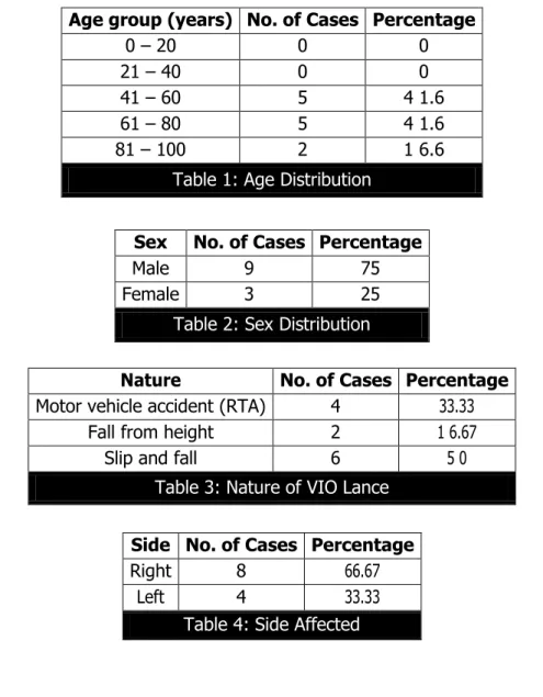
RESULTS: The following observations were made from data collected during study.
Age group (years) No. of Cases Percentage
0 – 20 0 0
21 – 40 0 0
41 – 60 5 4 1.6
61 – 80 5 4 1.6
81 – 100 2 1 6.6
Table 1: Age Distribution
Sex No. of Cases Percentage
Male 9 75
Female 3 25
Table 2: Sex Distribution
Nature No. of Cases Percentage
Motor vehicle accident (RTA) 4 33.33
Fall from height 2 1 6.67
Slip and fall 6 5 0
Table 3: Nature of VIO Lance
Side No. of Cases Percentage
Right 8 66.67
Left 4 33.33
J of Evidence Based Med & Hlthcare, pISSN- 2349-2562, eISSN- 2349-2570/ Vol. 2/Issue 43/Oct. 26, 2015 Page 7735
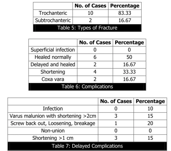
No. of Cases Percentage
Trochanteric 10 83.33
Subtrochanteric 2 16.67
Table 5: Types of Fracture
No. of Cases Percentage
Superficial infection 0 0
Healed normally 6 50
Delayed and healed 2 16.67
Shortening 4 33.33
Coxa vara 2 16.67
Table 6: Complications
No. of Cases Percentage
Infection 0 10
Varus malunion with shortening >2cm 3 15
Screw back out, Loosening, breakage 1 20
Non-union 0 0
Shortening >1 cm 3 15
Table 7: Delayed Complications
FOLLOW UP: All patients were followed at 2 months, 3 months, 6 months, At each follow up radiograph of operated hip with upper half femur was taken and assessed.
Functional results are assessed taking the 12 cases into consideration using salvati-wilson Hip Scoring System.5
Result No. of Cases Percentage
Excellent 0 0
Good 3 25
F air 6 50
Poor 1 8.33
Table 8: Functional Result of Trochanteric Fracture
Result No. of Cases Percentage
Fair 2 16.67
J of Evidence Based Med & Hlthcare, pISSN- 2349-2562, eISSN- 2349-2570/ Vol. 2/Issue 43/Oct. 26, 2015 Page 7736
SUMMARY: In the present study, 12 cases of comminuted peritrochanteric fracture of femur were managed by Locking Compression Plate for proximal femur. The data obtained was analysed and results evaluated.
Average age incidence in the present study was 62.7 years.
Predominantly males (75%) were affected.
Most cases occurred after a fall 10 (50%) cases which was statistically significant.
Right side involvement was more common.
Average post-operative stay was 13.5 days.
Out of the 12 cases, evaluated using Salvati-Wilson scoring: 3 cases (25%) had good, 8 cases (66.67%) fair, 1 case (8.33%) had poor score.
Average weight bearing time was14.5 weeks
Average union rate was 19.45 weeks.
The development of LCP-PF for peritrochanteric fractures is a complex system and requires proper selection of patient, careful attention of factors like understanding of the biomechanical principle of the plate, patient factor and definite selection of the patients for the treatment as there were high complication rates with respect to the implant.
REFERENCES:
1. Singh A K. Management of trochanteric fractures. Indian J Orthop 2006; 40: 100-2.
2. Koval KJ et al. Post –operative Weight Bearing after a Fracture of the Femoral Neck or an Intertrochanteric Fracture; Journal of Bone and Joint Surgery, 1998; 80A: 352-364.
3. Canale and Beaty: Campbell”s Operative Orthopaedics, 11th edition, (2007); P. 3237-3241.
4. Parker MJ, Maheshwar CB. The Use of Hip Scores in assessing the results of treatment of proximal femur fractures; International orthopaedics, Spinger.1997 Feb; 21: 262-264.
4. Civil Assistant Surgeon, Department of Orthopedics, Area Hospital, Srikala Hasthi.
NAME ADDRESS EMAIL ID OF THE CORRESPONDING AUTHOR: Dr. S. Hari Babu,
Associate Professor,
Department of Orthopedics, S V. Medical College, Tirupati. E-mail: [email protected]
Date of Submission: 04/10/2015. Date of Peer Review: 05/10/2015. Date of Acceptance: 12/10/2015. Date of Publishing: 24/10/2015.
AUTHORS:
1. S. Hari Babu 2. K. Satish 3. G. Suresh Babu 4. L. Anand
PARTICULARS OF CONTRIBUTORS: 1. Associate Professor, Department of
Orthopedics, S V. Medical College, Tirupati.
2. Associate Professor, Department of Orthopedics, S V. Medical College, Tirupati.