The outcome in early Cases of Treatment of
Subtrochanteric Fractures with Proximal Femur locking
Compression Plate
u Gunadham*, MD, J Jampa, MD, S Suntornsup, MD, b leewiriyaphun, MD
AbSTrACT
The objective of this study was to evaluate the outcome in early treatment of subtrochanteric fractures with proximal femur locking compression plate (PF-LCP).The patients included in this study were those with subtrochanteric fractures (AO type 32A-C) treated with PF-LCP (Synthes) between Jan 2009 and Jun 2011. The patient characteristics and details of clinical conditions were obtained from records. Clinical and radiographic follow-ups were done at one, two, four and 6 months intervals, and at one year. The primary outcome studied included fracture union and functional ambulatory status. Twenty-six patients were included in the study, 19 of whom were male with a mean age of 42.4 years. Fourteen patients (53.9%) had sustained AO type 32B fractures, the majority in motor vehicle accidents. Twenty-two fractures (84.6%) achieved union, while sixpatients (23.1%) had complications such as broken plate, varus collapse, and broken screw. Four patients (15.4%) underwent a second operation. At the end of the follow-ups, 25 patients (96.2%) were community ambulators. We conclude that PF-LCP is an effective alternative treatment for subtrochanteric fractures when properly performed.
KEYWORDS
Subtrochateric fracture, Proximal Femur Locking
Compression Plate (PF-LCP), Outcome, Complication, Cerclage wire
InTroDuCTIon
Subtrochanteric fractures account for 10-34% of all hip fractures 1, 2.They are due to high energy trauma in the young
patients, while in elderly patients, they are often caused by low energy trauma in osteoporotic bone.2 Surgical
treatment is the preferred method for subtrochanteric
femoral fractures and a variety of implants are used. These implants fall into two main categories, intramedullary and extramedullary. Intramedullary ixation is associated with short operative time and minimal blood loss and has better biomechanical properties when compared with extramedullary ixation. However, they have their own technical dificulties and complications 1, 3.
Extramedullary devices such as dynamic condylar screws and 95° condylar blade-plates provide strong ixation in the cancellous bone of the neck and head with considerable rotational stability. Their disadvantages are longer operating time, technically demanding -, extensive devascularization, higher infection rate-, delayed weight bearing, medial instability, refracture after plate removal and surgical approach 4. The introduction of biologic, soft
tissue sparing techniques has made plate ixation of femur a viable option. Indirect reduction and submuscular plating of subtrochanteric fractures produce good results 5.
The locking compression plate was introduced in the 21st
century as a new implant that allowed angular-stable plate ixation for the treatment of complex comminuted and osteoporotic fractures in different anatomic regions 6, 7.
Recently proximal femoral locking compression plate has been applied in the treatment of proximal femur fracture including subtrochanteric fracture. Locking plates have the advantage of allowing multiple angular-stable ixation points into the proximal femur, while leaving a smaller ‘foot print’ by preserving more bone stock after implantation compared with the use of large proximal lag screws 8.
The Proximal Femur Locking Compression Plate (PF-LCP) was introduced in Thailand around 2006 and had increased in popularity ever since. Initially it had been used mostly in complex and unstable intertrochanteric
Corresponding Author: Ukris Gunadham, Trang Hospital. 69 Kokkan Rd. Tubtiang, Muang, Trang, Thailand. 92000 E-mail: [email protected]
Department of Orthopedics, Trang Regional Hospital, Trang, Thailand
fractures and later in subtrochanteric fractures. Until recently, there have been few clinical studies of PF-LCP and the results are somewhat different 9, 10. From previous study,
PF-LCP provides stable ixation with high union rate and few complications 10, 11, while some studies found high rate
of failure even when performed by experienced surgeons 9.
This study was conducted to review the early outcome of - cases of PF-LCP use in subtrochanteric fractures in a government hospital in Southern Thailand.
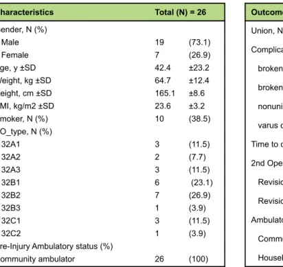
Characteristics Total (n) = 26
Gender, N (%)
Male 19 (73.1) Female 7 (26.9) Age, y ±SD 42.4 ±23.2 Weight, kg ±SD 64.7 ±12.4 Height, cm ±SD 165.1 ±8.6 BMI, kg/m2 ±SD 23.6 ±3.2 Smoker, N (%) 10 (38.5) AO_type, N (%)
32A1 3 (11.5) 32A2 2 (7.7) 32A3 3 (11.5) 32B1 6 (23.1) 32B2 7 (26.9) 32B3 1 (3.9) 32C1 3 (11.5) 32C2 1 (3.9) Pre-Injury Ambulatory status (%)
Community ambulator 26 (100)
Table I: Patient Characteristics
outcome Total (n) = 26
Union, N (%) 22 (84.6) Complication, N (%) 6 (23.1) broken plate 2 broken screw 1 nonunion 1 varus collapse 2 Time to complication, months ±SD 4.3 ±2.1 2nd Operation, N (%) 4 (15.4) Revision with PF-LCP 3 Revision with 135°ABP 1 Ambulatory status at end of follow-up, N (%) Community ambulator 25 (96.2) Household ambulator 1 (3.8)
Table II: outcomes
Table III: Comparative Analyses
Variables Cases without Cases with complication p-value
complication (n=6)
(n=20)
Gender, N
Male 14 5
Female 6 1 0.47 #
Age, y 46.0 ± 24.5 30.5 ± 19.1 0.16 * BMI, kg/m2 23.1 ± 3.3 25.3 ± 2.4 0.13 * Smoking, N
Smokers 7 3
Non-smokers 13 3 0.42 #
AO-OTA classiication, N
32A1-3 6 2
32B1-3 11 3
32C1-3 3 1 0.99 #
Kickstand screw, N
Used 17 6
Not used 3 0 0.44 #
Cerclage wire, N
Used 12 0
Not used 8 6 0.01 #
Good medial buttress, N
Present 16 3
Absent 4 3 0.18 #
Data are mean ± SD unless otherwise indicated. * p-value from unpaired t-test.
MATerIAlS AnD MeThoDS
This is a retrospective study of patients - aged 15 years or older, who had sustained subtrochanteric fractures (AO type 32A-C) and were treated with proximal femur locking compression plate (PF-LCP; Synthes) between Jan 2009 and Jun 2011. The patients with pathologic fractures (other than due to osteoporosis) were excluded. This study was approved by the institutional review board.
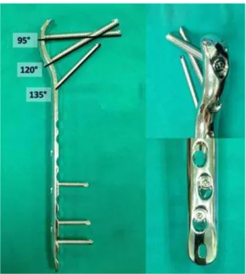
The PF-LCP is an angular-stable and limited contact plate speciically designed for treatment of complex, comminuted fractures of the per, inter and subtrochanteric femoral region. The plate is anatomically precontoured for the metaphysis of the proximal femur. The irst two proximal threaded holes of the plate are designed for cannulated 7.3-mm locking head screws that are inserted at 95° and 120° in relation to the shaft of the femur. The third threaded round hole is for a cannulated 5.0-mm locking head screw that is inserted at the level of the calcar at 135° angle, and this screw intersects with the most proximal 7.3-mm screw, serving as a so-called “kickstand screw”. (Figure 1.)
The remaining screw holes, which range from 4 to 16 in the PF-LCP, are LCP-combi-holes that allow the placement of either a conventional (4.5 mm) or a locking head screw (5.0 mm) at the level of the shaft. The most distal hole allows the use of a Kirschner wire for temporary ixation to achieve correct positioning of the plate.
Fig. 1: The locking compression plate for the proximal femur is a precontoured, angular stable, with large fragment screw (7.3/5.0/4.5mm).
Fig. 2: Example of case which the “kickstand” screw was not applied. Pre-operative (A), Immediate post-operative (B), 2-month post-post-operative (C), 4-month post-operative (D).
Surgery is performed with the patient supine under general anesthesia with traction on a fracture table. Through a standard lateral approach luoroscopically guided reduction and distal submuscular plate advancement were feasible. Proximal ixation was obtained at least with the two most proximal screws (locking or conical/ nonlocking). A “kickstand screw” was used routinely. Cerclage wiring was used to aid fracture reduction in some cases. Distal ixation was obtained with a minimum of three locking screws.
Postoperatively, patients were allowed - toe-touch weight bearing for the irst eight weeks, partial weight bearing after callus formation in radiographs, and weight as tolerated at 4 months. Clinical and radiographic follow-ups were done at one, two, four and sixmonth intervals, and at one year.
Radiographs of hip and femur were reviewed to obtain the AO-OTA classiication 12, ixation characteristics, medial
contact stability, and the use of cerclage wire and kickstand screw. Patient charts were reviewed to collect baseline characteristics and clinical outcomes. Primary outcome included fracture union and functional ambulatory status. Pre- and post-fracture ambulations were classiied based on standard deinitions of community and household ambulators 13. All community ambulators were able to
walk indoors and outdoors either independently or with assistive devices. Household ambulators were limited to indoor walking either independently or with assistive devices. Non-functional ambulators were either bed-bound or limited to bed-to-chair transfers with assistance.
Statistical analyses were performed with Stata version 10.0 (StataCorp, College Station, TX). Continuous data were analyzed using mean ± standard deviation (SD) and unpaired t-test. Non-continuous data were analyzed using proportion, percentage, and Fisher’s exact test. The p-value < 0.05 was considered as a statistical signiicance.
reSulTS
Twenty-six consecutive patients were included in this study. Nineteen patients (73.1%) were male and 7 patients (26.9%) were female. Mean age was 42.4 ±23.2 years, mean weight was 64.7 ±12.4 kilograms, mean height was 165.1 ±8.6 centimeters, and mean BMI was 23.6 ±3.2 kg/m2. Ten patients (38.5%) were smokers. Fourteen patients (53.9%) had AO type 32B fracture, while eight patients (30.8%) and four patients (15.4%) were classiied as AO type 32A and 32C respectively (Table I). Motor vehicle accidents - accounted for 19 patients (73.1%), low energy injuries - in four patients (15.4%), and falls from height - in three patients (11.5%). All patients were community ambulators prior to injury.
Average time to ixation was 7.7 ±3.9 days and average hospital stay was 14.5 ±5.6 days. Mean operative time was 109 ±22 minutes and mean blood loss was 619 ±276 ml. Cerclage wires were used in 12 patients (46.2%), while kickstand screws were used in 23 patients (88.5%). Nineteen patients (73.1%) achieved good medial buttress. Average of 2.7 proximal screws and 3.3 distal screws were applied (Table II).
Mean follow-ups was 11 ±6 months ( range 6 to 25 months). Twenty-two fractures (84.6%) achieved union, while six patients (23.1%) had complications. In two patients the plate had broken, two patients had varus collapse and one had broken screw and non-union of the fracture. Complications occurred in average of 4.3 ±2.1 months (range 2 to-7 months). Four patients (15.4%) underwent a second operation. Three patients had revision with PF-LCP and one patient underwent revision with 135° angled blade plate. At the end of the follow-ups, 25 patients (96.2%) were community ambulators, while one patient was household ambulator (Table II).
Comparative analyses between group of patients with and without complication were performed. No statistical signiicance was found among gender, age, BMI, history of smoking, AO-OTA classiication, the use of kickstand screw, or the presence of good medial buttress. However, it was discovered that cases with the use of cerclage wire had less complications than those without cerclage wire (0/12 in former group and 6/14 in latter group) with statistical signiicance (p-value = 0.01) (Table III).
DISCuSSIon
Stable subtrochanteric fracture can be treated successfully with conventional implants, such as sliding hip screws, cephalomedullary nails, and angular blade plates. However, comminuted and unstable subtrochanteric fractures are challenging injuries that are prone to complications 1,2.
Intramedullary device such as cephalomedullary nails showed increased fracture stability when compared to extramedullary devices. 14 In cadaveric study, cephalomedullary nail construct in the treatment of subtrochanteric fractures would be consistently superior biomechanically to either a PF-LCP construct or a 95°angled blade plate construct when a considerable fracture gap persists. Furthermore, in the insertion of cephalomedullary nail a large portion of bone had to be removed from the proximal femur with unknown long-term effects 15.
to prevent complication from subtrochanteric fracture, current trends are moving forward to biological ixation and subcutaneous insertion which PF-LCP 5,11,17. Until
recently, there are few clinical results of PF-LCP from previous studies which would be expected -for a newly developed implant.9, 10 Our study attempted to collect data
of early cases of PF-LCP in subtrochanteric fractures in a level-II trauma center in Southern Thailand.
From previous studies, the so-called kickstand screw played an important role in preventing varus collapse of the construct
9, 18.In most of our cases, the kickstand screw was utilized.
In cases where the 95° screw was not in the most superior position, the kickstand screw could not be applied (Figure 2.) However, the absence of kickstand screw was not associated with complications in our study. Besides the use of fracture table, indirect reduction of the fracture can be achieved with the supplement of circumferential wire.19, 20 Unlike
cephalomedullary nail, PF-LCP allows circumferential wire without additional incision. Cerclage wiring is an alternative technique to achieve reduction in dificult fractures 21, 22
From recent cadaveric study, cerclage wiring resulted in only minimal disruption of femoral blood supply 23.In this study,
cerclage wiring was used to achieve near-anatomic reduction in some cases and had less complication statistically compared with the group without cerclage wire (Figure 3). This may be attributable to the near perfect anatomical reduction and good medial buttress of the fracture site as well.
From the results of our study, although PF-LCP has theoretical advantages superior to other implants, the outcome of treatment was not promising. While 84.6% of fractures achieved union, 23.1% of fractures had complications and 15.4% underwent second operation. Compare with excellent results from previous studies (95-100% union rate) with less complication (1.8-12.5%) 10, 11, 18, the complications in this study may be due to patient
factors (i.e., poor bone quality, patient noncompliance) as well as technical factors (i.e., inadequate plate length, improper plate placement or screw ixation, lack of kickstand screw, short screw length, possibly lack of medial buttress of the fracture site) and also the lack of surgeon’s familiarity with the implant. At the end of the follow- up, 25 patients (96.2%) had returned to their pre-morbid level as community ambulators which were comparable with previous study.11
The authors propose- that PF-LCP may be suitable implant in unstable and comminuted subtrochanteric fractures, fractures with extension to greater trochanter which non-feasible with cephalomedullary nail. In order to achieve promising result from PF-LCP, the surgeon should focus on adequate plate length, near-anatomical reduction with or without circumferential wire, good medial buttress of fracture site, use of kickstand screw, biologic friendly or subcutaneous insertion when possible. In developing countries such as Thailand, there are many considerations regarding implant choices. Although there is universal coverage through the National Health Care System, locking compression plates are among the high-cost implants which need cost-beneit considerations before application.
Limitation of this study are the small number of patients and less surgeon’s expertise with the implants which may affect the outcome. Further studies are needed to show the outcome and the effectiveness of this method of ixation in future cases after the have surgeons gained experiences with using the implants.
ConCluSIon
The PF-LCP represents a feasible alternative for the treatment of subtrochanteric fractures when properly performed. Further clinical studies are necessary to show its role in the treatment of these fractures.
ACKnoWleDGeMenT
We thank Dr. Patarawan Woratanarat, MD, PhD (Clinical Epidemiology) from Faculty of Medicine Ramathibodi Hospital for her kind review of our manuscript.
ConFlICT oF InTereST STATeMenT DeClArATIonS
reFerenCeS
1. Craig NJ, Maffulli N. Subtrochanteric fractures: current management options. Disabil Rehabil. 2005
;27(18-19):1181-90.
2. Robinson CM, Court-Brown CM, McQueen MM, Christie J. Hip fractures in adults younger than 50 years of age.
Epidemiology and results. Clin Orthop Relat Res. 1995 (312): 238-46.
3. Bojan AJ, Beimel C, Taglang G, Collin D, Ekholm C, Jonsson A. Critical factors in cut-out complication after
gamma nail treatment of proximal femoral fractures. BMC Musculoskelet Disord. 2013; 14(1): 1.
4. Burnei C, Popescu G, Barbu D, Capraru F. Intramedullary osteosynthesis versus plate osteosynthesis in
subtrochanteric fractures. J Med Life. 2011 14; 4(4): 324-9.
5. Celebi L, Can M, Muratli HH, Yagmurlu MF, Yuksel HY, Bicimoglu A. Indirect reduction and biological internal ixation of comminuted subtrochanteric fractures of the femur. Injury. 2006; 37(8): 740-50.
6. Egol KA, Kubiak EN, Fulkerson E, Kummer FJ, Koval KJ. Biomechanics of locked plates and screws. J Orthop
Trauma. 2004; 18(8): 488-93.
7. Gautier E, Sommer C. Guidelines for the clinical application of the LCP. Injury. 2003; 34(2): B63-76.
8. Streubel PN, Moustoukas MJ, Obremskey WT. Mechanical failure after locking plate ixation of unstable intertrochanteric femur fractures. J Orthop Trauma. 2013; 27(1): 22-8.
9. Glassner PJ, Tejwani NC. Failure of proximal femoral locking compression plate: a case series. J Orthop Trauma.
2011; 25(2): 76-83.
10. Zha GC, Chen ZL, Qi XB, Sun JY. Treatment of pertrochanteric fractures with a proximal femur locking compression
plate. Injury. 2011; 42(11): 1294-9.
11. Saini P, Kumar R, Shekhawat V, Joshi N, Bansal M, Kumar S. Biological ixation of comminuted subtrochanteric fractures with proximal femur locking compression plate. Injury. 2012; 29: 345.
12. Muller ME. [Classiication and international AO-documentation of femur fractures]. Unfallheilkunde. 1980 ;83(5):251-9.
13. Koval KJ, Skovron ML, Aharonoff GB, Meadows SE, Zuckerman JD. Ambulatory ability after hip fracture. A
prospective study in geriatric patients. Clin Orthop Relat Res. 1995; (310): 150-9.
14. Kummer FJ, Olsson O, Pearlman CA, Ceder L, Larsson S, Koval KJ. Intramedullary versus extramedullary ixation of subtrochanteric fractures. A biomechanical study. Acta Orthopaedica Scandinavica. 1998; 69(6): 580-4.
15. Forward DP, Doro CJ, O’Toole RV, Kim H, Floyd JC, Sciadini MF, et al. A biomechanical comparison of a locking
plate, a nail, and a 95 degrees angled blade plate for ixation of subtrochanteric femoral fractures. J Orthop Trauma. 2012; 26(6): 334-40.
16. Latii MH, Ganthel K, Rukmanikanthan S, Mansor A, Kamarul T, Bilgen M. Prospects of implant with locking plate in ixation of subtrochanteric fracture: experimental demonstration of its potential beneits on synthetic
femur model with supportive hierarchical nonlinear hyperelastic inite element analysis. Biomed Eng Online.
2012; 11: 23.
17. Neogi DS, Trikha V, Mishra KK, Rohilla N, Yadav CS. Biological plate ixation of comminuted subtrochanteric fractures with the Dynamic Condylar Screw: a clinical study. Acta Orthop Belg. 2009; 75(4): 497-503.
18. Hasenboehler EA, Agudelo JF, Morgan SJ, Smith WR, Hak DJ, Stahel PF. Treatment of complex proximal femoral
fractures with the proximal femur locking compression plate. Orthop 2007; 30(8): 618-23.
19. Ban I, Birkelund L, Palm H, Brix M, Troelsen A. Circumferential wires as a supplement to intramedullary
nailing in unstable trochanteric hip fractures: 4 reoperations in 60 patients followed for 1 year. Acta Orthop.
20. Muller T, Topp T, Kuhne CA, Gebhart G, Ruchholtz S, Zettl R. The beneit of wire cerclage stabilisation of the medial hinge in intramedullary nailing for the treatment of subtrochanteric femoral fractures: a biomechanical
study. Int Orthop. 2011; 35(8): 1237-43.
21. Apivatthakakul T, Phornphutkul C, Bunmaprasert T, Sananpanich K, Fernandez Dell’Oca A. Percutaneous cerclage
wiring and minimally invasive plate osteosynthesis (MIPO): a percutaneous reduction technique in the treatment
of Vancouver type B1 periprosthetic femoral shaft fractures. Arch Orthop Trauma Surg. 2012;132(6): 813-22.
22. Apivatthakakul T, Phornphutkul C. Percutaneous cerclage wiring for reduction of periprosthetic and dificult femoral fractures. A technical note. Injury. 2012; 43(6): 966-71.
23. Apivatthakakul T, Phaliphot J, Leuvitoonvechkit S. Percutaneous cerclage wiring, does it disrupt femoral blood