RESEARCH ARTICLE
TSPO, a Mitochondrial Outer Membrane
Protein, Controls Ethanol-Related Behaviors
in
Drosophila
Ran Lin1, Danielle Rittenhouse1, Katelyn Sweeney1, Prasanth Potluri1, Douglas C. Wallace1,2*
1Center for Mitochondrial and Epigenomic Medicine, Children’s Hospital of Philadelphia Research Institute, Philadelphia, Pennsylvania, United States of America,2Department of Pathology and Laboratory Medicine, Perelman School of Medicine, University of Pennsylvania, Philadelphia, Pennsylvania, United States of America
*wallaced1@email.chop.edu
Abstract
The heavy consumption of ethanol can lead to alcohol use disorders (AUDs) which impact patients, their families, and societies. Yet the genetic and physiological factors that predis-pose humans to AUDs remain unclear. One hypothesis is that alterations in mitochondrial function modulate neuronal sensitivity to ethanol exposure. UsingDrosophilagenetics we report that inactivation of the mitochondrial outer membrane translocator protein 18kDa (TSPO), also known as the peripheral benzodiazepine receptor, affects ethanol sedation and tolerance in male flies. Knockdown of dTSPO in adult male neurons results in increased sensitivity to ethanol sedation, and this effect requires the dTSPO depletion-mediated increase in reactive oxygen species (ROS) production and inhibition of caspase activity in fly heads. Systemic loss of dTSPO in male flies blocks the development of tolerance to repeated ethanol exposures, an effect that is not seen when dTSPO is only inactivated in neurons. Female flies are naturally more sensitive to ethanol than males, and female fly heads have strikingly lower levels of dTSPO mRNA than males. Hence, mitochondrial TSPO function plays an important role in ethanol sensitivity and tolerance. Since a large array of benzodiazepine analogues have been developed that interact with the peripheral benzodiazepine receptor, the mitochondrial TSPO might provide an important new target for treating AUDs.
Author Summary
Alcohol use disorders (AUDs) affect millions of patients worldwide and result in high social and economic burdens. Although environmental factors are involved, there are clear genetic components to AUDs. Both the acute sedating effect of alcohol exposure and alco-hol tolerance contribute to long term risk for alcoalco-hol dependence and addiction. Yet the genetic etiology of AUDs remains to be determined. The mitochondria play a central role in ethanol metabolism and are important in many aspects of cellular physiology such as
OPEN ACCESS
Citation:Lin R, Rittenhouse D, Sweeney K, Potluri P, Wallace DC (2015) TSPO, a Mitochondrial Outer Membrane Protein, Controls Ethanol-Related Behaviors inDrosophila. PLoS Genet 11(8): e1005366. doi:10.1371/journal.pgen.1005366
Editor:Bingwei Lu, Stanford University School of Medicine, UNITED STATES
Received:February 27, 2014
Accepted:June 17, 2015
Published:August 4, 2015
Copyright:© 2015 Lin et al. This is an open access article distributed under the terms of theCreative Commons Attribution License, which permits unrestricted use, distribution, and reproduction in any medium, provided the original author and source are credited.
Funding:This work was supported by NIH Grants NS21325, DK73691 and R33CA182384 plus Simons Foundation Grant 205844 awarded to DCW. The funders had no role in study design, data collection and analysis, decision to publish, or preparation of the manuscript.
REDOX and ROS regulation, and apoptosis. The mitochondrial outer membrane translo-cator protein 18 kDa (TSPO) binds the benzodiazepines and perhaps other addictive drugs, and thus may play a role in AUDs. SinceDrosophilais a well-established model for ethanol-related behaviors, we have developed systems for manipulating theDrosophila tspogene and protein. With these systems, we have discovered that neuronal TSPO con-trols sensitivity to ethanol sedation via ROS and caspase-mediated signaling and that sys-temic TSPO levels are important in the development of tolerance to repeated ethanol exposure. Given the variety of known TSPO ligands, and the common mechanisms of var-ious abusive substances, our studies suggest that TSPO might be a promising target to combat alcoholism as well as addiction to other drugs.
Introduction
Alcohol is one of the most widely used drugs worldwide, but long term consumption leads to its abuse and dependence. An estimated 17.6 million adults in the United States have AUDs with associated health concerns of alcohol dependence, liver cirrhosis, cancer, and injuries. From 2006 through 2010, this generated an annual average of about 88,000 alcohol-related deaths and 2.5 million years of potential life lost [1,2].
To develop therapeutic strategies for alcoholism it will be necessary to determine the molec-ular and cellmolec-ular mechanisms underlying AUDs. Considerable effort has been invested in determining the role of the central nervous system in the etiology of AUD [3–5] but many fea-tures of the AUDs remain unexplained.
Neuronal function is highly dependent on mitochondrial bioenergetics [6,7]. In addition to the direct metabolizing of ethanol, the mitochondria are central to a wide range of essential neuronal cell functions including ATP synthesis, ROS production and REDOX homeostasis, Ca2+ buffering, and the metabolic regulation of apoptosis [8–10]. In humans mitochondrial DNA (mtDNA) alterations have been correlated with alcoholism, involving both acute ethanol responses and chronic damage [11–16]. In rodents, hepatic mtDNA depletion is seen in alco-hol exposed mice [17] and mtDNA complex I gene variants have been correlated with“ non-drinker”versus“drinker”rat lines derived from the same founder strain [18]. Variation in the mtDNA genes have also been shown to have profound effects of nuclear gene expression [19]. In previous studies we showed that the nuclear DNA codedDrosophilatranslocator protein 18kDa (dTSPO, CG2789) is localized in outer mitochondrial membrane (OMM) and impor-tant for regulating mitochondria bioenergetics, ROS production, caspase activity, and apoptotic function [20]. In humans, TSPO ligands are widely used in neuroimaging for neurodegenera-tive diseases and neuronal injuries, both of which are associated with increased brain TSPO lev-els and distribution [21]. As the previous nomenclature (peripheral benzodiazepine receptor, PBR) implies, TSPO binds the benzodiazepines and other psychotrophic drugs associated with tolerance and addiction [22]. Thus we hypothesized the TSPO may be an important factor in addiction to ethanol.
Drosophila’ssensitivity and tolerance to ethanol are similar to humans and rodents. Ethanol results in biphasic locomotor alterations. At lower doses ethanol acts as a stimulant, but at higher doses it acts as a depressant [23]. After repeated alcohol stimulation, tolerance is devel-oped, defined as acquired resistance. Tolerance is thought to be an intermediate step to alcohol dependence and addiction [24].
Furthermore, systemic but not neuronal loss of dTSPO inhibits the development of tolerance. By contrast, females are constitutively more sensitive to ethanol sedation than males and they have much lower dTSPO mRNA in their brains. Therefore, the mitochondrial TSPO is an important mediator of ethanol sensitivity and tolerance and contributes to gender-specific dif-ferences in alcohol sensitivity.
Results
Neuronal depletion of dTSPO increases ethanol sensitivity in adult male
flies
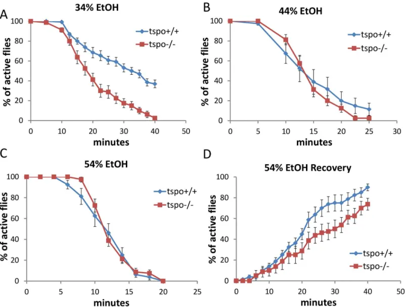
Acute ethanol sensitivity was analyzed by placing flies in vials closed by cotton clogs soaked with varying concentrations of ethanol thus exposing the flies to ethanol vapor. During initial exposure the flies flew to the top of the vial, and exhibited hyperactivity for a few minutes. With longer exposure, the flies became sedated and remained at bottom of the vial without locomotion. Wild type flies became comparably sedated whether the ethanol-soaked clogs were at the top of the vials or the vials were inverted with clogs at the bottom (S1 Fig). More-over, theDrosophilashowed a dose-dependent response to ethanol using this protocol (Fig 1). Therefore, the ethanol effects observed in the following experiments were due to the ethanol concentration rather than an environmental factor such as hypoxia due to ethanol vapor exclu-sion of air.
Thetspo[EY00814] mutantDrosophilahas a P-element inserted into thetspogene leading to loss of dTSPO expression [20]. Maletspo-/- flies exhibited higher sedation sensitivity than
tspo+/+ flies when exposed to ethanol vapor from 34% ethanol solution (Fig 1A) while at 44% or 54% ethanol vapor both thetspo-/- andtspo+/+ flies exhibited the same sensitivity (Fig 1B and 1C). Post sedation, we tested for the recovery from ethanol sedation by replacing the etha-nol-soaked clogs with normal clogs. This revealed that at 54% ethanol exposuretspo-/- males were slower to recover than thetspo+/+ male flies (Fig 1D). Thus,tspo-/- male flies are more sensitive to ethanol sedation than theirtspo+/+ counterparts. While therempAgene overlaps with thetspogene,rempA-/- deficiency is not the cause of the ethanol phenotypes since
rempA-/- flies exhibit comparable sensitivity to 34% ethanol vapor as wild type flies (S2 Fig). Since thetspomutation is present in all developmental stages of the fly, it could act through creating a developmental abnormality. However, Hematoxylin-Eosin histological comparison of the brains of male tspo-/- and +/+ flies did not reveal any gross anatomical differences (S3 Fig).
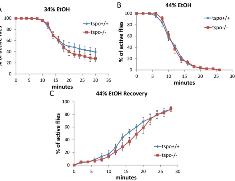
To determine whether the increased ethanol sensitivity was attributable to dTSPO function in neurons, we depleted dTSPO in neurons by inducing dsRNA (RNAi) to knockdown dTSPO mRNA in adult flies following eclosion (days after eclosion, dae). We used the Gal4-GeneS-witch/UAS system [25] in which Gal4 is activated within the flies when fed with mifepristone (RU486). The activated Gal4 binds to the UAS of the UAS-dTSPO-RNAi which induces the dsRNA expression and inhibition of the dTSPO mRNA. Since the Gal4 element is expressed under the neuronal specific elav promoter (elav-GeneSwitch), this switch was restricted to neu-rons. In this way, the flies were permitted to progress through larval and pupal development with normal TSPO activity, and following eclosion, the dTSPO RNAi was induced in neurons by exposure to RU486. Male flies harboring both elav-GeneSwitch and UAS-dTSPO-RNAi (elav-GS/+; TSPO-IR/+)(GS means‘Gene Switch’and IR means‘Inverted Repeats’) cassettes that were exposed to RU486 post eclosion had reduced head dTSPO mRNA as quantified by RT-PCR (Fig 2A). Therefore, activation of the elav-GS/+; TSPO-IR/+ system with RU486 spe-cifically depletes dTSPO mRNAs in the neurons.
In parallel with the whole body knockouts, the elav-GS/+; TSPO-IR/+ RU486 knockdown male flies exhibited faster ethanol sedation in the presence of 44% ethanol vapor than did flies who were not exposed to RU486 (Fig 2B). RU486 exposure of flies harboring only the neuronal elav-GeneSwitch (elav-GS/+) or the UAS-dTSPO-RNAi (TSPO-IR/+) cassette had no effect on the ethanol sensitivity (Fig 2C and 2D). Similarly, elav-GS/+; TSPO-IR/+ male flies exposure to 34% ethanol also showed increase sedation after RU486 induction relative to uninduced flies (S4A Fig). After 55% ethanol sedation (S4B Fig), the RU486-induced flies were slower to recover (S4C Fig). The difference between the RU486 induced and uninduced flies was not due to differential alcohol absorption or metabolism since after a brief exposure to 44% ethanol vapor both groups of fly heads (with and without RU486) had the same ethanol concentration Fig 1. Increased ethanol sensitivity in maletspomutant flies.Sensitivity to acute ethanol sedation was increased intspo-/- flies compared withtspo
+/+ flies (A-C). (A) With 34% ethanol solution, half sedation time fortspo+/+ was 30.6±1.9 min and fortspo-/- was 17.8±1.5 min, p = 0.01, n = 13 vials tested.
(B) With 44% ethanol solution, half sedation time fortspo+/+ was 14.3±2.2 min and fortspo-/- was 13.8±0.6 min, p>0.05, n = 8 vials tested. (C) With 54% ethanol solution, half sedation time fortspo+/+ was 11.3±0.8 min and fortspo-/- was 11.3±0.3 min, p>0.05, n = 8. (D). The rate for recovery after ethanol withdraw was slower intspo-/- thantspo+/+ flies, half recovery time fortspo+/+ was 20.4±2.1 min and fortspo-/- was 28.1±3.6 min, p>0.05, n = 8. Data
presented as mean±SEM.
(S4D Fig). Hence, dTSPO inactivation in adult neurons is sufficient to sensitize male flies to ethanol exposure.
Male and female flies exhibit sexual dimorphic response to ethanol exposure [4] and this sexual dimorphism was also observed in the brains of the TSPO knockout and knockdown flies. Maletspo-/- flies showed an increased sensitivity to ethanol sedation relative totspo
+/+ flies with 34% ethanol exposure and delayed recovery from 54% sedation (Fig 1A and 1D) while femaletspo-/- andtspo+/+ flies showed no difference in their response to 34% ethanol exposure (Fig 3A–3C).
In elav-GS/+; TSPO-IR/+ female flies, after neuronal inactivation of dTSPO by dsRNA expression, there was no effect on the sedation rate with exposure to 34% or 44% ethanol solu-tion (S5A and S5B Fig). Furthermore, only slightly delayed recovery was seen for female flies Fig 2. High male brain expression of TSPO associated with increased ethanol sensitivity in male neuronal dTSPO knockdown flies.(A) Levels of dTSPO mRNA in the heads of elav-GS/+; TSPO-IR/+ male and female flies with or without RU486 dsRNA induction, n = 3 groups of flies tested. Data presented as mean±SEM.***p<0.001. (B-D) Gene switch and control flies with or without RU486 (elav-GS/+ = elav-GeneSwitch and TSPO-IR/+ = UAS-dTSPO-RNAi). To induce gene switch, the flies were raised on regular food with 50μl of 4 mg/ml RU486 added on the surface of the food in vials for three days. (B) Sensitivity of elav-GS/+;TSPO-IR/+ flies to 44% ethanol vapor with and without RU486, half sedation time with RU486 was 16.0±0.6 min and without RU486 was 23.3±1.5, p<0.001, n = 10. (C) Sensitivity of flies harboring elav-GS/+ with or without RU486 exposed to 44% ethanol vapor, half sedation time with RU486 was 14.0±0.5 min and without RU486 was 15.3±0.9, p>0.05, n = 13, vials tested. (D) Sensitivity of flies harboring only TSPO-IR/ + exposed to 44% ethanol and with and without RU486, half sedation time with RU486 was 25.0±2.0 min and without RU486 was 22.5±1.0, p>0.05, n = 10.
doi:10.1371/journal.pgen.1005366.g002
after 44% vapor sedation (S5C Fig). The marked difference between male and female elav-GS/+; TSPO-IR/+ flies’sensitivity to ethanol following dTSPO inactivation by RU486 induction correlated with male fly heads having about four times the level of dTSPO mRNA as female heads. Moreover, neuronal knockdown of dTSPO reduced male head TSPO mRNA level but had no effect on female head TSPO mRNA level (Fig 2A). Therefore, the lack of sensitivity of female flies to neuronal inactivation of dTSPO is likely do to a gender-specific lack of TSPO in female fly brains.
Increased ROS mediated sensitivity to ethanol in dTSPO-depleting flies
To determine what might be the physiological basis of the neuron-specific effects of dTSPO deficiency on male ethanol sedation, we examined the effects on ROS production, which we previously found was increased in dTSPO deficient mitochondria [20]. ROS has been identified Fig 3. Comparable ethanol sensitivity in femaletspo-/- andtspo+/+ flies.(A) With 34% ethanol vapor, half sedation time fortspo+/+ was 23.0±2.5 minand fortspo-/- was 20.0±1.7 min, p>0.05, n = 14 vials tested. (B) With 44% ethanol vapor half sedation time fortspo+/+ was 11.0±0.5 min and fortspo-/- was 11.0±0.5 min, p>0.05, n = 10. (C) Recovery after withdraw from exposure to 44% ethanol vapor, half recovery rate fortspo+/+ was 16.0±1.2 min and for
tspo-/- was 18.0±1.2 min, p>0.05, n = 8. Data presented as mean±SEM.
as modulator of neuronal activity [26]. Using Amplex Ultrared to determine the amount of H2O2in fly heads, we found that H2O2levels were higher in male elav-GS/+; TSPO-IR/+ flies
treated with RU486 than untreated flies (Fig 4A). Hence, neuronal dTSPO knockdown increased fly head H2O2production. When these flies were fed with N-Acetyl-L-Cysteine
(NAC), an efficient antioxidant, the enhanced sedation effect of the dTSPO knockdown flies to 44% ethanol vapor was negated (Fig 4B). Intspo+/+ male flies, exposure of 44% ethanol vapor for 20 minutes resulted in sedation of most of the flies but did not significantly alter H2O2
con-tent in fly heads (Fig 4C). Hence, the increase in ROS is not caused by ethanol exposure. Fig 4. Depletion of dTSPO in neurons increases ROS which is necessary to sensitize flies to ethanol sedation.(A) H2O2content was higher in heads
of elav-GS/+; TSPO-IR/+ flies with RU486 induction of dTSPO dsRNA, n = 8 groups of flies tested. (B) Inhibition of increased sedation sensitivity to 44% ethanol vapor of elav-GS/+; TSPO-IR/+ flies treated with RU486 when treated together with the antioxidant N-Acetyl-L-Cysteine (NAC) (red versus blue curve). To induce gene switch, the flies were raised on regular food with 50μl of 4 mg/ml RU486 added on the surface of the food in vials for three days. To inhibit ROS production, 20μl of 500 mM NAC was premixed with the 50μl RU486 solution and the mixture added to the surface of the food in vials for five days. Half sedation time for control feeding flies was 17.6±1.3 min, for RU486-only feeding flies was 12.9±1.2 min, for NAC-only feeding flies was 15.9±1.5 min, for RU486 and NAC feeding flies was 17.6±1.6 min. Control versus RU486-only flies, p = 0.0182; NAC versus NAC+RU486 flies, p = 0.4292, n = 8, vials
tested. (C) H2O2content of heads of male wild type flies after 20 minutes exposure to 44% ethanol vapor (n = 3 groups of flies tested). Data presented as
mean±SEM.***p<0.001.
doi:10.1371/journal.pgen.1005366.g004
Rather, dTSPO inactivation in neurons up-regulates ROS and the increased ROS is responsible for the enhanced ethanol sensitivity of the dTSPO-depleted flies.
Aging-associated sensitivity to ethanol sedation is attenuated in
dTSPO-depleting flies
Since ethanol sedation sensitivity was controlled by TSPO and TSPO expression declines in
tspo+/+ flies to a minimum at 30 dae (S5B Figof [20]), we determined whether ethanol sensi-tivity changes during aging. Maletspo+/+ andtspo-/- flies were tested with 44% and 54% etha-nol sedation at different ages i.e. young (about 5 dae), mid-age (about 20 dae), and old (about 35 dae). Wild type (tspo+/+) flies exhibited increased sedation as they aged, with the effect already evident by 20 dae.tspo-/- flies also displayed and increased predilection to sedation with age (S6C Fig), but they were initially significantly more sensitive to ethanol. This is consis-tent with their higher level of oxidative stress as demonstrated by the marked reduction in their ROS-sensitive mitochondrial aconitase activity (Fig 6of [20]).
Neuronal dTSPO depletion suppresses caspase activity which is
sufficient to produce ethanol sensitivity
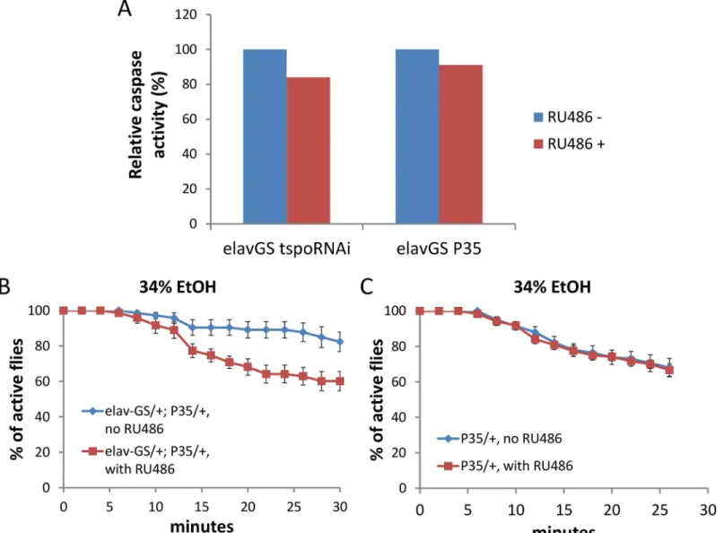
Depletion of dTSPO in flies suppresses caspase activation and impedes apoptosis [20]. How-ever, caspase also has cell death-independent functions which might be involved in neuronal control [27]. The activity of caspase 3/7, the most downstream caspase in the intrinsic apopto-sis pathway, was decreased in heads of flies with dTSPO-depleted neurons (Fig 5A). Neuronal expression of the caspase inhibitor protein, p35, also reduced caspase 3/7 activity to a similar degree as induction of the TSPO dsRNA (Fig 5A). The level of caspase reduction in TSPO knockdown and p35 induced neurons is likely to be much greater than shown inFig 5Awhere whole brain homogenates were assayed. Whole brain homogenates mix the enzymes of all cell types most of which are not neurons and thus not subject to dTSPO knockdown. Supporting this speculation, caspase 3/7 activity of whole body homogenatetspo-/- flies was tenfold lower than that oftspo+/+ flies (Fig 5, legend). Neuronal expression of the caspase inhibitor protein, p35, also increased sensitivity of male flies to ethanol sedation when exposed to 34% ethanol vapor (Fig 5B and 5C). This phenocopyied the dTSPO knockdown flies and confirmed the importance of reduced neuronal caspase 3/7 in ethanol sensitivity. Hence, both increased ROS production and decreased caspase activity in neurons are important in enhanced ethanol sensitivity.
Systemic but not neuronal depletion of dTSPO prevents flies from
ethanol tolerance
To determine if the effect of dTSPO on tolerance is attributable to neurons, we compared elav-GS/+; TSPO-IR/+ flies with or without RU486 to induce TSPO dsRNA. Knockdown of dTSPO in adult male neurons had no effect on the development of tolerance following a second ethanol exposure to 54% ethanol vapor (Fig 6B). Hence the suppression of tolerance in
dTSPO-depleting flies was not driven by neuronal dTSPO levels.
Since there might be other cell types in which dTSPO functions in tolerance formation, we isolated the heads and bodies of maletspo+/+ flies to examine the expression of dTSPO during tolerance. Within 4 hours after first exposure to 54% ethanol vapor, the amount of dTSPO mRNA in heads was decreased while the dTSPO mRNA in bodies was markedly increased. Fig 5. Depletion of dTSPO in neurons suppresses caspase activity which is sufficient to increase ethanol sensitivity.Gene switch was
accomplished as inFig 2. (A) Caspase 3/7 activity was moderately reduced in heads of elav-GS/+; TSPO-IR/+ or elav-GS/+; p35/+ (elav-GeneSwitch plus UAS-p35) flies when the dTSPO dsRNA or p35 were induced with RU486. All groups were measured twice, and data presented as mean. Since the TSPO-IR and P53 products are only expressed in neurons, but the caspase activity was assayed in whole heads, the ~20% decrease in caspase 3/7 underrepresents the extent of caspase reduction in neurons. This is demonstrated by whole body knockout of TSPO in which the relative whole body caspase 3/7 activity oftspo+/+ flies was 1.000±0.008 and oftspo-/- flies was 0.078±0.015, p<0.001, n = 4. (B) Induction of caspase inhibitor p35 with
RU486 significantly increased sensitivity to 34% ethanol vapor, Chi Square log rank test, p = 0.0006, n = 8 vials tested. (C) Flies harboring only UAS-p35 (p35/+) exposed to 34% ethanol vapor with or without RU486 were not different, Chi Square log rank test, p = 0.846, n = 12. Data presented as mean±SEM.
doi:10.1371/journal.pgen.1005366.g005
Both head and body dTSPO mRNA levels began to normalize at 6 hours post exposure (Fig 6C and 6D). Therefore, tolerance is associated with the induction of dTSPO in fly bodies, which is consistent with the loss of the capacity to develop tolerance intspo-/- flies but not in elav-GS/+; TSPO-IR/+ induced flies.
Discussion
We have found that TSPO is a mitochondrial modulator of ethanol sensitivity and tolerance in
Drosophila. Inactivation of dTSPO in either the whole body or in adult fly neurons conferred increased sensitivity of males but not females to ethanol exposure. The increased ethanol sensi-tivity associated with dTSPO deficiency is a product of increased ROS production and
Fig 6. Systemic loss of dTSPO inhibits the development of ethanol tolerance.Tolerance is revealed as a longer period required for sedation at a second exposure to 54% ethanol solution vapor. (A) Differential sedation from first versus second ethanol exposure oftspo+/+ versustspo-/- flies: Fortspo+/+ flies half sedation time for first exposure was 12.0±0.5 and for second exposure was 26.4±1.9, p<0.001. Fortspo-/- flies half sedation time for first exposure was 12.3±0.3 and for second exposure was 15.0±1.6, p = 0.11. Difference between second exposure sedation oftspo+/+ andtspo-/- flies, p<0.001, n = 8, vials tested. (B) Differential sedation from first versus second exposure of elav-GS/+; TSPO-IR/+ flies with or without RU486 induction, n = 12. Gene switch was induced as inFig 2. (C) dTSPO mRNA levels in the heads of tspo+/+ male flies after exposure to 54% ethanol vapor showing progressive loss of dTSPO mRNA for the first 4 hours after exposure followed by partial recover to original levels, n = 3 groups of flies tested. (D) dTSPO mRNA levels in the bodies of maletspo+/+ flies after exposure to 54% ethanol vapor showing a progressive increase up to 4 hours after exposure followed by a decline (n = 3). Data presented as mean±SEM.
decreased caspase activity in neurons. However, inhibition of the development of ethanol toler-ance was related to systemic but not neuronal TSPO levels and this correlated with the induc-tion of TSPO mRNA in fly bodies on ethanol exposure. Hence, our results show that TSPO is an essential mediator of alcohol sensitivity and tolerance, though not involving all the same tis-sue types.
Involvement of ROS and caspase in dTSPO-modulated ethanol
sensitivity
The involvement of TSPO-mediated increased neuronal ROS production and decreased cas-pase activity in the sensitivity to ethanol sedation is consistent with reports that oxidative stress and caspase-mediated apoptosis contribute to brain pathology [28]. Since TSPO controls mito-chondrial ROS production and caspase activation [20], it follows that modulation of ROS levels and caspase activity could mediate ethanol sensitivity.
Inactivation oftspoincreased ROS production and NAC negated the enhanced sensitivity to ethanol demonstrating that increased neuronal ROS is related to increased ethanol sedation sensitivity. Given the short exposure period of the flies to ethanol, the ROS effect is most likely due to its second messenger action [26] rather than due to a cell death mechanism. This is con-sistent with the recent report showing that expression of oxidative stress genes can be altered by ethanol exposure and their functions are essential for ethanol sensitivity [29,30]. It is possi-ble that TSPO-deficiency induced ROS production could also participate in development of tolerance, but this effect must be mediated by cells other than neurons.
Inactivation of dTSPO also inhibits caspase activity[20] and inhibition of neuronal caspase activity also sensitized flies to ethanol sedation. This was confirmed by expression of the cas-pase inhibitor p35 resulting in increased ethanol sensitivity. Since cascas-pase has been shown to function in neuronal apoptosis-independent pathways to control neuronal activity in both developmental and adult stages[27], it is reasonable to conclude that dTSPO depletion in fly neurons activates such pathways thus altering neuronal activity and ethanol response.
Gender-specificity of TSPO modulation on ethanol response
The male-specific effects of TSPO inactivation were particularly striking. Previous studies have shown that male flies are more resistant to ethanol-induced sedation than females [31], which we also observed. Inactivation of dTSPO in males increased their sensitivity to ethanol, bring-ing their sensitivity close to that of females. Furthermore, female flies were found to have much lower dTSPO mRNA in their heads than males and knockdown of neuronal dTSPO in male heads reduced dTSPO mRNA about 20% while having no effect on the dTSPO levels of female fly heads. Thus, female flies have inherently low expression of dTSPO in their neurons and this may account for to their increased sensitivity to ethanol sedation.
In humans, men and women also exhibit different responses to acute and long-term ethanol exposure [32,33]. Men are at higher risk of AUD than women, but once AUD develops, women are more susceptible to ethanol-induced damage in multiple organs. Perhaps differ-ences in TSPO expression contribute to human gender differdiffer-ences as well.
The molecular basis for the differences in dTSPO expression in flies is unknown. Male flies express a male specific splicing isoform of neuronal sex determination gene fruitless (fru), FruM. This may control the gender-specific production of neurotransmitters and neuropep-tides [31]. Such a system might also regulate dTSPO expression. Additional environmental factors to which male and female animals are differentially exposed may also affect dTSPO expression.
TSPO modulates ethanol sensitivity in adult flies
A variety of genes have been reported to control fly brain development and impact ethanol responses [5]. Since thetspomutation affects all developmental stages in fly, it’s deficiency could create a developmental abnormality that alter ethanol sensitivity. However, Hematoxy-lin-Eosin histological staining oftspo-/- brains did not reveal any gross anatomical defect com-pared withtspo+/+ brains. Furthermore, by using the RU486-inducible gene switch system to knockdown dTSPO only after eclosion, we avoided any alterations in fly anatomy demonstrat-ing that only physiological changes were important in ethanol sensitivity of adults. Hence, the ethanol sensitivity induced in male flies by the knockdown of dTSPO cannot be due to develop-mental alterations, but must be the product of the physiology of the adult neurons. This means that physiological modulation should be able to treat alcoholism.
The knockdown of dTSPO in neurons demonstrates that neuronal expression of TSPO is important in determining ethanol sensitivity. This neuronal action of TSPO is at variance with reports in mammals that TSPO probes co-localize primarily with glial [34,35]. That dTSPO must be expressed in neurons is not only confirmed by the current ethanol studies but also by our previous observations that systemic depletion of dTSPO protects flies from toxicity of neu-ronally-expressed Aβ42 [20]. Unfortunately, our current data do not indicate if the ethanol sensitivity effects of dTSPO knockdown are related to a specific group of neurons. Mammalian TSPO has been reported to function in hippocampal neurons to affect long-term potentiation and learning[36]. Also, ethanol effects have been reported for the KCNQ channel expressed in dopaminergic neurons[37] and PKA expressed in insulin-producing neurons [38].
Possible involvement of TSPO in addiction of benzodiazepines
Benzodiazepines are widely used for treatment of anxiety, insomnia, seizures and other neural disorders, and are known to enhance the effects of GABA at the GABAAreceptor. However,
long-term use of these drugs is controversial due to decreasing effectiveness, physical depen-dence, and withdrawal [39,40]. TSPO is also a target of benzodiazepines and our results suggest that benzodiazepine-derived antagonists might increase sensitivity to ethanol and decrease neurological damage [20] while benzodiazepine-derived agonists could have the opposite effects. Consequently, the TSPO may provide an important drug target for treatment of drug abuse and alcoholism [36] which could be conveniently investigated with the current system.
Conclusion
Our data demonstrate that the mitochondrial TSPO protein, also known as the peripheral ben-zodiazepine receptor, is important in determining both ethanol sensitivity and the develop-ment of ethanol tolerance. Given the existing of a broad range of benzodiazepine analogues, these compounds may provide a novel approach for treating AUDs.
Materials and Methods
Fly stocks and culture
backcrossed to w1118(isoCJ1) background. UAS-p35 stock was kindly provided by Dr. Nancy Bonini in University of Pennsylvania. TherempA[e02928] strain was also obtained from Bloomington Drosophila Stock Center.
To induce gene switch, flies combining elav-GeneSwitch with UAS-dTSPO-RNAi (elav-GS/+;TSPO-IR/+) or UAS-p35 (elav-GS/+;UAS-p35) were raised in regular food with 50μl
4 mg/ml ethanol solution of mifepristone (RU486, Sigma-Aldrich, St Louis, MO, USA) added on the surface of food in vials for 3 days. As control, the flies were raised in regular food with 50μl ethanol. The food vials were changed every 24 hours. In N-Acetyl-L-Cystein (NAC)
experiments, 20μl 500 mM NAC (Sigma-Aldrich) water solution or pure water was pre-mixed
with 50μl RU486 solution or ethanol solvent and then added on the surface of food in vials. In
NAC experiments, the drug feeding was extended to 5 days.
Ethanol sedation assay
Flies at 4–7 dae age were used for all sedation, recovery and tolerance assays, except for the NAC experiment where 6–9 dae flies were used. In the aging experiments, 19–22 or 34–37 dae flies were studied. Flies were sorted under CO2and loaded into empty narrow (25×95mm)
vials. Ten flies were loaded into a vial as a single trial and allowed to recover for at least 2 hours before use. Ethanol solutions of 34%, 44%, 54% (weight/vol) were made by mixing absolute ethanol (Sigma-Aldrich, catalog number E7023, for molecular biology) and ultrapure distilled water (Gibco, Grand Island, NY, USA) at the ratio (vol/vol) of 4:6, 5:5, and 6:4, respectively. For each vial, regular cotton clog was replaced with clog added with 1 ml ethanol solution at the vial-side surface. Recording to number of sedated flies started immediately. The interval for recording was 2 or 5 minutes.
To monitor the recovery, the ethanol-containing clog was replaced with regular clog imme-diately after all flies were sedated. The number of flies remaining sedated was counted every 2 or 5 minutes. For tolerance assays, the flies were transferred into regular food vial with regular clog after all flies were sedated. Four hours later, the flies were transferred back into empty vial and the recording for sedation was performed as same as in naïve flies.
Internal ethanol content assay
Internal ethanol content was measured with Abcam Ethanol Assay Kit (Ab65343, Abcam, Cambridge, MA, USA). In brief, twenty flies at 4–7 dae age were CO2anesthetized and loaded
into empty narrow vial. After 2 hours recovery, flies were exposed to ethanol vapor from cotton clog soaked with 44% ethanol solution for 6 minutes when>90% flies were inactive. Then flies were quickly frozen in liquid nitrogen and homogenized in lysis buffer provided by kit and then centrifuged for 14000 g for 10 min in 4°C. The diluted sample together with standard eth-anol samples were incubated in 96-well plate wells with etheth-anol oxidation reaction mix to pro-duce H2O2which further reacts with the probe in the mix to generate color. The absorbance at
570 nm was measured with a plate reader (SpectraMax Paradigm, Molecular Devices, Sunny-vale, CA, USA). The content of ethanol was calculated based on the standard curve, and finally normalized by the total protein concentration measured by the Bradford method.
Quantitative RT-PCR
Total RNA was extracted from bodies of 20–40 flies or 100 fly heads using RNeasy Mini Kit (Qiagen, Valencia, CA, USA). The RNA was converted to cDNA using oligo(dT)15 (Invitro-gen, Grand Island, NY, USA) and SuperScript II reverse transcriptase (Invitrogen). After reverse transcription, PCR reactions were performed using a ViiA7 Real-Time PCR System (Applied Biosystems, Grand Island, NY, USA) with SYBR Green Master Mix (Applied
Biosystems) and primers for rp49 (forward, 5- gctaagctgtcgcacaaatg -3, and reverse, 5- ccag-gaacttcttgaatccg -3) or dTSPO (forward, 5- ctcttcgtaccctacgtcgc -3, and reverse, 5- ctggttcga-taggtcggaaa -3). The PCR protocol involved denaturation at 95°C for 15 seconds and combined annealing and extension at 60°C for 1 min over 40 cycles. The melting curve was generated after these cycles to ensure that the amplification in each reaction was specific.
Caspase 3/7 activity
Isolated fly heads or whole bodies were homogenized in Homogenization Buffer (225 mM mannitol, 75 mM sucrose, 10 mM MOPS, 1 mM EGTA, pH 7.2) on ice, then centrifuged at 300 g for 5 min. The supernatant was collected and added in 96-well plate wells together with an equal volume of reaction buffer (ApoONE kit, Promega, Fitchburg, WI, USA). The plate was shaken gently for 5 min, and then incubated in dark for 15 hours in room temperature. Fluo-rescence was measured with a plate reader (SpectraMax Paradigm, Molecular Devices) with the excitation at 499 nm and emission at 521 nm. The fluorescent values were normalized by total protein concentration measured by the Bradford method, and the relative activity was cal-culated based on the ratio of normalized fluorescent signals between samples.
Hydrogen peroxide content
Isolated fly heads were homogenized in Homogenization Buffer on ice. The samples were then centrifuged at 14000 g for 10 min in 4°C to collect the supernatant. The standard reaction solu-tion containing 0.1 mM Amplex UltraRed, Invitrogen and 0.2 U/L horseradish peroxidase (Thermo Scientific, Pittsburgh, PA, USA) diluted in Homogenization Buffer was placed in 96-well plate wells. Then the fly extract samples or standard H2O2samples were added to the
plates and incubated for 15 min in the dark at room temperature. The fluorescence was mea-sured with a plate reader (SpectraMax Paradigm, Molecular Devices) with the excitation at 530 nm and emission at 590 nm. The H2O2content was calculated based on standard and
normal-ized to the total protein concentration measured by the Bradford method.
Histological staining
Fly heads were fixed in standard Bouin's Fixative, embed in paraffin blocks, and sectioned at a thickness of 6μm. Sections were placed on slides, stained with haematoxylin and eosin
(Vec-tor), and examined by bright-field microscopy.
Supporting Information
S1 Fig. Equal ethanol sedation sensitivity in male flies when ethanol vapor was given bot-tom-up or top-down.Wild type (tspo+/+) male flies were tested for ethanol sedation in vials with 44% ethanol solution-soaked cotton clog closed at top (top-down vapor) or bottom (bot-tom-up vapor). N = 7 vials tested. Data presented as mean ± SEM.
(TIFF)
S2 Fig. Comparable ethanol sedation sensitivity ofrempA-/- and +/+ male flies.Sedation sensitivity to acute exposure of 34% ethanol vapor was comparable inrempA-/- and wild type flies.tspo-/- male flies exhibited increased sedation sensitivity under same condition. Data pre-sented as mean ± SEM. N = 11.
S3 Fig. Similar gross anatomical appearance of the brains of tspo-/- and +/+ flies.Head sec-tions of male adult flies (5–8 dae) were stained with haematoxylin and eosin.
(TIFF)
S4 Fig. Increased ethanol sensitivity in male neuronal dTSPO knockdown flies.Gene switch was induced as inFig 2. (A-C) Male elav-GS/+; TSPO-IR/+ flies with and without RU486. (A) Sensitivity to acute sedation from 34% ethanol vapor, n = 4 vials tested. (B) Sensitivity to acute sedation from 54% ethanol vapor, n = 12. (C) Delayed recovery following sedation with 54% ethanol vapor, n = 6. (D) Internal ethanol content in whole bodies of elav-GS/+; TSPO-IR flies with or without RU486, n = 3 groups of flies tested. Data presented as mean ± SEM.
(TIFF)
S5 Fig. Comparable ethanol sensitivity in female neuronal dTSPO knockdown flies to con-trol flies.Female elav-GS/+; TSPO-IR/+ flies with or without RU486. Gene switch was induced as inFig 2. (A) Sensitivity to acute sedation from 34% ethanol vapor, n = 4, number of vials tested. (B) Sensitivity to acute sedation from 44% ethanol vapor, n = 8. (C) Slight delay in recovery following sedation with 44% ethanol vapor, n = 8. Data presented as mean ± SEM. (TIFF)
S6 Fig. Aging-associated increase in sensitivity to ethanol sedation is attenuated in male tspo-/- flies.Young (4–7 dae), mid-age (19–22 dae) and old (34–37 dae) maletspo+/+ and
tspo-/- flies were exposed to (A) 44% ethanol vapor or (B) 54% ethanol vapor. The differential increased resistance seen intspo+/+ flies relative totspo-/- flies is lost when thetspo+/+ flies exceed 20 dae. N = 2~5 vials tested for each trace. Data presented as mean ± SEM.
(TIFF)
S7 Fig. Ethanol tolerance is not altered in femaletspo-/- flies compared withtspo+/+ flies.
Femaletspo-/- andtspo+/+ flies showed similar increased resistance to first and second expo-sure to 44% ethanol vapor indicating that a systemic lack of dTSPO does not show the same loss of tolerance as seen in male dTSPO deficient flies. First exposure, n = 10 vials tested; sec-ond exposure, n = 4. Data presented as mean ± SEM.
(TIFF)
Acknowledgments
The authors would like to recognize the contributions of Dr. Nancy Bonini of University of Pennsylvania, the Vienna Drosophila RNAi Center (VDRC), and the Bloomington Stock Cen-ter for generously providing fly stocks.
Author Contributions
Conceived and designed the experiments: RL DCW. Performed the experiments: RL DR KS PP. Analyzed the data: RL DCW. Wrote the paper: RL DCW. Conceived, initiated, and super-vised TSPO project: DCW.
References
1. Grant BF, Dawson DA, Stinson FS, Chou SP, Dufour MC, et al. (2004) The 12-month prevalence and trends in DSM-IV alcohol abuse and dependence: United States, 1991–1992 and 2001–2002. Drug and Alcohol Dependence 74: 223–234. PMID:15194200
2. Stahre M, Roeber J, Kanny D, Brewer RD, Zhang X (2014) Contribution of excessive alcohol consump-tion to deaths and years of potential life lost in the United States. Preventing Chronic Disease 11: E109. doi:10.5888/pcd11.130293PMID:24967831
3. Devineni AV, Eddison M, Heberlein U (2013) The novel gene tank, a tumor suppressor homolog, regu-lates ethanol sensitivity in Drosophila. The Journal of Neuroscience 33: 8134–8143. doi:10.1523/ JNEUROSCI.3695-12.2013PMID:23658154
4. Devineni AV, Heberlein U (2013) The evolution of Drosophila melanogaster as a model for alcohol research. Annual Review of Neuroscience 36: 121–138. doi:10.1146/annurev-neuro-062012-170256 PMID:23642133
5. Kaun KR, Devineni AV, Heberlein U (2012) Drosophila melanogaster as a model to study drug addic-tion. Human Genetics 131: 959–975. doi:10.1007/s00439-012-1146-6PMID:22350798
6. Mattson MP, Liu D (2002) Energetics and oxidative stress in synaptic plasticity and neurodegenerative disorders. Neuromolecular Medicine 2: 215–231. PMID:12428812
7. Li Z, Okamoto K, Hayashi Y, Sheng M (2004) The importance of dendritic mitochondria in the morpho-genesis and plasticity of spines and synapses. Cell 119: 873–887. PMID:15607982
8. Wallace DC (2005) A mitochondrial paradigm of metabolic and degenerative diseases, aging, and can-cer: a dawn for evolutionary medicine. Annual Review of Genetics 39: 359–407. PMID:16285865
9. Wallace DC, Fan W (2010) Energetics, epigenetics, mitochondrial genetics. Mitochondrion 10: 12–31. doi:10.1016/j.mito.2009.09.006PMID:19796712
10. Wallace DC, Fan W, Procaccio V (2010) Mitochondrial energetics and therapeutics. Annual Review of Pathology 5: 297–348. doi:10.1146/annurev.pathol.4.110807.092314PMID:20078222
11. Lease LR, Winnier DA, Williams JT, Dyer TD, Almasy L, et al. (2005) Mitochondrial genetic effects on latent class variables associated with susceptibility to alcoholism. BMC Genetics 6 Suppl 1: S158. PMID:16451619
12. Tsuchishima M, Tsutsumi M, Shiroeda H, Yano H, Ueshima Y, et al. (2000) Study of mitochondrial DNA deletion in alcoholics. Alcoholism, Clinical and Experimental Research 24: 12S–15S. PMID:10803772
13. von Wurmb N, Oehmichen M, Meissner C (1998) Demonstration of the 4977 bp deletion in human mito-chondrial DNA from intravital and postmortem blood. Mutation Research 422: 247–254. PMID: 9838148
14. Fromenty B, Carrozzo R, Shanske S, Schon EA (1997) High proportions of mtDNA duplications in patients with Kearns-Sayre syndrome occur in the heart. American Journal of Medical Genetics 71: 443–452. PMID:9286453
15. Mansouri A, Fromenty B, Berson A, Robin MA, Grimbert S, et al. (1997) Multiple hepatic mitochondrial DNA deletions suggest premature oxidative aging in alcoholic patients. Journal of Hepatology 27: 96– 102. PMID:9252080
16. Cahill A, Cunningham CC, Adachi M, Ishii H, Bailey SM, et al. (2002) Effects of alcohol and oxidative stress on liver pathology: the role of the mitochondrion. Alcoholism, Clinical and Experimental Research 26: 907–915. PMID:12068261
17. Demeilliers C, Maisonneuve C, Grodet A, Mansouri A, Nguyen R, et al. (2002) Impaired adaptive resyn-thesis and prolonged depletion of hepatic mitochondrial DNA after repeated alcohol binges in mice. Gastroenterology 123: 1278–1290. PMID:12360488
18. Sapag A, Gonzalez-Martinez G, Lobos-Gonzalez L, Encina G, Tampier L, et al. (2009) Polymorphisms in mitochondrial genes encoding complex I subunits are maternal factors of voluntary alcohol consump-tion in the rat. Pharmacogenet Genomics 19: 528–537. doi:10.1097/FPC.0b013e32832dc12aPMID: 19494790
19. Picard M, Zhang J, Hancock S, Derbeneva O, Golhar R, et al. (2014) Progressive increase in mtDNA 3243A>G heteroplasmy causes abrupt transcriptional reprogramming. Proceedings of the National Academy of Sciences of the United States of America 111: E4033–4042. doi:10.1073/pnas. 1414028111PMID:25192935
20. Lin R, Angelin A, Da Settimo F, Martini C, Taliani S, et al. (2014) Genetic analysis of dTSPO, an outer mitochondrial membrane protein, reveals its functions in apoptosis, longevity, and Aβ42-induced neuro-degeneration. Aging Cell 13: 507–518. PMID:24977274
21. Rupprecht R, Papadopoulos V, Rammes G, Baghai TC, Fan J, et al. (2010) Translocator protein (18 kDa) (TSPO) as a therapeutic target for neurological and psychiatric disorders. Nature Reviews Drug Discovery 9: 971–988. doi:10.1038/nrd3295PMID:21119734
22. Dell'osso B, Lader M (2013) Do benzodiazepines still deserve a major role in the treatment of psychiat-ric disorders? A critical reappraisal. European Psychiatry 28: 7–20. doi:10.1016/j.eurpsy.2011.11.003 PMID:22521806
24. Ghezzi A, Atkinson NS (2011) Homeostatic control of neural activity: a Drosophila model for drug toler-ance and dependence. International Review of Neurobiology 99: 23–50. doi: 10.1016/B978-0-12-387003-2.00002-1PMID:21906535
25. Osterwalder T, Yoon KS, White BH, Keshishian H (2001) A conditional tissue-specific transgene expression system using inducible GAL4. Proceedings of the National Academy of Sciences of the United States of America 98: 12596–12601. PMID:11675495
26. Mehta A, Prabhakar M, Kumar P, Deshmukh R, Sharma PL (2013) Excitotoxicity: bridge to various trig-gers in neurodegenerative disorders. European Journal of Pharmacology 698: 6–18. doi:10.1016/j. ejphar.2012.10.032PMID:23123057
27. D'Amelio M, Sheng M, Cecconi F (2012) Caspase-3 in the central nervous system: beyond apoptosis. Trends in Neuroscience 35: 700–709.
28. Brocardo PS, Boehme F, Patten A, Cox A, Gil-Mohapel J, et al. (2012) Anxiety- and depression-like behaviors are accompanied by an increase in oxidative stress in a rat model of fetal alcohol spectrum disorders: Protective effects of voluntary physical exercise. Neuropharmacology 62: 1607–1618. doi: 10.1016/j.neuropharm.2011.10.006PMID:22019722
29. Awofala AA, Davies JA, Jones S (2012) Functional roles for redox genes in ethanol sensitivity in Dro-sophila. Functional and Integrative Genomics 12: 305–315. doi:10.1007/s10142-012-0272-5PMID: 22430022
30. Logan-Garbisch T, Bortolazzo A, Luu P, Ford A, Do D, et al. (2014) Developmental ethanol exposure leads to dysregulation of lipid metabolism and oxidative stress in Drosophila. G3 (Bethesda) 5: 49–59.
31. Devineni AV, Heberlein U (2012) Acute ethanol responses in Drosophila are sexually dimorphic. Pro-ceedings of the National Academy of Sciences of the United States of America 109: 21087–21092. doi:10.1073/pnas.1218850110PMID:23213244
32. Miller MA, Weafer J, Fillmore MT (2009) Gender differences in alcohol impairment of simulated driving performance and driving-related skills. Alcohol and Alcoholism 44: 586–593. doi:10.1093/alcalc/ agp051PMID:19786725
33. Ceylan-Isik AF, McBride SM, Ren J (2010) Sex difference in alcoholism: who is at a greater risk for development of alcoholic complication? Life Sciences 87: 133–138. doi:10.1016/j.lfs.2010.06.002 PMID:20598716
34. Bae KR, Shim HJ, Balu D, Kim SR, Yu SW (2014) Translocator protein 18 kDa negatively regulates inflammation in microglia. Journal of Neuroimmune Pharmacology 9: 424–437. doi: 10.1007/s11481-014-9540-6PMID:24687172
35. Imamoto N, Momosaki S, Fujita M, Omachi S, Yamato H, et al. (2013) [11C]PK11195 PET imaging of spinal glial activation after nerve injury in rats. NeuroImage 79: 121–128. doi:10.1016/j.neuroimage. 2013.04.039PMID:23611861
36. Tokuda K, O'Dell KA, Izumi Y, Zorumski CF (2010) Midazolam inhibits hippocampal long-term potentia-tion and learning through dual central and peripheral benzodiazepine receptor activapotentia-tion and neuroster-oidogenesis. The Journal of Neuroscience 30: 16788–16795. doi:10.1523/JNEUROSCI.4101-10. 2010PMID:21159950
37. Cavaliere S, Gillespie JM, Hodge JJ (2012) KCNQ channels show conserved ethanol block and func-tion in ethanol behaviour. PLoS One 7: e50279. doi:10.1371/journal.pone.0050279PMID:23209695
38. Corl AB, Rodan AR, Heberlein U (2005) Insulin signaling in the nervous system regulates ethanol intox-ication in Drosophila melanogaster. Nature Neuroscience 8: 18–19. PMID:15592467
39. Farb DH, Ratner MH (2014) Targeting the modulation of neural circuitry for the treatment of anxiety dis-orders. Pharmacological Reviews 66: 1002–1032. doi:10.1124/pr.114.009126PMID:25237115
40. Starcevic V (2014) The reappraisal of benzodiazepines in the treatment of anxiety and related disor-ders. Expert Review of Neurotherapeutics 14: 1275–1286. doi:10.1586/14737175.2014.963057 PMID:25242262