Yazışma Adresi /Correspondence: Sevda Söker, MD, PhD, Dept. Histology and Embryology,
Medicine Faculty of Dicle University, Diyarbakır, Turkey, Email: [email protected] Geliş Tarihi / Received: 14.03.2011, Kabul Tarihi / Accepted: 11.04.2011
Copyright © Klinik ve Deneysel Araştırmalar Dergisi 2011, Her hakkı saklıdır / All rights reserved ORIGINAL ARTICLE / ÖZGÜN ARAŞTIRMA
The efect of extremely low frequency magnetic ield on heart tissue iron
density
Aşırı düşük frekanslı manyetik alanın kalp dokusu demir yoğunluğu üzerine
etkisi
Sevda Söker1, Cemil Sert2, Mustafa Deniz3, Ercan Ayaz1, Selçuk Tunik1,Yusuf Nergiz1
1Medicine Faculty of Dicle University, Department of Histology and Embryology, Diyarbakır, Turkey 2Department of Biophysics, Medicine Faculty of Harran University, Şanlıurfa, Turkey
3Department of Anatomy, Medicine Faculty of Harran University, Şanlıurfa, Turkey
ÖZET
Amaç: Bu histolojik çalışmanın amacı; aşırı düşük frekans-lı, düşük yoğunluklu manyetik alanın kalp dokusu demir yoğunluğu üzerindeki etkilerini araştırmaktır.
Gereç ve yöntem: Bu çalışmada; sham, kontrol ve deney grubu olarak üçe bölünen 45 Spraque Dawley erkek rat kullanıldı. Deney grubu günde üç saat 14 gün boyunca metakrilat kutularda 0.25 mT oldukça düşük frekans-lı manyetik alana (ELF-MF) maruz bırakıldı. Sham grubu, ELF-MF uygulaması dışında deney grubuna benzer şekil-de uygulamaya maruz bırakıldı. Kontrol grubu ratlara hiç-bir şey uygulanmadı ve fareler çalışma peryodu boyunca yaşam sikluslarını kafes içinde tamamladı. Yapılan işlem sonrasında, 50 mg/kg intramuskuler ketalar anestezisi uy-gulanarak ratlar sakriiye edildi Kalp dokuları hemen %10 nötral formalinde ikse edilerek parain bloklara gömüldü. Kalp dokusunun histolojik örnekleri Hematoksilen-Eosin ve demir pigmentleri için Perls’ Prussian blue boyası ile boyandı. Nikon DS-2MV fotomikroskopta histolojik pre-paratlar fotoğralandı.
Bulgular: Kontrol, sham ve deney grubumuzun histolo-jik yapısı normal gözlendi. Kontrol, sham ve deney grubu arasında kalp dokusu demir boyaması açısından farklılık gözlenmedi.
Sonuç: Çalışmamız sonucunda, kontrol ve ELF-MF (de-ney) grubu arasında farklılık saptamadık. Bu çalışmada, ELF-MF’ye maruz kalan farelerin kalp dokusunda demir boyaması ile değişiklik olmadığı saptandı. Klin Deney Ar Derg 2011;2(2):144-8
Anahtar Kelimeler: Aşırı düşük frekanslı elektromanyetik
alan, kalp, demir.
ABSTRACT
Objectives: The aim of this histological study was to vestigate the effects of extremely low frequency, low in-tensity magnetic ield on the heart muscle ıron density. Materials and methods: In this study, 45 male Spraque Dawley rats were introduced and were divided into three groups as sham, control and experiment group. The ex-perimental group was exposed to a 0.25 mT to Extremely Low Frequency Magnetic Field (ELF-MF) for 14 days, 3h a day in metacrylate boxes. The sham group was treated like the experimental group, except for ELF-MF exposure.
For control, nothing applied to rats in this group and they completed their life cycle in the cage during the study pe
-riod. After exposure period, the rats were sacriiced under ketalar anesthesia (50 mg / kg, intramuscularly). Heart tis-sues were immediately ixed in 10% neutral formaldehyde and embedded in parafin blocks. Histological sections from cardiac tissue stained by hematoxylin-eosin, Perls’ Prussian blue for iron pigments. Histological slides were photographed under a Nikon DS-2MV photomicroscope. Results: The architecture and histology of the control, sham and experimental group were observed as normal. No differences were observed between the control, sham
and experimental rat groups in the iron stain of heart tis -sues.
Conclusion: As a result of our study, we did not ob-serve differences between the control and ELF-MF (ex-perimental) group. In this investigation we demonstrated that the exposure of cardiac tissue of rats to the ELF-MF
did not change in the iron stain study. J Clin Exp Invest 2011;2(2):144-8
Key words: Extremely low frequency magnetic ield,
INTRODUCTION
In modern societies, humans are frequently exposed to magnetic ields, including extremely low frequen -cy, low intensity and high intensity magnetic ields. Low intensity magnetic ields are generally pro -duced by power lines and many kinds of electrical appliances. High intensity magnetic ields are pro -duced by the apparatus such as magnetic resonance imaging equipment. An increased health risk due to exposure to electromagnetic ields (EMF) at 50 and 60 Hz has been reported by several authors.1-4
Iron is an essential element in a variety of vital processes, including respiratory electron transfer, oxygen transport.5 However, when iron reacts with H2O2, hydroxyl radicals are produced via a Fenton-type reaction. The hydroxyl radical is the most powerful oxidizing species among several reactive-oxygen radicals, and is capable of oxidizing most macromolecules including nucleic acids, lipids, and proteins. Ferritin is the protein responsible for iron storage.6 Cellular uptake of circulating excess
iron results in increased formation of ferritin and hemosiderin found in highest concentrations in pa-renchymal tissue of several organs. As long as iron is bound to ferritin, cytotoxic reactions are not ex-pected to occur. When iron is released from ferritin, low molecular iron complexes may undergo redox reactions, thus inlicting cytotoxic damage upon macromolecules.7-8 Iron ions are strong catalysts for
the peroxidation of membrane lipids, and give rise to membrane damage.7,9,10
Electromagnetic ields have adverse effects as a result of widespread use of electromagnetic energy on biological systems. Several experimental and bi-ological studies have dealt especially with increased incidence of various types of cancer, including childhood leukemia, lymphomas, brain tumours and breast cancers, effects on reproduction and develop -ment and behavioral changes.1,11 In vivo and in vitro
investigations claim that extremely low-frequency magnetic ield produced a genotoxic effect, origi -nating from types of free radicals.12,13 For biological
effects of free radicals, especially reactive oxygen species (ROS) may produce cellular and toxic ef-fects such as lipid peroxidation in cell membrane, protein degradation, enzyme inactivation.12,13
In-creased iron content of cells and tissue may increase the risk of cancer. In particular, high available iron
cancer. Iron can catalyze the production of oxygen radicals.
Among the possible health effects of exposure to extremely low frequency magnetic ields, cardio -vasculer effects have been reported in the database of EMF bioeffects.14, 15 Recent studies have inves -tigated whether exposure to a magnetic ield poses a risk for cardiovascular morbidity and mortality.16, 17 A statistically signiicant relationship was found
between exposure to a magnetic ield and reduced heart rate variability, which leads to certain disor -ders such as acute myocardial infarctus and cardiac arrhythmia.18 The effects of ELF-MF have been in
-vestigated on heart muscle tissue limited number of histological studies. The aim of this study was to investigate the possible effects of ELF-MF on heart, and heart muscle tissue of the intended relationship with iron status.
MATERIALS AND METHODS
Animals
The research was designed and implemented ac -cording to the principles of the Declaration of Hel -sinki. This experimental research was performed with the approval of the ethics committee of Har -ran University. Spraque Dawley rats were obtained from the Medical Science Application and Research Center of Dicle University.
All animals were 2 months old at the beginning of the study, weighing 250-280 g, and were fed with Standard pellet food (Tavas Inc, Adana, Turkey). The rats were divided three groups of ifteen: Control, sham, and experimental groups. The experimental group was exposed to a 0.25 mT to Extremely Low Frequency magnetic ield (ELF-MF) in metacrylate boxes (17x17x25 inches). The experimental group (n=15) was exposed to ELF-MF for 14 days, 3h a day. The sham group was treated like the experimen -tal group, except for ELF-MF exposure. Treatment of the control group differed from the experimental and sham groups. Animals in the control group were kept in a 14/10 hour light/dark environment at con -stant temperature of 22±10% humidity.
Magnetic ield generation and exposure to magnetic ield
Helmholtz coils of 25 cm diameter. The frequency of sinusoidal current was 50 Hz. This magnet was constructed by winding 300 turns of insulated soft copper wire with a diameter of 0.85 mm. Coils were placed vertically and horizontally, facing one an-other. The distance between coils was 25 cm. The average MF intensity was measured as 0.25 mT ± 0.01 mT at 12 different points both transverse and axial within the metacarylate cage by using a digital hall effect Gauss meter (Bell 5170, SYPRIS, USA). The measurements were made by an independent researcher who was not involved in the animal ex -periment. No temperature difference was observed between exposure and sham coils during the expo -sure.
Histological examinations
The animals were euthanized after the inal expo -sure by anesthesia with ketalar (50 mg/kg, intra -muscularly). In the end of experiment, rats were sacriiced; cardiac tissue was ixed by 10% neutral formaldehyde and embedded in parafin blocks. Histological sections from cardiac tissue stained by Hematoxylin-Eosin, Perls’ Prussian blue for iron pigments. Nikon DS-2MV camera and NIS ele -ments 3.00 software were used for observation and documentation.
RESULTS
In the end of this study was to investigate the effect of electromagnetic ield on heart muscle. It is com -monly believed that more than a few ferritin parti-cles in cell cytoplasm is a predictor of iron overload. Histological sections were stained by Perl’s Prus -sian blue method for the observation of “ hemosid -erin granules”. Pearl’s Prussian blue (ferrocyanide method) staining made Fe+3 apparent. Histological examination in all three groups was carried out by light microscope. In this investigation we demon -strated that the exposure of cardiac tissue of rats to the ELF-MF did not change in the light microscopic study.
The control group and other groups in the archi -tecture and histology were observed as normal (Fig -ure 1, 2, 3). No differences between control, sham and experimental groups rat have been observed in the iron stain of heart tissues (Figure 4,5,6).
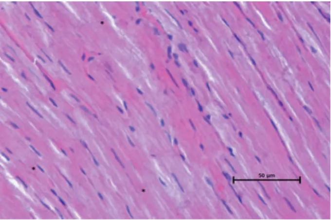
Figure 1. Photo micrograph of Experimental (Exposed to
magnetic ield) group. Note: Myoibrils (arrowhead) are seen (Hematoxylin-eosin staining, original magniication X 40).
Figure 2. Photo micrograph of Sham group. Note:
Myo-ibrils (arrowhead) are seen (Hematoxylin-eosin staining, original magniication X 40).
Figure 3. Photo micrograph of Control group. Note:
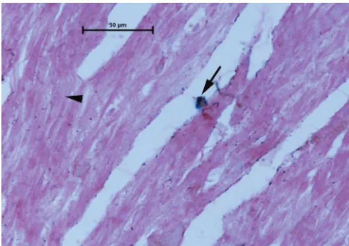
Figure 4. Photo micrograph of Experimental (Exposed to
magnetic ield) group. Note: Myoibrils (arrowhead) and iron accumulation (arrow) are seen (Perls’ Prussian blue staining, original magniication X 40).
Figure 5. Photo micrograph of Sham group. Note:
Myoi-brils (arrowhead) and iron accumulation (arrow) are seen (Perls’ Prussian blue staining, original magniication X 40).
Figure 6. Photo micrograph of Control group. Note: Iron
accumulation (arrow) are seen (Perls’ Prussian blue stain-ing, original magniication X 40).
DISCUSSION
Low frequency magnetic ields are widely applied in electrical appliances and different equipment such as television sets, computers and kitchen ap-pliances. Recently, low frequency magnetic ield has been considered to be a therapeutic agent and it has started to be more and more commonly used in medicine.14 Electromagnetic ields have adverse
effects as a result of widespread use of electromag-netic energy on biological systems. In recent years, a large number of multidisciplinary investigations led to the increasing awareness of the existence of multiple effects of MF in biological systems.19 The
responds of acute cardiovascular system to an elec-tric and magnetic ield is still being analyzed.
Several experimental and biological studies have dealt especially with increased incidence of various types of cancer.1,11 In vivo and in vitro in
-vestigations claim that extremely low-frequency magnetic ield produced a genotoxic effect, origi -nating from types of free radicals.12,13 For biologi
-cal effects of free radi-cals, especially ROS may produce cellular and toxic effects such as lipid per-oxidation in cell membrane, protein degradation, enzyme inactivation.12,13 Iron is an essential element
in a variety of vital processes, including respira -tory electron transfer, oxygen transport.5 However, when iron reacts with H2O2, hydroxyl radicals are
produced via a Fenton-type reaction. So, iron can catalyze the production of free radicals. Iron ions are strong catalysts for the peroxidation of mem-brane lipids, and give rise to memmem-brane damage.7,9,10 Ferritin is the protein responsible for iron storage.6
Cellular uptake of circulating excess iron results in increased formation of ferritin and hemosiderin found in highest concentrations in parenchymal tis-sue of several organs. Increased iron content of cells and tissue may increase the risk of cancer. In par -ticular, high available iron status may increase the risk of a radiation-induced cancer. Biological stud -ies have not yet provided direct evidence for a link between electromagnetic ields and cancer.19 Shao T
said the reasonable to link the biological effects of electromagnetic ields with ferritin gene expression and ferritin synthesis which are mainly regulated by iron, hormones and cAMP and eventually the EMF-cancer link.20
-mediating role in some biological effects of nonion-izing electromagnetic ields.12 Certainly, cellular
iron metabolism affects these processes. Genetic damage in cells can lead to malignancy and cancer. However, excessive cumulative genetic damages could also result in cell death.12 An epidemiologic
study by Savitz et al. on cardiovascular disease mor -tality in a cohort of electric utility workers showed an association between exposure to extremely low frequency (ELF) magnetic ields and cardiovascular disease mortality.18
In conclusion, extremely low frequency mag -netic ields are used in daily life. The effects of ELF-MF have been investigated on heart muscle tissue limited number of histological studies. In our study was to investigate the possible effects of EMF on heart, and cardiac muscle tissue of the intended re-lationship with iron status. As a result of our study, in the cardiac tissue of rat exposed to extremely low frequency magnetic ields, there was no change detected in the light microscopic study. In this in -vestigation we demonstrated that no differences between control, sham and experimental group rats have been observed in the iron stain of heart tissues. Administered dose and duration for the magnetic ields effects on the heart, although important, fur -ther studies are needed to demonstrate whe-ther the ELF-MF exposure can induce adverse effects on the cardiac tissue.
REFERENCES
1. Zecca L, Mantegazza C, Margonato V, et al. Biological ef
-fects of prolonged exposure to ELF electromagnetic ields in rats: III. 50 Hz Electromagnetic Fields. Bioelectromag
-netics 1998;19(1):57-66.
2. Coelho AM, Roger WR Jr, Smith HD. Effects of current ex
-posure to 60 Hz electric and magnetic ields on the social behavior of baboons. Bioelectromagnetics 1995; Suppl 3: 71-92.
3. Seegal RF, Wolpaw JR, Dowman R. Chronic exposure of pri
-mate to 60 Hz electric and magnetic ields: II. Neurochemi
-cal effects. Bioelectromagnetics 1989;10(3):289-301. 4. Svedenstal BM, Johanson KJ. Fetal loss in mice exposed to
magnetic ields during early pregnancy. Bioelectromagnet
-ics 1995; 16(5): 284-9.
5. Papanikolaou, G. and Pantopoulos, K. Iron metabolism and toxicity. Toxicol Appl Paharmacol 2005; 202 (2) : 199- 211.
6. Harrison PM, Arosio P. Ferritins molecular properties, iron storage function and cellular regulation. Biochim Biophys Acta 1996;1275 (3): 161-203.
7. Yoon JH, Lee MS, Kang JH. Reaction of ferritin with hy
-drogen peroxide induces lipid peroxidation. BMB Rep 2010;43(3):219-24.
8. Puntarulo S. Iron, oxidative stress and human health. Mol Aspects Med 2005; 26 (4-5): 299-312.
9. Valko M, Morris, H, Cronin MT. Metals, toxicity and oxida
-tive stress. Curr Med Chem 2005; 12 (10):1161- 208. 10. Ganguli A, Kohli HS, Khullar M, Lal Gupta K, Jha V,
Sakhuja V. Lipid peroxidation products formation with var
-ious iron preparations in chronic kidney disease. Ren Fail 2009; 31(2): 106-10.
11. Feychting M, Forssén U, Floderus B. Occupational and res
-idential magnetic ield exposure and leukemia and central nervous system tumors. Epidemiology 1997;8(4):384-9. 12. Lai H, Singh NP. Magnetic-ield-induced DNA strand
breaks in brain cells of the rat. Environ Health Perspect 2004 ;112(6):687-94.
13. Wolf FI, Torsello A, Tedesco B et al. 50-Hz extremely low frequency electromagnetic ields enhance cell proliferation and DNA damage: possible involvement of a redox mecha
-nism. Biochim Biophys Acta. 2005;1743(1-2):120-9. 14. Goraca A, Ciejka E, Piechota A. Effects of extremely low
frequency magnetic ield on the parameters of oxidative stress in heart. J Physiol Pharmacol 2010 61(3):333-8. 15. Borjanovic SS, Jankovic SM, Pejovic Z. ECG changes in
humans exposed to 50 Hz magnetic ields. J Occup Health 2005 ; 47(5):391-6.
16. Hakansson N, Gustavsson P, Sastre A, Floderus B. Occupa
-tional exposure to extremely low-frequency magnetic ields and mortality from cardiovascular disease. Am J Epidemiol 2003;158 (6): 534-42.
17. Sorahan T, Nicols L. Mortality from cardiovascular disease in relation to magnetic ield exposure: Findings from a study of UK electricity generation and transmission work
-ers, 1973-1997. Am J Ind Med 2004; 45 (1): 93-102. 18. Savitz DA, Liao D, Sastre A, et al. Magnetic ield exposure
and cardiovascular disease mortality among electric utility workers. Am J Epidemiol 1999;149(2):135-42.
19. Ventura C, Maioli M, Pintus G, Gottardi G, Bersani F. Elf-pulsed magnetic ields modulate opioid peptide gene expression in myocardial cells. Cardiovasc Res 2000 45 (4):1054-64.
20. Shao T. EMF-cancer link: the ferritin hypothesis. Med Hy