Today the cause of reproductive disorders in women frequently is the inflammation of pelvic organs. Chronic inflammation of the ovaries (oophoritis) is a common disease, accompanied by various gynecological dis-orders, and occurs independently as well. According to the Ministry of Healthcare of Ukraine, the frequency of inflammatory diseases of pelvic organs in the structure of pathology in gynecological practice reaches 60–65% [1]. As a consequence, prolonged and recurrent course of the disease in 60% of cases can lead to infertility. It should be noted that oophoritis largely occurs due to the presence of primary immunodeficiency, which facilitates the transition of acute inflammatory process at a chronic stage. At the same time prolonged inflammation causes a decrease in immunological reactivity of an
organism. In chronic inflammatory processes the change of cytokine profile is natural, which leads to statistically significant decline in the efficiency of infertility treatment [2].
During the treatment of uterine appenda ges’ inflammation the patients receive medicines of almost all pharmacological groups, inclu-ding the reserve antibiotics and non-steroidal anti-inflammatory drugs that lead to forma-tion of antibiotic-resistant strains, activaforma-tion of opportunistic flora, increased allergization, dysfunction of the immune system [1, 2]. As a rule the effectiveness of oophoritis treatment is low, and therefore the researches of alternative methods of treatment are intensively carried out, particularly the use of cell therapy. Taking into account the abovementioned it is reasonable to search for new medicines having the ability to impact on the chronic phase UDC 611.018.2/.6:615.014.41:618.11-002.2 doi 10.15407/biotech7.05.035
MULTIPOTENT MESENCHYMAL STROMAL CELLS
OF BONE MARROW IN THERAPY OF CHRONIC
INFLAMMATION OF MURINE OVARIES
Key words: ch ronic inflammation of ovaries, bone marrow multipotent mesenchymal stromal cells, cell therapy.
N. А. Volkova
M. S. Yukhta Institute for Problems of Cryobiology and Cryomedicine Т. А. Yurchuk of the National Academy of Sciences of Ukraine, Kharkiv E. D. Ivanova
L. V. Stepanuk E. V. Pavlovich L. V. Sokol
Email: [email protected]
Received 23.05.2014
of inflammation. The analysis of available reports indicates the relevance of research purposely to explore opportunities to improve reproductive function in animals and humans using cell-tissue therapy. For example, the cryopreserved placenta extract is used as a stimulant of reproductive function [3]. The question about the effectiveness of medicines, which include the cryopreserved components, such as stem cells or biologically active components, is timely and appropriate one for solving the task of correction of hormonal and reproductive dysfunction.
There is an evidence of positive impact of multipotent mesenchymal stromal cells (MMSCs) in treatment of gynecological diseases [4–7]. Carrying out the research, we used bone marrow derived MMSCs. It is known that MMSCs are identified by their ability to adhesion in vitro, differentiation into various types of tissues and the expression of several cell surface markers, among which the most typical are CD 105, CD 73, CD 90, at the absence of CD 34, CD 45 markers [8]. Modern technologies of culture and cryopreservation make possible receiving the stock of autological stem cells with a subsequent long-term storage at low temperatures without significant changes in functional status.
Existing data about the introduction of MMSCs into intact animals indicate immune modulating effect and diffuse distribution of donor’s material in the tissues of mesoderm origin [6, 9]. For assessment the impact of transplanted cellular material to restore the damaged tissue or organ histological, biochemical, biophysical, immunological methods are used, however, the location of the introduced cells is not always determined. According to this a simultaneous assessment of the ability of cryopreserved MMSCs to homing and stimulation of regenerative processes is important. This allows determining the term of a cell keeping in a tissue being restored, and the regenerative potential of the cells themselves.
The aim of this work was to investigate the effect of cryopreserved bone marrow derived multipotent mesenchymal stromal cells on reparative regeneration and to determine their localization under intravenously administration in the animals with chronic inflammation of the ovaries.
Materials and Methods
MMSCs of the femur bone marrow of mice (n = 5) was used in the study. The cells were isolated by washing out with Hanks solution
(PAA, Austria) followed by flushing through a needle with gradually decreased diameter. The next step was centrifugation at 1500 rpm (834 g) for 5 min. The cell suspension was re-suspended in culture medium and plated on a culture flask (PAA) with 103 cells per cm2 density. Cultural medium contained: Iskove’s Modified Dulbecco’s Mediume (PAA), 10% fetal bovine serum (FBS) (HyClone, USA), gentamicin (150 mg / ml) (Farmak, Ukraine) and amphotericin B (10 mg / ml) (PAA). Cultural medium was changed every 3 days. In the study we used standard conditions of culture at 37 °C in the atmosphere of 5% CO2. Cell culture was passaged after reaching a confluent monolayer.
Cell cryopreservation was carried out in cultural medium supplemented with 10% DMSO (PAA) and 20% FBS. 1 ml of suspension was placed in cryovials (Nunc, USA). Cryopreservation was performed with a programmable freezer ZPM-1 (SDTB with PP IPC&C NAS of Ukraine). Cooling rate was 1 deg/min down to –80 °C, with following transfer into liquid nitrogen [8]. Samples were stored under the low temperature bank conditions for 4 months, thawed on water bath at 40 °C up to the liquid phase. Removing of cryoprotectant was performed by slow addition of 1:9 Hanks solution (PAA), followed by centrifugation at 1500 rpm (834 g) for 5 min. Cell viability was assessed by exclusion of supravital trypan blue dye (Sigma-Aldrich, USA).
As experimental objects we used 50 outbred mature female white mice weighing 18–20 g. In order to cause chronic inflammation of the ovaries an inactivated vaccine of Staphylococcus aureus strain 209 was once injected intraperitoneally with insulin syringe and then mice were kept in the animal house conditions for 21 days without treatment. For vaccine we prepared a daily culture of Staphylococcus aureus by the standard method [10]. The resulted culture was washed out from agar with 5 ml of saline solution with titer determination by the method of standard dilutions and inactivated by incubation at 75 °C during 1h. After incubation the suspension of inactivated bacterial cells was diluted to 50×106 microbes’ bodies in 0.3 ml of saline solution per animal.
(CrMMSCs); experimental group 2 (n = 10) — bone marrow derived CrMMSCs labeled PKH-26. The volume of administrated liquid in all the experimental groups was 0.2 ml per animal (0.5×10⁵ viable CrMMSCs); control group 2 — intact animals (n = 10) of similar weight and age without manipulation. Insertion of PKH-26 (Sigma-Aldrich) in CrMMSCs was carried out according to the instructions of the manufacturer.
The animals were removed from the experiment to the 10th and 21st day after cell therapy by dislocation of the cervical vertebrae. For histological study and detection of entered cell location, ovaries were cut off with the surrounding tissue of omentum. Material for histological study was fixed in the 10% aqueous neutral formalin and then the serial paraffin sections with 4–5 µm thickness were done and stained with hematoxylin-eosin. The number of follicles and their development stages were classified as reported [11], modified by Gougeon [12] with the microscope. To assess the state of the oocytes they were mechanically obtained from the ovaries of animals of the control groups and experimental group 1 to the 21st day of therapy. Presence/ absence of apoptosis in the oocytes was assessed with Annexin-V (Becton-Dickinson, USA). Staining was performed according to the standard procedure of the manufacturer. Oocyte microimages were performed with the fluorescent microscope (MIKMED-2, Russia).
To investigate the labeled cell localization in the animal organism, the ovaries were isolated and the cryostat sections (7 µm) were prepared. The cryostat samples were investigated with fluorescent microscope. Autofluorescence was blocked with 0.3M glycine solution (PAA) by 20-min incubation and followed by microscopy [13]. For detection of labeled PKH-26 MMSCs frozen slices were additionally stained with 1 µg/ml DAPI (Sigma-Aldrich) for nuclei visualization. Results were fixed by photographing.
All the manipulations with the animals were carried out in accordance to the requirements of the “European Convention for the Protection of Vertebrate Animals used for Experimental and other Scientific Purposes” (Strasbourg, 1986), “General Principles of Animal Experiments”, approved by II National Congress of Bioethics (Kyiv, 2004), and the rules of the Commission of Bioethics of the IPC&C NAS of Ukraine.
In statistical analysis we used a single-factor analysis of variance and Student’s t-test using Statistica 8 software.
Results and Discussion
At the first stage the viability of MMSCs were assessed by trypan blue staining, the results were 95.2 ± 4.6% and 78.5 ± 6.2% for native and cryopreserved MMSCs, respectively.
The results of macroscopic research of an abdominal cavity showed that animals of the control and experimental groups had a normal blood supply in the vessels, a moderate amount of fat around the ovaries without visual signs of inflammatory reaction and exudation at all stages of observation.
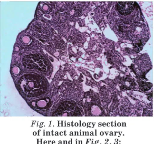
Histological structure of intact animals’ ovaries corresponded to the standards of mature mice with no signs of inflammatory changes (Fig. 1). Ovarian surface was covered with a single layer of cubical epithelium, under which tunica albuginea was located. Follicular structure of the samples was presented by primordial, primary, preantral and antral follicles that were separated by a stroma. The environment of antral follicles was homogeneous with weak eosin staining; oocytes were without signs of degeneration with evenly surrounded granulosa cells. Single atretic follicles with typical histological structure were defined on the periphery of sections. Corpora lutea were filled with radial strands of luteum cells.
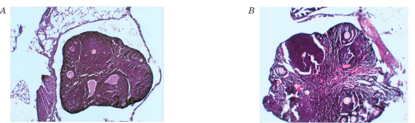
In the central part of histological sections of the ovaries of animals from group 1 with chronic inflammation after injection of saline solution the stromal fibroblast-like cells which were surrounded by single developing follicles were observed to the 10th day (Fig. 2, A). The leukocyte infiltration was observed at the edges and in the center of all the samples. It should be noted a significant amount of atretic follicles, which were characterized by the contour deformation. Atresia occurred according to the
Fig. 1. Histology section of intact animal ovary.
type of productive process with a multilayer membrana granulosa forming. Single primary follicles with the signs of degeneration were detected along the section periphery. In such follicles the layers of cuboidal granulosa cells were not only compressed but also separated and were less closely adjacent to an oocyte. It is known that disorder in a contact between oocyte and granulosa cells is an irreversible process, adversely affecting an oocyte metabolism and development [14].
Further degradation of the structural components of ovarian tissue was occurred with the increase in observation period to the 21th day in the animals with oophoritis after injection of saline solution (Fig. 2, B). Thus, the area of ovarian section was filled with stromal interstitial cells. Primordial and primary follicles in most cases were not available, only a few preantral follicles with signs of degeneration were noted on the periphery of histological section.
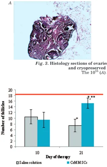
Compared with the control group 1 the analysis of histological research of the ovaries of the animals with CrMMSC therapy showed that the morphological structure of the ovaries had a positive recovery dynamics to the 10th day after the cell injection (Fig. 3, A). Along the periphery of histological sections of ovaries, amount increasing of follicles of both early and late stages of follicle genesis was observed. Follicular profile was presented by the primordial, primary and preantral stages of oocytes. The intensity of leukocyte infiltration in ovarian tissue was significantly lower compared with the control group 1.
The results of histological examination of the ovaries in animals with the oophoritis and CrMMSC therapy to the 21st day (Fig. 3, B) showed a reparative regeneration with a tendency to normalization of morphological parameters of ovarian tissue. Slight leukocyte infiltration was observed on the surface of ovarian cortical layer that pointed to
the reduction of inflammation intensity in experimental preparations relative to the control group 1. The follicular profile was characterized by the presence of primordial, primary, preantral and single antral follicles, which were located at the edge of sections. In the ovaries of the animals with bone marrow CrMMSC therapy as well as extinction inflammatory signs the activation of regenerative processes took place, while in the control animals with administered saline solution they were not observed.
The results of total number of follicles per ovary counting in histological sections (Fig. 4) to the 10th day showed statistically significant reduction (P < 0.05) of this parameter both in the control and experimental groups up to the values of 10.5 ± 2.48 and 9.4 ± 2.63 respectively compared with intact animals (18.3 ± 4.52). With the increasing of observation period up to the 21th day, the number of follicles in the control group 1 continued decreasing (7.4 ± 2.18), while in the experimental group this parameter significantly increased (P < 0.05) up to 15.3 ± 1.8, although it did not reach the values of the control group 2.
It is known that the oocyte quality affects significantly on the viability of embryos at pre-implantation stages of development, the probability of pregnancy and fetal development. Therefore, it is important to determine the presence/absence of apoptotic features in oocytes. These results are shown in Fig. 5. In the oocytes of the control group 2 the signs of apoptosis were not fixed. In the oocytes of the control group 1 together with the decrease of total number of oocytes 85.3 ± 5.2% ones had the apoptosis signs (Fig. 5, A, B). Number of oocytes with apoptotic signs was 5.7 ± 0.8% in the animals with CrMMSC therapy (Fig. 5, C, D). Thus, there was detected a tendency to restore the total number of oocytes in the ovaries of experimental animals after 3 weeks of intravenous injection of
A B
Fig. 2. Histology sections of ovaries of animals with chronic inflammation and administration of saline:
CrMMSCs, that can indicate the activation of folliculogenesis after cell therapy.
At the final stage we determined the lo ca-li zation of CrMMSCs after their intravenous injection into mice with oophoritis. It is known that the detection of transplanted cells in the recipient’s organism is performed with the help of histochemical, radioimmunological methods and fluorescent microscopy. For this purpose the nanoparticles, fluorescent dyes, embedded genetic fluorescent probes and other agents that promote detection of the introduced cell location are injected into the cells prior to their application. We have chosen the probe PKH-26, which according to the information of developer is a lipophilic dye, non-radioactive substance that binds to cell membranes and shows no toxic effects [15]. Fig. 6 shows micrographs of bone marrow MMSC culture labeled with PKH-26 dye. There
was observed the presence of luminescence in a red region of the spectrum in the cryostat sections of ovaries to the 10th and 21st day after CrMMSC therapy. To the 10th day the emission intensity was weak and had the shape of small-sized conglomerates, diffusely located in the center of the samples (Fig. 7, A). To the 21st day the luminescent objects appeared and looked like a small sized conglomerates in the center and edge part of the sections (Fig. 7, B). It should be noted that the number of luminescent objects in cryostat sections of ovaries was lower to the 21st day compared to the previous period. It is known that PKH-26 dye does not pass from a cell to cell, but has the ability to transition into daughter cells by mitosis. Thus, the luminescenting cells in the examined tissues can be directly introduced cells as well as daughter ones.
The phenomenon of directed migration of stem cells to the area of injury, ischemic or neoplastic lesions was an important step in understanding the mechanisms of regeneration tissue processes. Due to homing, which existence was proven in many studies, there is a migration of cells to the damaged and ischemic areas [16–18] to implement the cell therapy effects at an organism level. To date, 79 cytokines, growth factors and hemoatractants and over 20 types of receptors have been identified to involve in the directed migration processes of various types of stem cells in a norm and pathology [19]. The central role in this process belongs to the interaction between SDF-1α factor (its content increases in the areas of injury) and receptor CXCR4 on the MMSCs [20, 21]; moreover there is discussed the importance of ligand-receptor interactions of SCFc-Kit [22], HGF/c-met [23] and others.
The studies indicate that injected in the tail vein MMSCs have been determined in ovarian tissue to the 10th and 21st day after treatment. This is one more proof of the hypothesis Fig. 3. Histology sections of ovaries of animals with chronic inflammation
and cryopreserved MMSC administration:
The 10th (A) and 21st (B) days
A B
Fig. 4. Changes in number of follicles in ovaries of experimental animals:
line — index of intact animals;
* — P < 0.05 compared to previous observation period; ** — P < 0.05 compared with the corresponding
that MMSCs are not only passively carried throughout the body by a blood flow, but also perform a directed migration to the area of inflammation of damaged tissues.
According to the date [24, 25], the bone marrow MMSCs have immune regulatory effect except their regenerative properties. Due to the last administrated cells indirectly influence the course of inflammation in the ovaries. The results of cryopreserved MMSC influence on the process of ovarian recovery allow concluding about their possible usage in therapy of oophoritis.
It should be noted that intravenous cell injection, including MMSCs, is considered to be safe and non-invasive method of therapy. For this reason the systemic MMSCs injection may be the most appropriate in the treatment of considered pathology. However, there remains open the question about more remote effects of cell therapy, including effects on fertility, reduced under chronic inflammation.
The results in animals suggest that intra-venous MMSC injection can be effective for treatment of inflammation in human ovaries. Perhaps the mechanisms of directed migration and stem cells homing to the source of injury belong to the complex of tissue homeostasis system, which regulates the effector functions of damaged cells modulating the survival processes and apoptosis, proliferation and differentiation.
The development of regenerative cell therapy is the direction of medical biotechnology, which is rapidly growing and getting usage in the treatment of gynecological pathology [4–6]. Based on the results of this study, we pay attention of researchers to the fact that bone marrow MMSCs can be considered as a potential object of cell therapy of gynecological diseases. However, this issue requires further study during longer period of time. In addition, it is important to identify the fertility characteristics of control animals with oophoritis and animals with CrMMSC therapy alone and in combination with standard anti-inflammatory drugs.
A B C D
Fig. 5. Oocytes of animals with chronic ovarian inflammation (A, B) and cryopreserved MMSC administra-tion (C, D) to the 21st day:
light (A, B) and fluorescent (B, D) microscopy. ×150
Fig. 6. MMSC culture labeled PKH-26:
fluorescent microscopy. ×150
Fig. 7. Localization of labeled cryopreserved MMSC in ovaries to the 10th (A) and 21st (B) day:
fluorescent microscopy. ×100 B
Thus, it can be argued that intravenous injection of cryopreserved bone marrow multipotent mesenchymal stromal cells into mice with chronic inflammation of the ovaries produces a modulatory effect on the course of inflammation and restoration of
folliculogenesis without generating apoptosis in oocytes, as well as that cryopreserved bone marrow multipotent mesenchymal stromal cells at intravenous injection are detected in the animal ovaries to the 10th and 21st day.
REFERENCES
1. Dubossarskaja Z. M., Miljanovskij A. I., Koljadenko V. G. Chronic inflammation of internal female genitals. Kyiv: Zdorov’ja. 2003, P. 115–118. (In Russian).
2. Drannik G. N. Clinical Immunology and Allergology. Moskow: OOO Med. inform. agentstvo, 2003. 604 p. (In Russian).
3. Grishhenko N. G., Klimenko N. A., Gorgol’ N. I. Tatarko S. V Еffect of placental cryoextract on chronic inflammation of the ovaries in mice. Medycyna s’ohodnі і zavtra. 2010, N 2–3, P. 7–17. (In Russian).
4. Takehara Y., Yabuuchi A., Ezoe K., Kuroda T. The restorative effects of adipose-derived mesenchymal stem cells on damaged ovarian function. Labor. Invest. 2013, 93 (2), 181–193. 5. Fu X., He Y., Xie C., Liu W. Bone marrow
mesenchymal stem cell transplantation improves ovarian function and structure in rats with chemotherapy-induced ovarian damage. Cytotherapy. 2008, 10 (4), 353–363. 6. Weina L., Qixuan X., Junwen Q. Effect of
mesenchymal stem cell transplantation on immunological injury of the ovary in mice. J. South Med. Univ. 2011, 31 (5), 825–829. 7. Komarova S., Roth J., Alvarez R., Curiel D. T.,
Pereboeva L. Targeting of mesenchymal stem cells to ovarian tumors via an artificial receptor. J. Ovar. Res. 2010, 3 (12), 12–18. 8. Haack-Sorensen M., Bindslev L., Mortensen S.,
Friis T., Kastrup J. The influence of freezing and storage on the characteristics and functions of human mesenchymal stromal cells isolated for clinical use. Cytotherapy. 2007, 9 (4), 328–337. 9. Dittmar T., Entschladen F. Migratory properties
of mesenchymal stem cells. Adv. Biochem. Eng. Biotechnol. 2013, V. 129, P. 117–136.
10. Egorov N. S. Practical work on Microbiology. Mosсow: MGU. 1976. 307 p. (In Russian). 11. Oktay K., Newton H., Mullan J. Development
of human primordial follicles to antral stages in SCID/hpg mice stimulated with follicle stimulating hormone. Hum. Reprod. 1998, 13 (5), 1133–1138.
12. Gougeon A. Dynamics of follicular growth in the human: a model from preliminary results. Hum. Reprod. 1986, 1 (2), 81–87.
13. Baschong W., Suetterlin R., Laeng R. H. Control of autofluorescence of archival formaldehyde-fixed, paraffin-embedded tissue in confocal la-ser scanning microscopy (CLSM). J. Histochem. Cytochem. 2001, 49 (12), 1565–1572.
14. Kiroshka V. V., Medinec E. A., Tishhenko Ju. O., Bondarenko T. P. Dynamics of changes in
morphology neonatal ovarian tissue during cold storage, depending on the composition of the incubation medium. Problemy kriobiologii. 2012, 22 (1), 61–70. (In Russian).
15. Johnsson C., Festin R., Tufveson G., Totter-man T. H. Ex vivo PKH26-labelling of lym-phocytes for studies of cell migration in vivo. Scand. J. Immunol. 1997, 45 (5), 511–514. 16. Le Blanc K., Mougiakakos D. Multipotent
mesenchymal stromal cells and the innate immune system. Nat. Rev. Immunol. 2012, 12 (5), 383–396.
17. Karp J. M., Leng G. S. Mesenchymal stem cell homing: the devil is in the details. Stem Cells. 2009, 4 (3), 206–216.
18. Lee J. S., Hong J. M., Moon G. J., Lee P. H., Ahn Y. H., Bang O. Y. A long-term follow-up study of intravenous autologous mesenchymal stem cell transplantation in patients with ischemic stroke. Stem Cells. 2010, 28 (6), 1099–1106.
19. Horuk R. Chemokines receptors. Cytokine and growth factor review. 2001, 12 (4), 313–335. 20. Kucia M., Reca R., Miecus K., Wanzeck J.,
Wojakowski W, Janowska-Wieczorek A., Ra-ta jczak J., RaRa-tajczak M. Z. Trafficking of normal stem cell and metastasis of cancer stem cells involve similar mechanisms: Pivotal role of the SDF-1-CXCR4 axis. Stem Сells. 2005, 23 (7), 879–894.
21. Bhakta S., Hong P., Koc O. The surface adhe-sion molecule CXCR4 stimulates mesenchy-mal stem cell migration to stromesenchy-mal cell-de-rived factor-1 in vitro but does not decrease apoptosis under serum deprivation. Cardio-vasc. ReCardio-vasc. Med. 2006, 7 (1), 19–24.
22. Erlandson A., Larsson J., Forsberg-Nilsson K. Stem cell factor is a chemoattractant and a survival factor for the CNC stem cell. Exp. Cell Res. 2004, 301 (2), 201–210.
23. Wondergem R., Ecay T. W., Mahieu F. HGF\ SF and menthol increase human glioblastoma cell calcium and migration. Biochem. Bio-phys. Res. Commun. 2008, 372 (1), 210–215. 24. Krampera M., Cosmi L., Angeli R., Pasini A.,
Liot ta F., Andreini A., Santarlasci V., Mazzing-hi B., Pizzolo G., Vinante F., Romagnani P., Mag-gi E., Romagnani S., Annunziato F. Role for interferon-gamma in the immunomodulatory activity of human bone marrow mesenchymal stem cells. Stem Cells. 2006, 24 (2), 386–398. 25. Tabera S., Perez-Simon J. A., Diez-Campelo M.
МУЛЬТИПОТЕНТНІ МЕЗЕНХІМНІ СТРОМАЛЬНІ КЛІТИНИ КІСТКОВОГО
МОЗКУ В ТЕРАПІЇ ХРОНІЧНОГО ЗАПАЛЕННЯ ЯЄЧНИКІВ У МИШЕЙ
Н. О. Волкова, М. С. Юхта, Т. О. Юрчук, О. Д. Іванова, Л. В. Степанюк, О. В. Павлович,
Л. В. Сокол
Інститут проблем кріобіології і кріомедицини
НАН України, Харків
Е-mail: [email protected]
Метою роботи було дослідити вплив на хро-нічне запалення яєчників мишей та визначити локалізацію мультипотентних мезенхімних стромальних клітин кісткового мозку за вну-трішньовенного введення. Результати гістоло-гічного дослідження засвідчили, що в експери-ментальних тварин в умовах клітинної терапії відбувалась активізація репаративного проце-су з тенденцією до нормалізації морфологіч-них параметрів оваріальної тканини на фоні згасання запальних проявів. На 21-шу добу в контрольній групі з уведенням фізіологічно-го розчину загальна кількість фолікулів була знижена (7,4 ± 2,18%) порівняно з інтакт ними тварин (18,3 ± 4,52%), а 85,3 ± 5,2% ооцитів мали ознаки апоптозу (Аnnехіn+). В експери-ментальній групі кількість фолікулів досто-вірно збільшувалась до величини 15,3 ± 1,8%, а кількість апоптозних ооцитів зменшувалась (5,7 ± 0,8%) порівняно з контролем. Люмінес-центна мікроскопія кріостатних зрізів яєчни-ків тварин після терапії міченими РКН-26 клі-тинами дала змогу виявити наявність дифуз-ного розподілу люмінесціюючих об’єктів, які мали вигляд невеликих за розміром конгломе-ратів клітин. Встановлено, що кріоконсерво-вані мультипотентні мезенхімні стромальні клітини кісткового мозку за умов внутрішньо-венного введення тваринам із хронічним запа-ленням яєчників справляють модулювальну дію на перебіг запалення, сприяють відновлен-ню фолікулогенезу і визначаються в яєчниках на 10-ту та 21-шу добу після введення.
Ключові слова: хронічне запалення яєчників,
мультипотентні мезенхімні стромальні
клітини кісткового мозку, клітинна терапія.
МУЛЬТИПОТЕНТНЫЕ МЕЗЕНХИМНЫЕ СТРОМАЛЬНЫЕ КЛЕТКИ КОСТНОГО МОЗГА В ТЕРАПИИ ХРОНИЧЕСКОГО ВОСПАЛЕНИЯ ЯИЧНИКОВ У МЫШЕЙ
Н. А. Волкова, М. С. Юхта, Т. А. Юрчук, Е. Д. Иванова, Л. В. Степанюк, Е. В. Павлович,
Л. В. Сокол
Институт проблем криобиологии и криомедицины НАН Украины,
Харьков
Е-mail: [email protected]
Целью работы было исследование влияния на хроническое воспаление яичников мышей и определение локализации криоконсерви-рованных мультипотентных мезенхимных стромальных клеток костного мозга при вну-тривенном введении. Результаты гистологи-ческого исследования свидетельствовали, что у экспериментальных животных в условиях клеточной терапии происходила активизация репаративных процессов с тенденцией к нор-мализации морфологических параметров ова-риальной ткани на фоне угасания воспалитель-ных проявлений. На 21-е сутки в контроль-ной группе с введением физиологического раст вора общее количество фолликулов было снижено (7,4 ± 2,18%) относительно интакт-ных животинтакт-ных (18,3 ± 4,52%), а 85,3 ± 5,2% ооци тов имели признаки апоптоза (Аnnехіn+). В экспериментальной группе количество фол-ликулов достоверно увеличивалось до вели-чины 15,3 ± 1,8% а количество апоптозных ооцитов снижалось (5,7 ± 0,8%) по сравнению с контролем. Люминесцентная микроскопия криостатных срезов яичников животных сле терапии меченными РКН-26 клетками по-казала наличие диффузного распределения люминесцирующих объектов, которые имели вид небольших по размеру конгломератов кле-ток. Установлено, что криоконсервированные мультипотентные мезенхимные стромальные клетки костного мозга при внутривенном вве-дении животным с хроническим воспалением яичников оказывают модулирующее влияние на течение воспаления, способствуют восста-новлению фолликулогенеза и определяются в яичниках экспериментальных животных на 10- и 21-е сутки после введения.