Sequencing and Analysis of the
Pseudomonas fluorescens
GcM5-1A Genome:
A Pathogen Living in the Surface Coat of
Bursaphelenchus xylophilus
Kai Feng1☯, Ronggui Li2☯, Yingnan Chen1, Boguang Zhao3
*, Tongming Yin1*
1Co-Innovation Center for Sustainable Forestry in Southern China, College of Forestry, Nanjing Forestry University, Nanjing, 210037, China,2Department of Biology, Qingdao University, Qingdao, 266071, China,
3College of Forestry, Nanjing Forestry University, Nanjing, 210037, China
☯These authors contributed equally to this work. *[email protected](BZ);[email protected](TY)
Abstract
It is known that several bacteria are adherent to the surface coat of pine wood nematode (Bursaphelenchus xylophilus), but their function and role in the pathogenesis of pine wilt dis-ease remains debatable. ThePseudomonas fluorescensGcM5-1A is a bacterium isolated from the surface coat of pine wood nematodes. In previous studies, GcM5-1A was evident in connection with the pathogenicity of pine wilt disease. In this study, we report thede novo
sequencing of the GcM5-1A genome. A 600-Mb collection of high-quality reads was obtained and assembled into sequence contigs spanning a 6.01-Mb length. Sequence annotation predicted 5,413 open reading frames, of which 2,988 were homologous to genes in the other four sequencedP.fluorescensisolates (SBW25, WH6, Pf0-1 and Pf-5) and 1,137 were unique to GcM5-1A. Phylogenetic studies and genome comparison revealed that GcM5-1A is more closely related to SBW25 and WH6 isolates than to Pf0-1 and Pf-5 isolates. Towards study of pathogenesis, we identified 79 candidate virulence fac-tors in the genome of GcM5-1A, including theAlg,Fl,Waagene families, and genes coding the major pathogenic proteinfliC. In addition, genes for a complete T3SS system were iden-tified in the genome of GcM5-1A. Such systems have proved to play a critical role in sub-verting and colonizing the host organisms of many gram-negative pathogenic bacteria. Although the functions of the candidate virulence factors need yet to be deciphered experi-mentally, the availability of this genome provides a basic platform to obtain informative clues to be addressed in future studies by the pine wilt disease research community.
Introduction
Pseudomonas fluorescensare Gram-negative bacteria that have diverse lifestyles and versatile metabolisms. These bacteria are found in decaying and living plants, soil, and water. SomeP. fluorescensisolates benefit plants by suppressing pathogens, aiding in nutrient absorption, and OPEN ACCESS
Citation:Feng K, Li R, Chen Y, Zhao B, Yin T (2015) Sequencing and Analysis of thePseudomonas fluorescensGcM5-1A Genome: A Pathogen Living in the Surface Coat ofBursaphelenchus xylophilus. PLoS ONE 10(10): e0141515. doi:10.1371/journal. pone.0141515
Editor:Ulrich Melcher, Oklahoma State University, UNITED STATES
Received:July 28, 2015
Accepted:October 7, 2015
Published:October 30, 2015
Copyright:© 2015 Feng et al. This is an open access article distributed under the terms of the
Creative Commons Attribution License, which permits unrestricted use, distribution, and reproduction in any medium, provided the original author and source are credited.
Data Availability Statement:Sequence data of this study were deposited at DDBJ/EMBL/GenBank, under accession number JJOE00000000.
Funding:Funding for this work is provided by the Natural Science Foundation (31070575), the State Forestry Administration, People’s Republic of China (20070430), and
degrading environmental pollutants [1]. Other isolates produce compounds that negatively affect the plant’s growth [2].
To date, the genomes of four isolates ofP.fluorescenshave been sequenced and are publicly available, including SBW25, WH6, Pf0-1, and Pf-5 [3–5]. The published genomes revealed that P.fluorescenshas an open pan-genome of approximately 6 to 7 Mb. In addition to the core genes, each isolate possesses 1000 to 1500 unique genes. Previous phylogenetic studies catego-rized the four sequencedP.fluorescensisolates into two distinct clusters: SBW25 and WH6 formed one cluster, and Pf0-1 and Pf-5 formed a second cluster. In these sequenced isolates, Pf-5 and SBW25 are rhizosphere bacteria that promote plant growth and suppress plant patho-gens [3] [6]. WH6 produces a chemical compound called the germination-arrest factor (GAF) that specifically and irreversibly blocks the germination of the seeds of a large number of grassy weed species without significantly affecting the growth of established seedlings or mature plants [7]. And Pf0-1 is a bacterium that is well adapted to the soil environment and contrib-utes significantly to the turnover of organic matter [5].
Another isolate ofP.fluorescens, GcM5-1A, was reported to increase egg production, devel-opmental rate, body length and diameter of both male and female pine wood nematodes (PWN) [8]. GcM5-1A is one of the main bacteria isolated from the surface coat of Bursaphe-lenchus xylophilus(the nematode causing pine wilt disease), which has been implicated in con-nection with the pathogenesis of pine wilt disease (PWD) in several previous studies [9–12]. The devastating pine wilt disease spreads among pine trees in Asian countries, especially in Japan, China and Korea. This disease is caused by PWN (whose genome has been sequenced in 2011 [13]), and is transferred by pine sawyer beetles (Monochamus alternatus) [14]. PWN can reproduce quickly in the sapwood of the susceptible pine species, which causes wilting and death of the host in a short period of time. Recently, this disease has also been observed in European countries [15], and is gradually becoming a threat to pine forests worldwide.
The pathogenesis of PWD remains debatable. PWN was previously recognized as the only pathogen that caused this disease [16]. However, it was reported that PWN lost pathogenicity after surface sterilization [17], which led to the hypothesis that the pathogenicity might be a combined effect of the PWN and the microbes adherent to their surface coat. Various evidence supported this hypothesis. For instance, Shinyaet al.’s study indicated that the surface coat of PWN could protect the microbes and was essential for the infection of pine trees by PWN [9]. In addition, Guoet al. reported thatP.fluorescensGcM5-1A, isolated from the surface coat of PWN, could produce a flagellin, namelyfliC, that was able to increase populations of pine wood nematodes and their associated bacteria [10]. It was also found thatfliCcould induce the death ofPinus thunbergiisuspension cells in 24 h [11], and Xuet al. proved thatfliCincreased damage to the host pine through enhancing the oxidative stress [12].
Although several studies have been carried out testing the connection of the PWN surface coat microbes with the pathogenicity of PWD, their relationship remain debatable [18], and the underlying mechanisms need to be deciphered at the molecular level. Many virulence fac-tors may play a role in the infection process. For instance, it was found that many gram-nega-tive pathogenic bacteria employed a type III secretion system (T3SS) to subvert and colonize their host organisms. The T3SS injects effector proteins directly into the cytosol of eukaryotic cells, and thus allows the manipulation of host cellular activities to the benefit of the pathogen [19]. T3SS provides a unique virulence mechanism to infect host cells [20]. Among the pub-lished genomes of theP.fluorescensisolates, a complete T3SS was only identified in WH6 [4], which produces the GAF herbicide that helps to control grassy weeds [21]. Whether GcM5-1A possesses a complete T3SS remains unknown. Besides the T3SS system, other candidate viru-lence factors may exist and need to be addressed in understanding the role of PWN surface coat microbes in connection with the pathogenicity of PWD at the molecular level. Competing Interests:The authors have declared
Taken together, the perspectives of this study are to: 1) sequence the genome of GcM5-1A to provide a basic platform for the pine wilt disease research community; 2) compare the genome of GcM5-1A and study the phylogenetic relationship of GcM5-1A with that of the other four sequencedP.fluorescensisolates at genome-wide level; and 3) detect the candidate virulence factors, including the T3SS system, based on sequence annotation, to provide clues to be addressed in future studies.
Results and Discussion
Genome sequencing and assembling
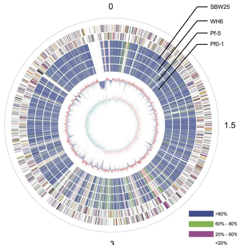
The GcM5-1A genome was sequenced using a 454 GS-FLX sequencer with a GS titanium XLR70 kit (Roche Inc.), which resulted in 1,534,199 sequencing reads with an average length of 409 bp. The total throughput exceeded 600 Mb. The genome sizes of the four sequencedP. fluorescensisolates ranged from 6.27 to 7.07 Mb. Using this range as a baseline, the sequencing depth of GcM5-1A was estimated to be approximately 100X. After filtering with the Newbler de novoAssembler (Roche Inc.), 98.23% of the total reads were selected and used in the final assembly. As a result, the genome assembly generated 263 contigs greater than 100 bp, among which 79 contigs were greater than 500 bp. The final assembly covered 6.01 Mb. Subsequently, all of the contigs were submitted to BLAST against the nr database using BLASTX [22]. No contigs exhibited high similarity with genes from organisms outside the bacterial kingdom, except for contig24, which shared a highly homologous DNA fragment with thePseudomonas phage phiPsa374, with 73% sequence identity. The homologous fragment of contig24 covered 34% of the genome of phiPsa374. Therefore, we placed contig24 at the terminus of chromo-some replication of the established circular graph (Fig 1). The sequence alignment revealed that the contig shared almost no sequence similarity with any of the otherP.fluorescens iso-lates. Further analysis revealed that contig24 had a size and GC content similar to those of sev-eralPseudomonasphages. We hypothesized that contig24 might represent a horizontally transferred phage sequence that diverged significantly from the archetype during the evolu-tionary process or an exogenous DNA segment from an unknown phage.
To order the obtained contigs, a high-quality reference genome was required. As revealed by BLASTX, GcM5-1A exhibited higher similarity with SBW25 and WH6 compared with Pf0-1 and Pf-5. Compared with SBW25, WH6 was incompletely sequenced [4]. Therefore, the genome assembly of SBW25 was selected as a reference to order the obtained sequence contigs. All contigs greater than 1 kb were ordered in line with the reference genome by the Mauve Aligner [23]. There were 188 contigs smaller than 1 kb. Together with contig24, these contigs only accounted for 2.1% of the GcM5-1A genome. Among the ordered contigs, contig4 con-tained the gene which is responsible for initiation of replication; thus, it was marked as the starting point of the GcM5-1A genome.
Genome comparison of the sequenced
P.
fluorescens
isolates
Fig 1. Circular graph ofP.fluorescensGcM5-1A.From the outer to the innermost circle, circle 1 presents the physical coordinates, in 100,000 bp per interval, as defined by the black sticks. The red sticks on circle 1 indicate the sites of the physical gaps. Circles 2 and 3 depict the CDS on the positive and negative strands. Circles 4, 5, 6 and 7 depict the orthologous similarity between GcM5-1A and WH6, between GcM5-1A and SBW25, between GcM5-1A and Pf-5, and between GcM5-1A and Pf0-1, respectively. Different colors indicate different similarity levels. Blue indicates similarity greater than 80%. Green indicates similarity within the range of 60 to 80%. Pink indicates similarity within the range of 20 to 60%. White reflects similarity less than 20%. Circle 8 indicates the GC content (red>60.5%, blue<60.5%). Circle 9 depicts the GC-skew (purple>0, cyan<0).
those of WH6 [4]. The core gene set accounted for 50.8%, 50.4%, 52.1%, and 48.7% of the total genes in the SBW25, WH6, Pf0-1, and Pf-5 genomes, respectively. The percentage of shared orthologous groups between GcM5-1A and SBW25 was 70%, between GcM5-1A and WH6 was 68%, between GcM5-1A and Pf0-1 was 62%, and between GcM5-1A and Pf-5 was 65%. By contrast, it was 82% between thePseudomonas syringaepv.syringaeB728a andP.syringaepv. tomatoDC3000 [26], and it was at least 86% among the 5 isolates (PA2192, C3719, PA01, PA14 and PACS5) ofP.aeruginosa[27]. Compared with the increased percentage of shared ortholo-gous groups between the isolates ofP.syringaeandP.aeruginosa, the lower percentage of shared orthologous groups between isolates ofP.fluorescensindicated that the genomes ofP.fluorescens isolates had undergone a heavier gene reshuffling than otherPseudomonasspecies.
Phylogenetic and syntenic analyses
To investigate the phylogenetic relationship among the five sequencedP.fluorescensgenomes, 2,935 single-copy orthologous groups were used to generate a multiple alignment using MUS-CLE [28] with default parameters. A phylogenetic tree was then constructed using MEGA5 [29] with the neighborhood-joining method and the bootstrap parameter set to 500. Previous studies categorized theP.fluorescensisolates into two distinct clades. SBW25 and WH6 formed one clade, and Pf0-1 and Pf-5 formed the second clade. In this study, GcM5-1A was evident in the SBW25 and Wh6 clade (Fig 4A).
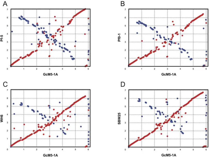
Long-range synteny of GcM5-1A with otherP.fluorescensisolates appeared more often at the origin and terminus of chromosome replication of the genome. Compared with Pf0-1 and Pf-5, fewer outliers were noted between the genome of GcM5-1A and those of SBW25 and WH6, indicating that the genome of GcM5-1A shares higher colinearity with that of the latter than the former (Fig 5). Thus, GcM5-1A was more closely related to SBW25 and WH6 than to Pf0-1 and Pf-5, which was consistent with the results obtained from the phylogenetic tree. Given that the genome assembly of GcM5-1A was ordered using SBW25 as reference, the syn-teny between GcM5-1A and SBW25 was expected to be better than for any other pair-wise comparison. In the genome of a bacterium, the leading strand often has an excess G content compared with C, whereas the lagging strand has excess C compared with G [30,31]. From the circular graph of GcM5-1A (Fig 1), the GC skew index (GC Skew = (G—C)/(G + C)) clearly demonstrated a G bias, with most of the GC skew indexes above 0 in the leading strand. C bias was observed in the lagging strand, which suggests that the established synteny was reliable and properly reflected the relationship between theseP.fluorescensisolates.
Candidate virulence factors
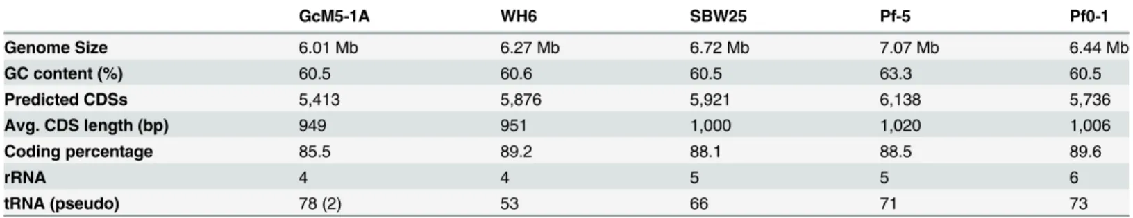
A previous study of theP.fluorescensGcM5-1A indicated that the isolate secretedfliCflagellin, which is deleterious to black pine seedlings [10]. When GcM5-1A and PWN were cultured Table 1. Genome features of GcM5-1A and other sequencedP.fluorescensisolates.
GcM5-1A WH6 SBW25 Pf-5 Pf0-1
Genome Size 6.01 Mb 6.27 Mb 6.72 Mb 7.07 Mb 6.44 Mb
GC content (%) 60.5 60.6 60.5 63.3 60.5
Predicted CDSs 5,413 5,876 5,921 6,138 5,736
Avg. CDS length (bp) 949 951 1,000 1,020 1,006
Coding percentage 85.5 89.2 88.1 88.5 89.6
rRNA 4 4 5 5 6
tRNA (pseudo) 78 (2) 53 66 71 73
together, the fecundity, egg-hatch rate and the development of PWN were significantly improved [32]. Xuet al. reported that treating the black pine cell suspension with the flagellin of GcM5-1A results in an oxidative burst of the pine cells and subsequent cell death, this indi-cating the host’s response to the attack of the pathogen [12]. However, the cell death of pine Fig 2. Shared and unique genes in each of the sequencedP.fluorescensisolates.
callus induced by flagellin promoted the proliferation of PWN and the associated GcM5-1A, which was feeding on the callus cells [11]. Liet al. further revealed that the flagellin of GcM5-1A would attach to the pine cell membrane and lead to shrinkage of the cell membrane, a con-centration of the cytoplasm, formation of micronuclei, degradation of cytoplasmic RNA, and breakage of genomic DNA. However, DNA ladder formation was not observed, which indi-cated that an unusual form of apoptosis occurred in the flagellin-treated pine cells [33]. The above evidence suggested that GcM5-1A potentially plays a role in the pathogenesis of pine wilt disease.
Alignment of the N-terminal sequences of thefliCgenes demonstrated that this gene shared high similarity between GcM5-1A and Pf-5 [12]. The expression thefliCgenes of these two iso-lates inEscherichia coliproduces recombinant flagellin that exhibit similar toxicity to pine sus-pension cells [10]. In a previous study, researchers observed that pine seedlings inoculated with the axenic PWN and transformedE.colithat produced secretive flagellin of Pf-5 resulted in symptoms of wilt. In contrast, pine seedlings inoculated with axenic PWN and untransformed E.colidid not cause symptoms of wilt [34]. Thus, thefliCgene is a crucial pathogenic factor in the genome of GcM5-1A and is located on contig57 in the genome assembly. Based on sequence similarity, we constructed a gene tree for thefliCgenes in the five sequenced isolates ofP.fluorescens(Fig 4B). The tree demonstrated that thefliCgenes from Pf0-1 and Pf-5 were closely related, as were those of SBW25 and WH6. Whereas thefliCgene in GcM5-1A was more phylogenetically diverged from others, but it was more close to Pf0-1 and Pf-5 than SBW25 and WH6.
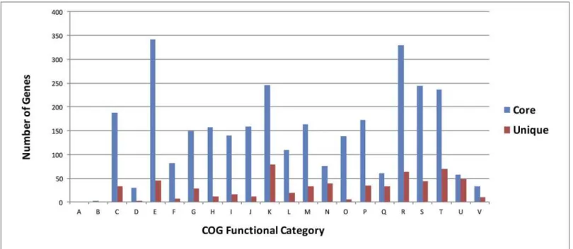
Fig 3. Functional classification of core and unique genes of GcM5-1A.A: RNA processing and modification; B: Chromatin structure and dynamics; C: Energy production and conversion; D: Cell cycle control, cell division, chromosome partitioning; E: Amino acid transport and metabolism; F: Nucleotide transport and metabolism; G: Carbohydrate transport and metabolism; H: Coenzyme transport and metabolism; I: Lipid transport and metabolism; J: Translation, ribosomal structure and biogenesis; K: Transcription; L: Replication, recombination and repair; M: Cell wall/membrane/envelope biogenesis; N: Cell motility; O: Posttranslational modification, protein turnover, chaperones; P: Inorganic ion transport and metabolism; Q: Secondary metabolites biosynthesis, transport and catabolism; R: General function prediction only; S: Function unknown; T: Signal transduction mechanisms; U: Intracellular trafficking, secretion, and vesicular transport; V: Defense mechanisms
In addition to thefliCgene, we searched for additional virulence factors that might relate to pathogenesis in the genome of GcM5-1A. For this purpose, all of the predicted CDSs were sub-mitted to BLAST against the VFDB, which was built for bacterial pathogens [35]. This analysis identified 79 CDSs as candidate virulence factors (S1 Table). Among these factors, theAlg genes encoding the polysaccharide alginate participate in the formation of biofilms, which play an important role in the plant-pathogen interaction [36]. TheFlgenes encode the bacterial flagella that affect the bacterial motility. These genes are also essential for the formation of bio-films inP.syringae[37], whereas theWaagenes are involved in the synthesis and transforma-tion of lipopolysaccharides in Gram-negative bacteria. Lipopolysaccharides play direct roles in the plant-pathogen interaction [38,39]. TheRhlgenes encode rhamno lipids that are crucial forP.syringaeswarming, which is an important factor in pathogen infection [40]. All of the candidate virulence factors that were identified in the genome of GcM5-1A suggested that this microbe plays a role in the pathogenesis of pine wilt disease.
Fig 4. Phylogenetic tree of the five sequencedP.fluorescensisolates and the phylogenetic tree offliCgenes.(a) the phylogenetic tree was generated based on the multiple alignments of amino acid sequences of 2,935 single-copy orthologous groups. (b) Gene tree constructed for the 5 sequencedP.
fluorescensisolates based on the amino acid sequence of thefliCgene.
The flagellum secretion system T3SS
InPseudomonas spp., the complete T3SS was first identified inP.syringaepv.tomatoDC3000. This microbe is the model system used to study T3SS, and it is pathogenic to tomato and Ara-bidopsis[41]. In pathogenic bacteria, T3SS uses its needle-like structure, known as the needle complex [42], to detect the presence of eukaryotic organisms. When eukaryotic organisms are detected, the T3SS injects bacterial proteins into the host cell and helps the bacteria colonize and multiply in the host cells [43,44]. In the previously sequenced isolates ofP.fluorescens, the complete T3SS was only identified in WH6. Using the T3SS genes from DC3000 and WH6 as queries, the complete T3SS was identified in the genome of GcM5-1A; thus, GcM5-1A appears to be the secondP.fluorescensisolate that possesses the complete T3SS. In the GcM5-1A genome, CDS00691~CDS00712 were identified as T3SS genes (Fig 6). Similarity analysis of the T3SS candidates of GcM5-1A and DC3000 was performed by using BLASTP with 30% sequence identity and 80% sequence overlap. GcM5-1A was found to possess all the T3SS Fig 5. Synteny plots of GcM5-1A and the other sequencedP.fluorescensisolates.Genomes of theP.fluorescensisolates Pf-5 (A), Pf0-1 (B), WH6 (C) and SBW25 (D) were compared with the WH6 genome sequence. Genome scales are indicated in 1-Mb increments.
genes except for the D, Z, A, and S genes when compared with DC3000. GcM5-1A lacked D and A genes compared with WH6. In T3SS, the A and F genes were homologous to each other [45,46]. The S and R genes shared high similarity and were functionally redundant [47]. Dele-tion of the Z gene did not affect T3SS funcDele-tion [48–50]. The function of the D gene remains unclear. Although upon consideration of the standard T3SS in DC3000, the genes that are lack-ing in GcM5-1A should have little effect on T3SS function. Based on the above analyses, we propose that the GcM5-1A genome possesses a fully functional T3SS.
Conclusions
Pine wilt disease is a disastrous for pine forests, and the pathogenesis of this disease remains debatable. Some pathologists have proposed that the surface coat of PWN shelters microbes that might play a role in the onset of this disease. To provide insight into the pathogenicity of such microbes, we sequenced a Gram-negative bacterial isolate ofP.fluorescens, GcM5-1A, which inhabits the surface coat of PWN. We obtained 1.5 million high-quality reads, which were assembled into sequence contigs 6.01 Mb in length. Sequence annotation predicated 5,413 CDSs. Among the five sequencedP.fluorescensisolates, GcM5-1A was more closely related to isolates SBW25 and WH6 than to Pf0-1 and Pf-5. We detected 79 virulence factors in the genome of GcM5-1A, including gene coding the major pathogenic proteinfliC. GcM5-1A also possessed a fully functional T3SS. The detected candidate virulence factors provide informative clues to be addressed on in elucidating the role of GcM5-1A in the pathogenesis of pine wilt disease at molecular level in future studies. The availability of this genome sequence alongside that of PWN provides essential platforms for further study of their interaction and their roles in the pathogenesis of pine wilt disease.
Materials and Methods
Bacterial culture and genomic DNA extraction
P.fluorescensGcM5-1A (CCTCC No: M 204065) was isolated from PWN obtained from wilted Pinus thunbergiiin Nanjing Forestry University, China. The isolation of GcM5-1A was per-formed as described by Zhaoet al. [51]. Field studies were conduct under the permission of Fig 6. Comparisons of T3SS in GcM5-1A with that inP.syringaeDC3000 and WH6.
Nanjing Forestry University China. This isolate was shake-cultured in Luria-Bertani (LB) medium at 28°C for 24 h, and bacterial cells were harvested by centrifugation at 5,000×g for 10 min at 4°C. Genomic DNA ofP.fluorescensGcM5-1A was isolated from the bacterial pellet according to the method described by Chenet al. [52]. DNA purity was examined by agarose gel electrophoresis, and the concentration was measured using a Nanodrop ND2000 (Thermo Scientific, Waltham, MA, USA).
Sequencing and assembly
The GcM5-1A genome was sequenced on a 454 GS-FLX sequencer (Roche, Inc, Basel, Switzer-land.) using the FLX Titanium Sequencing Kit (Roche, Inc, Basel, SwitzerSwitzer-land.). Sequence assembly was conducted with a Newblerde novoAssembler (version 2.5.3) with default param-eters. In the final assembly, contigs less than 100 bp in length were discarded. Sequence data from this study were deposited at DDBJ/EMBL/GenBank under accession number
JJOE00000000.
Gene annotation
The coding DNA sequences (CDSs) of GcM5-1A were predicted by Glimmer3 [53], which uses interpolated Markov models (IMMs) to identify the coding regions and distinguish them from noncoding DNA. Glimmer3 used the long-CDS program to find the long and non-over-lapping CDSs, and then set the long-CDS training system for the subsequent predictions. The predicted genes were annotated against the nr database and the COG database, with an e-value cut-off of 1e-5.
Non-coding RNA prediction
Transfer RNAs (tRNAs) were predicted using tRNAscan-SE [54]. This method first pre-filters the input sequence to identify candidate tRNAs; then, a highly selective tRNA“covariance model”is implemented for tRNA predictions and allows for the identification of 99 to 100% of tRNA genes in the DNA sequence with less than one false positive.
Ribosomal RNA (rRNA) was predicted using RNAmmer [55], which utilized two levels of hidden Markov models. An initial spotter mode searched both strands for detecting the approximate position of an rRNA gene. Flanking regions were then extracted and parsed to the full model, which matched the entire rRNA gene. By enabling this two-level approach, the pro-gram avoided running the entire genome sequence through the model and allowed faster predictions.
Phylogenetic tree construction
Synteny and the circular graph
Synteny between GcM5-1A and theP.fluorescensisolates was performed by MUMmer [56] and visualized byad hocPerl scripts. The circular graph of GcM5-1A, which was consistent with the other four isolates ofP.fluorescens, was constructed using DNAplotter [57].
Identifying the T3SS and candidate virulence factors
To identify the T3SS in GcM5-1A, we submitted the T3SS genes ofP.syringaepv. tomato DC3000 to BLAST against the entire gene set of GcM5-1A using BLASTP with parameters as follows: an e-value of 1e-5, 30% sequence similarity, 80% sequence length overlap and max_-target_seqs of 1.
All of the predicted CDSs of GcM5-1A were submitted to BLAST against the Virulence Fac-tor Database (VFDB) using BLASTP with an e-value of 1e-5 and max_target_seqs of 1. The BLAST results were then filtered using the criteria of sequence identity greater than 60% and sequence overlap greater than 90% between the query and the template.
Supporting Information
S1 Table. Candidate virulence factors identified in the genome of GcM5-1A. (XLS)
Acknowledgments
Special thanks go to the academic editor and anonymous reviewers for their help in editing the languages and in formulating the final revision.
Author Contributions
Conceived and designed the experiments: TY BZ. Performed the experiments: KF RL. Ana-lyzed the data: KF RL. Contributed reagents/materials/analysis tools: KF BZ RL. Wrote the paper: KF TY BZ RL YC.
References
1. Haas D, Défago G. Biological control of soil-borne pathogens by fluorescent pseudomonads. Nat Rev Microbiol. 2005; 3: 307–319. PMID:15759041
2. Li Y, Sun Z, Zhuang X, Xu L, Chen S, Li M. Research progress on microbial herbicides. Crop Prot. 2003; 22: 247–252.
3. Paulsen IT, Press CM, Ravel J, Kobayashi DY, Myers GSA, Mavrodi DV, et al. Complete genome sequence of the plant commensalPseudomonas fluorescensPf-5. Nat Biotechnol. 2005; 23: 873–878. PMID:15980861
4. Kimbrel JA, Givan SA, Halgren AB, Creason AL, Mills DI, Banowetz GM, et al. An improved, high-qual-ity draft genome sequence of the Germination-Arrest Factor-producingPseudomonas fluorescens
WH6. BMC Genomics. 2010; 11: 522. doi:10.1186/1471-2164-11-522PMID:20920191
5. Silby MW, Cerdeno-Tarraga AM, Vernikos GS, Giddens SR, Jackson RW, Preston GM, et al. Genomic and genetic analyses of diversity and plant interactions ofPseudomonas fluorescens. Genome Biol. 2009; 10: R51. doi:10.1186/gb-2009-10-5-r51PMID:19432983
6. Trippe K, McPhail K, Armstrong D, Azevedo M, Banowetz G.Pseudomonas fluorescensSBW25 pro-duces furanomycin, a non-proteinogenic amino acid with selective antimicrobial properties. BMC micro-biol. 2013; 13: 111. doi:10.1186/1471-2180-13-111PMID:23688329
7. Halgren A, Maselko M, Azevedo M, Mills D, Armstrong D, Banowetz D. Genetics of germination-arrest factor (GAF) production byPseudomonas fluorescensWH6: identification of a gene cluster essential for GAF biosynthesis. Microbiology. 2013; 159: 36–45. doi:10.1099/mic.0.062166-0PMID:23125119
9. Shinya R, Morisaka H, Takeuchi Y, Ueda M, Futai K. Comparison of the surface coat proteins of the pine wood nematode appeared during host pine infection and in vitro culture by a proteomic approach. Phytopathol. 2010; 100: 1289–1297.
10. Guo D, Zhao B, Li R, Kulinch Q, Ryss A. Purification of flagellin ofPseudomonas fluorescensGcM5-1A carried by the pine wood nematote,Bursaphelenchus xylophilus, and its in vitro toxicity to a suspension of cells ofPinus thunbergii. Russ J Nematol. 2008; 16:151–157.
11. Zhang L, Yue T, Zhao B, Guo D, Wu B, Wang T, et al. Flagellin promotes propagation of pine wood nematode and its carrying Pseudomonas fluorescens GcM5-1A in callus ofPinus thunbergiithrough inducing cell death. Afr J Microbiol Res. 2012; 6: 1322–1328.
12. Xu Z, Yu J, Cui L, Li M, Li R, Guo D. Effects ofPseudomonas fluorescensflagellin on physiological and biochemical characteristics in the suspension cells ofPinus thunbergii. Eur J Plant Pathol. 2013; 136: 729–736.
13. Taisei K, Cotton JA, Dalzell JJ, Hasegawa K, Kanzaki N, McVeigh P, et al. Genomic insights into the origin of parasitism in the emerging plant pathogen Bursaphelenchus xylophilus. Plos Pathogens. 2011; 7:e1002219–e1002219. doi:10.1371/journal.ppat.1002219PMID:21909270
14. Filipiak A. The pine wilt disease. Sylwan 2008; 152: 9–19.
15. Suzuki K. Pine wilt disease–a threat to pine forest in Europe. Dendrobiology. 2002; 48:71–74.
16. Mamiya Y. Pathology of the pine wilt disease caused byBursaphelenchus xylophilus. Annu Rev Phyto-pathol. 1983; 21: 201–220. doi:10.1146/annurev.py.21.090183.001221PMID:25946434
17. Ryss AY, Kulinich OA, Sutherland JR. Pine wilt disease. a short review of worldwide research. Forestry Studies in China. 2011; 13: 132–138.
18. Nascimento FX, Hasegawa K, Mota M, Vicente CS. Bacterial role in pine wilt disease development–
review and future perspectives. Env Microbiol Rep. 2015; 7:51–63.
19. Daniela B, Sheng YH. Type III Protein Secretion in Plant Pathogenic Bacteria. Plant Physiol. 2009; 150:1656–1664. doi:10.1104/pp.109.139089PMID:19458111
20. Coburn B, Sekirov I, Finlay BB. Type III secretion systems and disease. Clin Microbiol Rev. 2007; 20: 535–549. PMID:17934073
21. Banowetz GM, Azevedo MD, Armstrong DJ, Halgren AB, Mills DI. Germination-Arrest Factor (GAF): Biological properties of a novel, naturally-occurring herbicide produced by selected isolates of rhizo-sphere bacteria. Biol Control. 2008; 46: 380–390.
22. Altschul SF, Madden TL, Schäffer AA, Zhang J, Zhang Z, Miller W, et al. Gapped BLAST and PSI-BLAST: a new generation of protein database search programs. Nucleic Acids Res. 1997; 25: 3389–
3402. PMID:9254694
23. Rissman AI, Mau B, Biehl BS, Darling AE, Glasner JD, Perna NT. Reordering contigs of draft genomes using the Mauve aligner. Bioinformatics. 2009; 25: 2071–2073. doi:10.1093/bioinformatics/btp356 PMID:19515959
24. Chen F, Mackey AJ, Stoeckert CJ Jr, Roos DS. OrthoMCL-DB: querying a comprehensive multi-spe-cies collection of ortholog groups. Nucleic Acids Res. 2006; 34: D363–368. PMID:16381887
25. Tatusov RL, Galperin MY, Natale DA, Koonin EV. The COG database: a tool for genome-scale analysis of protein functions and evolution. Nucleic Acids Res. 2010; 28: 33–36.
26. Feil H, Feil WS, Chain P, Larimer F, DiBartolo G, Copeland A, et al. Comparison of the complete genome sequences ofPseudomonas syringaepv. syringae B728a and pv. tomato DC3000. Proc Natl Acad Sci. 2005; 102: 11064–11069. PMID:16043691
27. Mathee K, Narasimhan G, Valdes C, Qiu X, Matewish JM, Koehrsen M, et al. Dynamics of Pseudomo-nas aeruginosagenome evolution. Proc Natl Acad Sci. 2008; 105: 3100–3105. doi:10.1073/pnas. 0711982105PMID:18287045
28. Edgar RC. MUSCLE: multiple sequence alignment with high accuracy and high throughput. Nucleic Acids Res. 2004; 32: 1792–1797. PMID:15034147
29. Tamura K, Peterson D, Peterson N, Stecher G, Nei M, Kumar S. MEGA5: molecular evolutionary genet-ics analysis using maximum likelihood, evolutionary distance, and maximum parsimony methods. Mol Biol Evo. 2011; 28: 2731–2739.
30. Arakawa K, Tomita M. The GC Skew Index: A Measure of Genomic Compositional Asymmetry and the Degree of Replicational Selection. Evol Bioinform. 2007; 3: 159–168.
31. Marin A, Xia XH. GC skew in protein-coding genes between the leading and lagging strands in bacterial genomes: New substitution models incorporating strand bias. J Theor Biol. 2008; 253: 508–513. doi: 10.1016/j.jtbi.2008.04.004PMID:18486155
33. Li S, Guo D, Zhao B, LI R. Lethal effect of flagellin secreted byPseudomonas fluorescenson cells of
Pinus thunbergii. Acta Bot Boreali-Occidential Sinica. 2008; 28: 2154–2158.
34. Li S, Zhao B, Li R, Guo D. Construction of engineering bacterium expressing flagellin ofPseudomonas fluorescensand its toxicity toPinus thunbergii in vivo. J Qingdao Univ (Nat Sci Ed). 2010; 25: 35–40.
35. Yang J, Chen L, Sun L, Yu J, Jin Q. VFDB 2008 release: an enhanced web-based resource for compar-ative pathogenomics. Nucleic Acids Res. 2008; 36: D539–542. PMID:17984080
36. Gacesa P. Bacterial alginate biosynthesis-recent progress and future prospects. Microbiology. 1998; 144: 1133–1143. PMID:9611788
37. O'Toole GA, Kolter R. Flagellar and twitching motility are necessary for Pseudomonas aeruginosa bio-film development. Mol Microbiol. 2002: 30: 295–304.
38. Newman MA, Dow J, Daniels M. Bacterial lipopolysaccharides and plant-pathogen interactions. Eur J Plant Pathol. 2001; 107: 95–102.
39. Conrath U, Pieterse CMJ, Mauch-Mani B. Priming in plant-pathogen interactions. Trends Plant Sci. 2002; 7: 210–216. PMID:11992826
40. Xu J, Platt TG, Fuqua C. Regulatory linkages between flagella and surfactant during swarming behav-ior: lubricating the flagellar propeller? J Bacteriol. 2012; 194: 1283–1286. doi:10.1128/JB.00019-12 PMID:22267512
41. Blocker A, Jouihri N, Larquet E, Gounon P, Ebe F, Parsot C, et al. Structure and composition of the Shi-gella flexneri‘needle complex’, a part of its type III secreton. Mol Microbiol. 2001; 39: 652–663. PMID: 11169106
42. Ghosh P. Process of protein transport by the type III secretion system. Mol Biol Rev. 2004; 68: 771–
795.
43. Gophna U, Ron EZ, Graur D. Bacterial type III secretion systems are ancient and evolved by multiple horizontal-transfer events. Gene. 2003; 312: 151–163. PMID:12909351
44. Buell CR, Joardar V, Lindeberg M, Selengut J, Paulsen IT, Gwinn ML, et al. The complete genome sequence of theArabidopsisand tomato pathogenPseudomonas syringaepv.tomatoDC3000. Proc Natl Acad Sci. 2003; 100: 10181. PMID:12928499
45. Deng WL, Preston G, Collmer A, Chang CJ, Huang HC. Characterization of the hrpC and hrpRS oper-ons ofPseudomonas syringaepathovars syringae, tomato, and glycinea and analysis of the ability of hrpF, hrpG, hrcC, hrpT, and hrpV mutants to elicit the hypersensitive response and disease in plants. J Bacteriol. 1998; 180: 4523–4531. PMID:9721291
46. Lee YH, Kolade OO, Arvidson DN, He SY. Identification of HrpA mutants that block type III secretion in
Pseudomonas syringaepv.tomatoDC3000. Phytopathology. 2004; 94: S59–S59.
47. Wei Z, Kim JF, Beer SV. Regulation of hrp genes and type III protein secretion inErwinia amylovoraby HrpX/HrpY, a novel two-component system, and HrpS. Mol Plant Microbe In. 2000; 13: 1251–1262.
48. Alfano JR, Bauer DW, Milos TM, Collmer A. Analysis of the role of thePseudomonas syringaepv. syrin-gaeHrpZ harpin in elicitation of the hypersensitive response in tobacco using functionally non-polar hrpZ deletion mutations, truncated HrpZ fragments, and hrmA mutations. Mol Microbiol. 1996; 19: 715–728. PMID:8820642
49. Ortiz-Martin I, Thwaites R, Mansfield JW, Beuzon CR. Negative regulation of the Hrp type III secretion system inPseudomonas syringaepv.phaseolicola. Mol Plant Microbe In. 2010; 23: 682–701.
50. Preston G, Deng WL, Huang HC, Collmer A. Negative regulation of hrp genes inPseudomonas syrin-gaeby HrpV. J Bacteriol. 1998; 180: 4532–4537. PMID:9721292
51. Zhao BG, Wang HL, Han SF, Han ZM. Distribution and pathogenicity of bacteria species carried by Bur-saphelenchus xylophilus in China. Nematology. 2003; 5:899–906.
52. Chen WP, Kuo TT. A simple and rapid method for the preparation of gramnegative bacterial genomic DNA. Nucleic Acids Res. 1993; 21: 2260. PMID:8502576
53. Delcher AL, Bratke KA, Powers EC, Salzberg SL. Identifying bacterial genes and endosymbiont DNA with Glimmer. Bioinformatics. 2007; 23: 673–679. PMID:17237039
54. Schattner P, Brooks AN, Lowe TM. The tRNAscan-SE, snoscan and snoGPS web servers for the detection of tRNAs and snoRNAs. Nucleic Acids Res. 2005; 33: W686–W689. PMID:15980563
55. Lagesen K, Hallin P, Rodland EA, Staerfeldt HH, Rognes T, Ussery DW. RNAmmer: consistent and rapid annotation of ribosomal RNA genes. Nucleic Acids Res. 2007; 35: 3100–3108. PMID:17452365
56. Kurtz S, Phillippy A, Delcher AL, Smoot M, Shumway M, Antonescu C, et al. Versatile and open soft-ware for comparing large genomes. Genome Biol. 2004; 5: R12. PMID:14759262