from
Myoviridae
Phages OBP, 201
Q
2-1 and PVP-SE1
Maarten Walmagh1, Yves Briers1,2, Silvio Branco dos Santos3, Joana Azeredo3, Rob Lavigne1*
1Laboratory of Gene Technology, Katholieke Universiteit Leuven, Leuven, Belgium,2Institute of Food, Nutrition and Health, ETH Zurich, Zurich, Switzerland,3IBB -Institute for Biotechnology and Bioengineering, Centre of Biological Engineering, Universidade do Minho, Braga, Portugal
Abstract
Peptidoglycan lytic enzymes (endolysins) induce bacterial host cell lysis in the late phase of the lytic bacteriophage replication cycle. Endolysins OBPgp279 (fromPseudomonas fluorescens phage OBP), PVP-SE1gp146 (Salmonella enterica serovar Enteritidis phage PVP-SE1) and 201Q2-1gp229 (Pseudomonas chlororaphisphage 201Q2-1) all possess a modular structure with an N-terminal cell wall binding domain and a C-terminal catalytic domain, a unique property for endolysins with a Gram-negative background. All three modular endolysins showed strong muralytic activity on the peptidoglycan of a broad range of Gram-negative bacteria, partly due to the presence of the cell wall binding domain. In the case of PVP-SE1gp146, this domain shows a binding affinity forSalmonellapeptidoglycan that falls within the range of typical cell adhesion molecules (Kaff= 1.266106M21). Remarkably, PVP-SE1gp146 turns out to be thermoresistant up to temperatures
of 90uC, making it a potential candidate as antibacterial component in hurdle technology for food preservation. OBPgp279, on the other hand, is suggested to intrinsically destabilize the outer membrane ofPseudomonasspecies, thereby gaining access to their peptidoglycan and exerts an antibacterial activity of 1 logarithmic unit reduction. Addition of 0.5 mM EDTA significantly increases the antibacterial activity of the three modular endolysins up to 2–3 logarithmic units reduction. This research work offers perspectives towards elucidation of the structural differences explaining the unique biochemical and antibacterial properties of OBPgp279, PVP-SE1gp146 and 201Q2-1gp229. Furthermore, these endolysins extensively enlarge the pool of potential antibacterial compounds used against multi-drug resistant Gram-negative bacterial infections.
Citation:Walmagh M, Briers Y, Santos SBd, Azeredo J, Lavigne R (2012) Characterization of Modular Bacteriophage Endolysins fromMyoviridaePhages OBP, 201Q2-1 and PVP-SE1. PLoS ONE 7(5): e36991. doi:10.1371/journal.pone.0036991
Editor:Young-Hwa Song, Research Center Borstel, Germany
ReceivedFebruary 10, 2012;AcceptedApril 11, 2012;PublishedMay 15, 2012
Copyright:ß2012 Walmagh et al. This is an open-access article distributed under the terms of the Creative Commons Attribution License, which permits unrestricted use, distribution, and reproduction in any medium, provided the original author and source are credited.
Funding:This work was funded by a predoctoral fellowship of the ‘‘Instituut voor aanmoediging van Innovatie door Wetenschap en Technologie in Vlaanderen’’ (I.W.T., Belgium) (SBO 083179) (www.iwt.be), an EMBO Long-Term Fellowship (ALTF 104-2009) from the European Molecular Biology Organization in Heidelberg (Germany) (http://www.embo.org/programmes/fellowships), and two grants (SFRH/BD/32278/2006 grant and SFRH/BPD/75311/2010 grant) from the Fundac¸a˜o para a Cieˆncia e Tecnologia (FCT) in Portugal (www.fct.pt). The funders had no role in study design, data collection and analysis, decision to publish, or preparation of the manuscript.
Competing Interests:The authors have declared that no competing interests exist. * E-mail: [email protected]
Introduction
The recent genome analysis of theMyoviridae bacteriophages Pseudomonas fluorescensOBP [1] andPseudomonas chlororaphis201Q2-1 [2], both member of the ‘‘phiKZ-like viruses’’, and theSalmonella enterica serovar Enteritidis (further abbreviated as S. Enteritidis) PVP-SE1, a ‘‘rV5-like virus’’ [3] reveals an abundance of interesting enzymes and functions. Among those are the predicted peptidoglycan (PG) degrading hydrolases (endolysins) OBP gene product (gp) 279 (YP_004958186.1) [1], 201Q2-1gp229 (YP_001956952.1) [2] and PVP-SE1gp146 (YP_004893953.1) [3] involved in bacterial host lysis and progeny release at the end of the phage lytic replication cycle.
The use of externally applied endolysins from phages infecting Gram-positive bacteria as antibacterial compounds has proven successful in various applications [4]. However, the use of endolysins from Gram-negative origin against Gram-negative pathogens is an unexploited research focus as these bacteria possess an outer membrane (OM) leaflet which prevents efficient peptidoglycan lysis [5].
In this context,Pseudomonas aeruginosaphage endolysins KZ144 (from giant phage QKZ) and EL188 (giant phage EL) were recently shown to have lytic activity on the PG of P. aeruginosa
PAO1 [6]. KZ144 and EL188 possess a modular structure composed of an N-terminal cell wall binding domain and a C-terminal catalytic domain which is rare amongst endolysins from Gram-negative origin [6,7]. The presence of this cell wall binding domain is linked with the high muralytic activity of KZ144 and EL188 on OM permeabilizedP. aeruginosa[6].
In this paper we expand the pool of modular endolysins from Gram-negative infecting Myoviridae phages with three new candidates (OBPgp279, 201Q2-1gp229 and PVP-SE1gp146) and extensively describe and compare them at the biochemical and antibacterial level. These data help to elucidate their fundamental properties and emphasize their potential as antibacterial agents towards treatment of Gram-negative infections.
Materials and Methods
Bacterial strains
difference which has no further impact on the obtained results. For this reason, we consistently named the strain PAO1 throughout the manuscript. The clinicalP. aeruginosaBr667 strain was a burn wound isolate from the Military Queen Astrid Hospital (Brussels, Belgium) that is multidrug-resistant against 10 out of 11 tested antibiotics due to reduced OM permeability [8]. Escherichia coli XL1-Blue andE. coliBL21(DE3)pLysS are commercially available expression strains (Agilent Technologies, Santa Clara, CA, USA). Salmonella entericaserovar Typhimurium LT2 (further abbreviated as S. Typhimurium LT2), Staphylococcus aureus subsp. aureus Rosenbach ATCC 6538, Micrococcus lysodeikticus ATCC 4698, Bacillus subtilisPSB3 and Lactococcus lactissubsp.lactiswere kindly provided by the Centre of Food and Microbial Technology (University of Leuven, Belgium). All these strains were grown at 37uC in Lysogeny Broth (LB).
Isolation and cloning of endolysin open reading frames and derivatives
Open reading frame (ORF) 279 from phage OBP, ORF 229 from phage 201Q2-1, ORF 146 of PVP-SE1 and the sequences encoding the individual catalytic domains of each endolysin were all amplified (Pfu polymerase, ThermoFischer Scientific, Waltham, MA, USA, Table S1) using genomic DNA template isolated from the respective phages. All obtained PCR products were cloned into the pEXP5CT-TOPOH expression vector according to the manufacturer’s manual (Life Technologies, Carlsbad, CA, USA) and sequence-verified (Big Dye polymerase, ABI3130 Sequencher, Life Technologies). The pEXP5CT-TOPOHvector provides a C-terminal His6-tag for efficient Ni2+- NTA purification.
To obtain fusion proteins of the peptidoglycan binding domains (PBD) of endolysins OBPgp279, PVP-SE1gp146 and 201Q2-1gp229 to the Enhanced Green Fluorescent Protein (EGFP), the corresponding sequences encoding the different PG binding moieties of the endolysins were amplified (Pfu polymerase, Table S1) and spliced to the EGFP encoding sequence (Takara, Otsu, Japan) using aBamHI restriction site. The fusion fragments were cloned into the pEXP5CT-TOPOHexpression vector for protein expression and purification.
Large scale expression and purification of endolysins and their derivatives
All proteins were expressed for 4 hours at 37uC or for 18 hours at 16uC inE. coliBL21(DE3)pLysS cells (Life Technologies) upon induction of mid-exponential growing cells (optical density at 600 nm wavelength (OD600 nm) of 0.6) using 1 mM of
isopropyl-b-D-thiogalactopyranoside (IPTG). After expression, the bacterial cell pellet was resuspended in a lysis buffer (0.5 M NaCl, 20 mM of NaH2PO4/NaOH, pH 7.4), and disrupted with a combination
of freeze-thawing (280uC/room temperature) and sonication (10
cycles of 30 s pulse and 30 s rest, Vibra-CellTM Sonics and Materials, Newtown, CT, USA). For efficient lysis of expression strains producing the EGFP fusion proteins, 10 mg Hen Egg white Lysozyme (HEWL, Across Organics, Belgium) was added to the lysis buffer. Recombinant endolysin was purified on Ni2+-NTA resin stacked HisTrapTM HP 1 ml columns (GE Healthcare, Waukesha, WI, USA) using protein-dependent imidazole concen-trations in the washing buffer (0.5 M NaCl, 20 mM of NaH2PO4/
NaOH, 60–70 mM imidazole, pH 7.4) (Table S1). Purity of the recombinant proteins was estimated to be more than 90% by SDS-PAGE. Protein concentrations were determined spectropho-tometrically after dialyzing against a phosphate buffered saline (PBS) pH 7.4 buffer using Slide-A-LyzerH MINI dialysis units (ThermoFischer Scientific, Table S1).
Peptidoglycan binding capacity assay
The binding capacity of the PBDs in OBPgp279, PVP-SE1gp146 and 201Q2-1gp229 was experimentally verified using a fluorescence assay modified for Gram-negative cell walls [6]. For this assay,P. aeruginosaPAO1 orS.Typhimurium LT2 cells with permeabilized OM are used, according to [9]. Summarized, mid-exponentially growingP. aeruginosaPAO1 orS.Typhimurium LT2 cells (OD600 nm= 0.6) were spun down and subsequently
incubat-ed for 45 min in chloroform-saturatincubat-ed 0.05 M Tris/HCl (pH 7.7) at room temperature. Subsequently, cells were washed in a KH2PO4/K2HPO4buffer (ionic strength of 80 mM, pH 7.2) to
remove residual chloroform and concentrated to an OD600 nmof
1.5. The fusion proteins OBP1–117-EGFP, OBP7–54-EGFP,
OBP57–117-EGFP, PVP1–63-EGFP, 201Q2-18–63-EGFP and the
wild type (wt) EGFP (negative control) were incubated at a concentration of 2mM with the OM permeabilizedP. aeruginosa PAO1 andS.Typhimurium LT2 cells, in a total volume of 200ml for 5 min at room temperature. Unbound fusion proteins were removed with two consecutive washing steps in 5 mM HEPES/ NaOH buffer (pH 7.2). Cells were visualized using epifluorescence microscopy (Leica type DMLB DS 0.6.23./100 S/F, McBain instruments, Chatsworth, NJ, USA, equipped with a LED light source Fluo LED 4000, McBain Instruments and a GFP filter). At conditions where no binding occurred, epifluorescence was overlaid by phase contrast to visualize uncolored cells.
To quantify binding, cell suspensions (100ml) were incubated
with 2mM fusion protein in a total volume of 200ml. Subsequently, the labeled cells were washed twice with 5 mM HEPES/NaOH buffer (pH 7.2), transferred to microplate wells and placed in the Fluoroskan Ascent FL fluorescence reader (ThermoFischer Scien-tific). Fluorescence was measured at 520 nm, using an excitation wavelength of 480 nm. The OD600 nmwas measured in parallel and
fluorescence was expressed per unit of OD600 nm to take small
differences in cell density into account.
Peptidoglycan binding affinity measured by surface plasmon resonance
Surface plasmon resonance (BIAcore X, BIAcore AB, Uppsala, Sweden) was used to obtain sensorgrams for binding of the fusion protein PVP1–63-EGFP to the surface of immobilized, autoclavedS.
Typhimurium LT2 cells [10]. In a first step, the carboxymethylated surface of a BIAcore Pioneer C1 chip was coated with 0.5 mg/ml PVP1–63-EGFP (flow rate = 5ml/min; 10 mM sodium acetate
pH 5.0), using the amine coupling technique. Ethanolamine was added to block unbound positions on the chip. Secondly, autoclaved S.Typhimurium LT2 cells (density of 361010cells/ml) resuspended in HBS-T buffer (10 mM HEPES, 150 mM NaCl, 3.4 mM EDTA, 0.005% Tween20, pH 7.8) were applied to the chip surface containing covalently bound PVP1–63-EGFP (total volume = 30ml,
flow rate = 3ml/min). The obtained double layer served as the basis
for the binding affinity measurements, performed at 25uC with 2 different concentrations (5 and 10mM) of PVP1–63-EGFP for 3 min
(association phase) and 12 min (dissociation phase) (flow rate = 10ml/min). Each concentration was measured in three-fold. Regeneration of the coated chip in between different measurements takes place by several washing steps with increasing NaCl concentrations (2 M, 3 M, 4 M; 50ml; 10ml/min). Kinetic data are calculated based on the measurements using the BIA-evaluation software (version 3.0, BIAcore).
Typhimurium LT2 andE. coliXL1-Blue cells with a chloroform/ Tris permeabilized OM [9]. In this assay, 30ml of enzyme was added to 270ml of OM permeabilized cells, resulting in a decrease of the optical density over time, as measured spectrophotometri-cally at 655 nm using a Microplate Reader 680 (Bio-Rad, CA, USA). A standardized calculation method to quantify the muralytic activities of lytic enzymes using this assay has been optimized and extensively described [11]. Obtained values for the negative control (30ml of PBS pH 7.4) were subtracted from the
sample values.
The influence of pH on the muralytic activity was assessed using OM permeabilized cells resuspended in an universal pH buffer (150 mM KCl, 10 mM KH2PO4, 10 mM Na-citrate and 10 mM
H3BO4) adjusted to different pH’s between 3 and 10. To
investigate the thermal stability of OBPgp279, 201Q2-1gp229 and PVP-SE1gp146 after heat treatment, 5mM of each endolysin was incubated at 42uC and 50uC for different time-intervals (1, 2, 3, 4, 8 and 24 hours) in a Biometra T3000 Thermocycler (Go¨ttingen, Germany) followed by a cooling step to room temperature. Proteins showing full activity at 50uC were heated up to 60, 70, 80, 90 and 100uC during 20, 40 and 60 min time-intervals and cooled down again. The residual muralytic activity of each sample relative to the activity of unheated reference sample at time 0 ( = 100% activity) was determined.
In vitroantibacterial activity assay
Mid-exponentially growing P. aeruginosa PAO1, P. aeruginosa Br667, E. coli XL1-Blue and S. Typhimurium LT2 cells
(OD600 nm= 0.6) were 100-fold diluted in 5 mM HEPES/NaOH
(pH 7.2) to a final density of 106colony forming units/ml. Each cell culture (100ml) was incubated for 30 min at room
temper-ature with 50ml OBPgp279 (1.5mM final concentration),
PVP-SE1gp146 (5mM) or 201Q2-1gp229 (3mM) dialyzed against a PBS
buffer (pH 7.4) together with 50ml of 5 mM HEPES/NaOH (pH 7.2) buffer or 50ml of EDTA (final concentration of 0.5 mM) dissolved in the same buffer. As negative control, 100ml of cells were added to 50ml of 5 mM HEPES/NaOH (pH 7.2) and 50ml
of a PBS buffer (pH 7.4). After incubation, cell suspensions were diluted in three-fold to 105, 104and 103 cells/ml and 100ml of
each dilution was plated out on LB medium. Colonies were counted after an overnight incubation at 37uC. The antibacterial activity was quantified as the relative inactivation in logarithmic units ( = log10(N0/Ni) with N0= number of untreated cells (in the
negative control) and Ni= number of treated cells counted after
incubation).
Results
Predicted modular domain structures of OBPgp279, PVP-SE1gp146 and 201Q2-1gp229
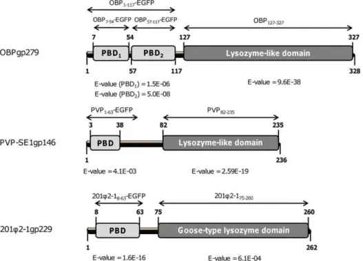
Using Blastp, Pfam and HHpred prediction tools [12–14], endolysins OBPgp279 (from P. fluorescens infecting phage OBP), 201Q2-1gp229 (from P. chlororaphisinfecting phage 201Q2-1) and PVP-SE1gp146 (from S. Enteritidis infecting phage PVP-SE1) were predicted to adopt a modular structure with an N-terminal PBD and a C-terminal catalytic domain (Figure 1). The catalytic domain of 201Q2-1gp229 showed similarity with a goose-type lysozyme domain, known for its three-dimensional structural correspondence with bacteriophage T4 lysozyme and HEWL [15] (Figure 1). The catalytic domains of OBPgp279 and PVP-SE1gp146 both contained a conserved sequence motif (Figure S1), which was typically found in members of the glycoside hydrolase 19 (GH19) family (general sequence motif = F/H/Y-G-R-G-A/P-X-Q-I/L-S/T-F/H/Y/W-H/N-F/Y-N/Y, X =
hydro-Figure 1. Schematic diagram of the domain organization of OBPgp279, 201Q2-1gp229 and PVP-SE1gp146.For each endolysin, the
predicted N-terminal PG binding domains (PBD) are marked in light grey, whereas the C-terminal catalytic domains (lysozyme-like or goose-like lysozyme domains) are in dark grey. E-values for OBP127–327(predicted by HHpred), PBD1and PBD2of OBPgp279 (both HHpred), PVP82–235(Blastp),
PBD of PVP-SE1gp146 (Pfam), 201Q2-175–210(HHpred) and PBD of 201Q2-1gp229 (Pfam) are all indicated beneath the respective domains. The names
philic amino acid) [16]. Other recently characterized endolysins like the Acinetobacter baumannii phage QAB2 lysin (LysAB2, ADX62345.1) [17] or the Salmonella enterica phage e15 lysin (NP_848233.1) [18] also belong to this GH19 family. Both the goose-type lysozyme and the GH19 family are members of the lysozyme-like superfamily which contains a broad range of lytic domains involved in hydrolysis of theb-1,4 linkages between the N-acetylmuramine and N-acetylglucosamine sugars of the PG backbone [19].
The N-terminal domains of OBPgp279, 201Q2-1gp229 and PVP-SE1gp146 possess specific repeated motifs which are conserved in the confirmed PBDs of the modular endolysins KZ144 and EL188 [6]. These repeated motifs largely matched the consensus sequence (D-G-(Pho)2-G-K/N-G/N-T, Pho =
hydro-phobic amino acid) proposed previously [6]. Two such motifs (D-G-L-F-G-E-K-C and D-G-K-W-G-G-T) were present in the N-terminal domain of OBPgp279 (one in each predicted sub-domain) and one in 201Q2-1gp229 (D-G-V-F-G-D-N-T) and PVP-SE1gp146 (D-G-L-Y-G-P-A-T) (Figure S1). This conserved feature is seen in many proteins that interact with repeated structures such as PG [20]. For OBPgp279, seven additional amino acids (L-A-X-Pho-X-L-Y, X = same hydrophilic amino
acid, Pho = hydrophobic amino acid) were detected in front of both repeated motifs with four amino acids in between, strengthening the idea that the PBDs of OBPgp279 were duplicated throughout evolution. The presence of these PG binding motifs inside the N-terminal domains strongly supported the predicted PG binding function. Furthermore, the repeated motif observation suggested the presence of a general sequence conserved among all PBDs of modular endolysins with a Gram-negative background.
N-terminal cell wall binding domains of OBPgp279, PVP-SE1gp146 and 201Q2-1gp229
Peptidoglycan binding capacity. To evaluate the PG binding capacity, we produced the fusion proteins OBP1–117
-EGFP, PVP1–63-EGFP and 201Q2-18–63-EGFP comprising the
predicted binding domains of OBPgp279 (amino acids 3 to 117), PVP-SE1gp146 (amino acids 3 to 38) and 201Q2-1gp229 (amino acids 8 to 63), N-terminally fused to EGFP. For this purpose, OM permeabilizedP. aeruginosaPAO1 andS.Typhimurium LT2 cells [9] were incubated with 2mM fusion protein. Cells were washed to remove unbound protein and the retained fluorescence of PG bound protein was quantified (Figure 2A). A negative wt EGFP
Figure 2. Peptidoglycan binding capacity of fusion proteins OBP1–117-EGFP, OBP7–54-EGFP, OBP57–117-EGFP, PVP1–63-EGFP and
201Q2-18–63-EGFP.(A) The specific fluorescence (i.e. the fluorescence of the treated cells subtracted by the fluorescence of untreated cells and cell
buffer) of 2mM of each fusion protein measured after incubation with OM permeabilizedP. aeruginosaPAO1 (dark grey bars) orS.Typhimurium LT2 cells (light grey bars) is shown here for the different EGFP fusion constructs. Obtained values were compared with the specific fluorescence of the unbound wt EGFP. 2mM of KZ-EGFP, the fusion protein of the PBD of KZ144 and EGFP, was used as positive control. Average and standard deviation values of three independent experiments are shown. (B) Epifluorescence microscopy of OM permeabilizedP. aeruginosaPAO1 cells treated with OBP1–117-EGFP (left) and PVP1–63-EGFP (right). Cells were incubated with 2mM of each fusion protein for 5 min. Cell pellets were washed twice and visualized using epifluorescence microscopy (with 5006magnification, size bar is indicated). Both fusion proteins are visualized in green targeted to the bacterial cell wall.
control and a positive KZ1–83-EGFP [6] control were included for
comparison. The results showed that the N-terminal domains of both OBPgp279 and PVP-SE1gp146 have PG binding activity, since the measured fluorescence levels under saturating concen-trations were comparable with KZ1–83-EGFP [6]. This
observa-tion also indicated that the same number of binding sites was occupied by the fusion proteins. For 201Q2-18–63-EGFP, no
fluorescence was detected, possibly due to improper folding of the fusion protein during expression. Binding capacity of OBP1–117
-EGFP and PVP1–63-EGFP was visually confirmed using
epifluor-escence microscopy (Figure 2B). The cell wall of the targetedP. aeruginosaPAO1 andS.Typhimurium LT2 cells became fluores-cent in less than 5 min after incubation with the fusion constructs
OBP1–117-EGFP and PVP1–63-EGFP. However, a single
subdo-main (either OBP7–54 or OBP57–117) of OBPgp279 was not
sufficient for cell wall binding since no fluorescence was retained with any of both fusion proteins (Figure 2A). This could be explained because of improper folding of the single subdomains in the absence of the other subdomain, or because both subdomains were needed simultaneously for effective PG binding.
Peptidoglycan binding affinity of PVP1–63-EGFP. For PVP1–63-EGFP, the interaction strength with the PG of S.
Typhimurium LT2 ( = ligand) was analyzed using surface plasmon resonance analysis [10]. This technique made it possible to follow the interaction between the fusion proteins and immobilized Gram-negative cells in real time and to determine the binding affinity constant of each interaction. For this, autoclaved S. Typhimurium LT2 cells were immobilized on a PVP1–63-EGFP
coated sensor chip. To this double layer, free PVP1–63-EGFP
proteins were added allowing us to observe the interaction between the fusion proteins and the immobilized cells.
To calculate the apparent affinity constantKafffor binding of the
fusion proteins to the Gram-negative cell wall, we used an indicative two-state model which assumes a 1:1 binding (AB) between fusion protein (A) and ligand (B) occurs followed by a conformational change in the complex (AB..):
AzB/
kd1? ka1
AB/
kd2? ka2
AB:
This proposed model enabled us to calculate the association (ka1),
dissociation (kd1), forward (ka2) and backward (kd2) rate constants
from which a Kaff constant of 1.26610 6
M21 (or a dissociation constant of 7.9361027M) was determined for fusion protein
PVP1–63-EGFP (Table 1). The PG binding affinity constant of the
PBD of PVP-SE1gp146 fell within the range of average SH2 domains and typical cell adhesion molecules [21].
Biochemical properties of OBPgp279, 201Q2-1gp229 and PVP-SE1gp146
Substrate specificity. To confirm the predicted catalytic activity and to determine the bacterial host substrate spectrum, each endolysin (1mM final concentration) was incubated with
three different OM permeabilized Gram-negative cell substrates (P. aeruginosaPAO1,S.Typhimurium LT2 andE. coliXL1-Blue) and four intact Gram-positive bacteria with different structural types of peptidoglycan (S. aureus subsp.aureus Rosenbach ATCC 6538, M. lysodeikticus ATCC 4698, B. subtilis PSB3 and L. lactis subsp. lactis) (Figure S2). All three OM permeabilized Gram-negative cell substrates were efficiently lysed by the tested endolysins as shown by the decrease in OD, demonstrating their muralytic properties. In contrast, all tested Gram-positive bacteria survived the treatment even with an excess of OBPgp279, PVP-SE1gp146 and 201Q2-1gp229 (Figure S2).
Quantification of muralytic activity. The muralytic activ-ities of OBPgp279, 201Q2-1gp229 and PVP-SE1gp146 were first quantified on OM permeabilizedP. aeruginosaPAO1 as described previously [11]. At the optimal pH for enzymatic activity of 7.2 which corresponded to the pH of the bacterial cytoplasm (Figure S3), lysis ofP. aeruginosaPAO1 could be detected upon addition of 10 to 20 nM endolysin. Enzymatic activity further increased linearly dependent on the endolysin concentration. A saturated enzymatic activity was reached at concentrations of 50, 200 and 100 nM for OBPgp279, 201Q2-1gp229 and PVP-SE1gp146, respectively (data not shown). Based on the linear relation between lysis and concentration, muralytic activity was expressed in enzymatic units (Table 2) [11]. OBPgp279 possessed a 1.5 to 6 times higher muralytic activity value than PVP-SE1gp146 and 201Q2-1gp229, respectively, and a 5 to 10 times higher value compared to the previously reported modular endolysins EL188 and KZ144 [6]. As expected, muralytic activity values determined on OM permeabilizedS.Typhimurium LT2 andE. coliXL1-Blue were consistent with those obtained forP. aeruginosa PAO1 (data not shown).
In addition, we tested the necessity of the peptidoglycan binding domain for proper muralytic activity. Therefore, the individual C-terminal catalytic domains of OBPgp279 (OBP127–327),
PVP-SE1gp146 (PVP82–235) and 201Q2-1gp229 (201Q2-175–260) were
cloned and purified to compare their activity with the obtained value for the full length endolysin. In the case of PVP82–235, no
soluble protein could be obtained despite changes in expression
Table 1.Apparent association and dissociation kinetic data for the binding of PVP1–63-EGFP to immobilizedS.Typhimurium LT2 cells.
Analyte concentration A+B«AB AB«AB..
(M) (mg ml21)
Association rate constant
Dissociation rate constant
Forward rate constant
Backward rate constant
Apparent affinity constant
ka1(M21s21) kd1(s21) ka2(s21) kd2(s21) (M21)
5.0061026 176 6.78
6103 3.3461022 9.64
61022 2.25
61023 1.09
6106
10.061026 352 1.146103 5.9461022 3.94
61023 6.07
61024 1.43
6106
Average 1.266106
Two different concentrations (5 and 10mM) were measured in three-fold and the corresponding average is represented. The association rate (ka1), dissociation rate (kd1), forward rate (ka2) and backward rate (kd2) constants are calculated according to a two-state model that describes a 1:1 binding (AB) of the fusion protein analyte (A) to the immobilized ligand (B), followed by a conformational change in the complex (ABRAB..).
parameters. Expression of both OBP127–327 and 201Q2-175–260
yielded pure protein in large amounts for activity testing (Table S1). In the absence of the PBD, the individual catalytic domains of OBPgp279 and 201Q2-1gp229 were still active, retaining 62 and 44% of the muralytic activity of the full length endolysins on OM permeabilized P. aeruginosa PAO1, respectively (Table 2). For OBP127–327, the residual value was significantly higher than the
full length modular endolysins 201Q2-1gp229, KZ144 and EL188. These results indicated that both catalytic domains could work independently from the PBD part and emphasized that the PBD also had an important contribution to the total enzymatic activity of the full length modular endolysins.
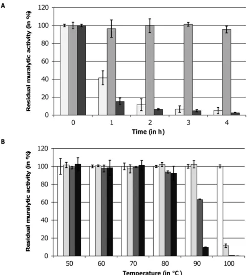
Thermoresistance. All three endolysins could be stored at 4uC in elution buffer for several weeks and months without loss of activity. The residual muralytic activity of each endolysin on OM permeabilized P. aeruginosa PAO1 was determined after a heat treatment at 42uC and 50uC and a subsequent cooling step to room temperature. No reduction of enzymatic activity was observed after prolonged incubation of the enzymes at 42uC (data not shown). When heated at 50uC, the muralytic activity of OBPgp279 and 201Q2-1gp229 dropped below 10% of the initial activity after 2 and 1 h incubation, respectively (Figure 3A). In contrast, PVP-SE1gp146 remained fully active, even after 24 h of incubation. At higher temperatures, PVP-SE1gp146 retained its maximal activity at 80uC incubation for 1 h and was only completely inactivated after 40 min at 100uC (Figure 3B).
In vitroantibacterial activity with and without the OM permeabilizer EDTA
The antibacterial activity of OBPgp279, 201Q2-1gp229 and PVP-SE1gp146 was investigated on the Gram-negative bacteriaP. aeruginosa PAO1, P. aeruginosa Br667, E. coli XL1-Blue and S. Typhimurium LT2, and compared with endolysins KZ144 and EL188 (Table 3A). To overcome the OM barrier, 0.5 mM EDTA was added (Table 3B). EDTA is extensively described as an effective OM permeabilizer [22,23] and was shown to promote the penetration of endolysin EL188 fromP. aeruginosainfecting phage
EL through the OM of clinical P. aeruginosa isolates in mM concentrations [24].
The addition of OM permeabilizer EDTA (Table 3B) caused a significant increase in antibacterial activity of OBPgp279 on all four strains. This observation was more pronounced for P. aeruginosaPAO1 (3 additional log units) and P. aeruginosa Br667 (2 additional log units) than for E. coli XL1-Blue and S. Typhimurium LT2 (both 0.70 additional log units). For endolysins PVP-SE1gp146 and 201Q2-1gp229, onlyP. aeruginosaPAO1 was efficiently reduced in presence of EDTA.
Remarkably, OBPgp279 reduced P. aeruginosa PAO1 and the multi-drug resistant P. aeruginosa Br667 with 1.1060.07 and 1.0860.08 log units, respectively, in the absence of OM destabilizing agents. Contrarily,E. coli XL1-Blue and S. Typhi-murium LT2 showed no sensitivity to the endolysins action. Both PVP-SE1gp146 or 201Q2-1gp229 did not influence the two P. aeruginosastrains, neither did KZ144 and EL188 (Table 3A).
Discussion
Modularity feature in endolysins of Gram-negative origin Based on their structural and biochemical characteristics, OBPgp279, PVP-SE1gp146 and 201Q2-1gp229 are assumed to play an important role in the bacterial host lysis at the end of the phage replication cycle.In silico, these three endolysins were shown to share a modular structure with a PBD at the N-terminal side of the protein and a catalytic domain at the C-terminal side. For OBPgp279 and PVP-SE1gp146, the presence of the PBD was experimentally confirmed. This modular composition is rather rare among endolysins with Gram-negative background, as the majority of these endolysins are globular with only a catalytic domain [25]. Even though they share the same general function and modular composition, OBPgp279, PVP-SE1gp146 and 201Q2-1gp229 are unrelated at the sequence level. This observa-tion emphasizes the relevance of the biochemical and antibacterial characterization of these enzymes in order to discover new and desirable traits for use as bio-control tools.
We may question why some endolysins of Gram-negative infecting phages have a PBD contained within their structure. For Gram-positive infecting phages, the presence of a PBD is speculated to be necessary to keep the enzyme bound to the cell wall debris. In this way, surrounding bacteria (which are potential candidates for a new infection) remain protected from the hydrolyzing activity of newly released endolysin [25]. In this respect, Loessner and coworkers [26] demonstrated that theListeria monocytogenesphage endolysins Ply118 and Ply500 have a binding affinity (3–66108M21) which is comparable with the antigen-ligand affinity in a secondary immune response and can be considered as irreversible. Contrarily, Gram-negative bacteria possess an OM surrounding their PG layer, which eliminates the risk of premature lysis before a new infection can occur. The binding affinities of the Gram-negative phage endolysins KZ144 (2.956107M21) [10] and PVP-SE1gp146 (1.26
6106M21) are 10 to 100 times lower but are still relatively high: for PVP-SE1gp146 in equilibrium, only 1 in a million molecules is in the unbound state.
Not only endolysins of phiKZ-related phages feature a modular composition [6], but also members of other unrelated and smaller myoviruses as shown here forS.Enteritidis phage PVP-SE1 (‘‘rV5-like virus’’) and as described in literature forP. aeruginosa phage QCTX (‘‘P2-like virus’’) [27] and Burkholderia cepacia phages Bcep781 and Bcep1 (‘‘Bcep789-like viruses’’) [28]. Modular endolysins are assumed to exert a higher enzymatic activity than non-modular or globular ones due to the presence of an additional
Table 2.Comparison of muralytic activities of OBPgp279, PVP-SE1gp146, 201Q2-1gp229, the catalytic domains of OBPgp279 (OBP127-327) and of 201Q2-1 (201Q2-175–260), with phage endolysins KZ144 and EL188.
Muralytic activity (in units/mM)
OBPgp279 19,979,151 (0.983)
PVPSE1gp146 13,613,960 (0.992)
OBP127–327 12,361,837 (0.981) EL188 *4,735,148 (0.983)
201Q2-1gp229 4,468,638 (0.995) KZ144 *2,058,435 (0.992)
201Q2-175–260 1,950,766 (0.970)
*[6] Brierset al.
The muralytic activities of the endolysins were calculated from the slope of the best linear regression of the corresponding saturation curves, according to the definition for enzyme unit adapted from Briers and coworkers [11]. OM permeabilizedP. aeruginosaPAO1 cell substrate resuspended in the optimal KH2PO4/K2HPO4buffer (pH 7.2) was used to test the enzymatic activity. The endolysins were dialyzed against a PBS buffer (pH 7.4). Activity values of KZ144 and EL188 [6] were marked with an asterisk. R-square values for each slope are indicated between brackets. Constructs were ranked from the highest to the lowest muralytic activity.
Figure 3. Thermoresistance of OBPgp279, PVP-SE1gp146 and 201Q2-1gp229.(A) The residual muralytic activity of OBPgp279 (1mM, light grey bars), PVP-SE1gp146 (5mM, intermediate grey bars) and 201Q2-1gp229 (3mM, dark grey bars) on OM permeabilizedP. aeruginosaPAO1 cell substrate after 1, 2, 3 and 4 h heat treatment at 50uC is shown. (B) For PVP-SE1gp146 (5mM), the residual activity on OM permeabilizedP. aeruginosa PAO1 after incubation for 0 (white bars), 20 (light grey bars), 40 (dark grey bars) and 60 (black bars) min on different temperatures between 50 and 100uC was determined. For each curve, averages and standard deviations of three repeated and independent experiments are given.
doi:10.1371/journal.pone.0036991.g003
Table 3.In vitroantibacterial activity of OBPgp279, PVP-SE1gp146 and 201Q2-1gp229 without (A) and with (B) the OM permeabilizer EDTA against different Gram-negative species, compared toP. aeruginosaphage endolysins KZ144 and EL188.
A OBPgp279 201Q2-1gp229 PVP-SE1gp146 KZ144 El188
P. aeruginosaPAO1 1.1060.07 0.1760.10 0.1960.01 0.5560.14 0.3260.06
P. aeruginosaBr667 1.0860.08 0.0860.03 0.1060.03 0.3460.07 0.1160.04
E. coliXL1-Blue 0.3860.03 0.0460.04 0.1460.01 0.1460.03 0.2160.12
S.Typhimurium LT2 0.0860.01 0.0760.04 0.1360.02 0.1260.04 0.0960.02
B EDTA OBPgp279 201Q2-1gp229 PVP-SE1gp146 KZ144 El188
EDTA EDTA EDTA EDTA EDTA
P. aeruginosaPAO1 0.5860.02 4.3660.29 3.2560.10 3.6860,13 3.0560.11 3.3060.12
P. aeruginosaBr667 0.0360.02 3.1460.22 0.1760.20 0.1260,02 0.8860.04 0.7560.12
E. coliXL1-Blue 0.2960.11 1.1860.20 0.5860.25 0.3460,02 0.4560.13 0.5960.17
S.Typhimurium LT2 0.0560.05 0.7760.05 0.2660.02 0.1460,21 0.3360.14 0.2460.15 Cell cultures (initial cell density of 106cells/ml) were incubated for 30 min with 1.5
PG binding domain which keeps the enzyme in close proximity to the PG substrate [25]. This assumption is confirmed here: the muralytic activity of the modular endolysins OBPgp279, PVP-SE1gp146 and 201Q2-1gp229 is 160, 104 and 30 times higher compared to the value for the globular phage T4 lysozyme [29]. For an unknown reason, some unrelated phages possess strong and fast-acting modular endolysins instead of less active, globular ones.
Biochemical activity of modular endolysins
Based on their biochemical activity, OBPgp279 and PVP-SE1gp146 degrade the PG layer of the tested Gram-negative species with the highest efficiencies compared to 201Q2-1gp229, EL188 and KZ144. In contrast to the catalytic transglycosylase specificity of KZ144 and EL188 [6,30], OBPgp279, PVP-SE1gp146 and 201Q2-1gp229 are predicted to have lysozyme-like activity. Despite the similar catalytic function, a large variation in muralytic activity values is observed for the individual catalytic domains of OBPgp279 and 201Q2-1gp229, due to differences in the primary protein structure. This variation only partially explains the large differences in muralytic activity values of the full length endolysins, since the PBD part also influences the muralytic activity in a protein dependent manner.
Previously, Briers and co-workers [6] revealed the insensitivity of Gram-positive bacteria for the muralytic action of modular endolysins KZ144 and EL188. We found that also OBPgp279, PVP-SE1gp146 and 201Q2-1gp229 leave the Gram-positive bacteria S. aureus subsp. aureus Rosenbach ATCC 6538, M. lysodeikticus ATCC 4698,L. lactissubsp.lactisandB. subtilisPSB3 unharmed. An explanation must be found in the structure of their PG layer. Sensitive Gram-negative species share a common A1c chemotype of PG with a direct peptide bond between adjacent muropeptides, whereas insensitiveS. aureussubsp.aureusRosenbach ATCC 6538,M. lysodeikticusATCC 4698 andL. lactissubsp.lactis have a PG of the A3a, A2a and A4a chemotype, respectively [31,32]. These last 3 chemotypes show resistance for the mode of action of muralytic enzymes, like HEWL, due to the absence of a direct inter-peptide cross-link [33].Bacillus subtilisPSB3, however, possesses the A1cchemotype of its Gram-negative counterparts but 17.3% of the glucosamine sugars in its PG are N-deacetylated [34]. As previously shown for commercial HEWL, this species-specific modification explains for the resistance against PG lytic enzymes [33]. Therefore, a direct cross-linked A1cchemotype of PG without substantial N-deacetylation is an essential prerequisite for efficient hydrolysis by endolysins from Gram-negative infecting phages.
Thermoresistance of PVP-SE1gp146
An unexpected result is the observed thermoresistance of endolysin PVP-SE1gp146 at temperatures up to 90uC. To our knowledge, PVP-SE1gp146 is the first Gram-negative phage derived endolysin described so far to show thermoresistance. For the structural lysin gp36 ofP. aeruginosaphageQKMV (KMV36C) only the catalytic domain-containing C-terminus was thermo-resistant [35]. Heat-inactivated, unfolded KMV36C has the opportunity to refold back again into its (partially) active form after cooling and therefore retains its complete enzymatic activity. Recently, this mechanism was also suggested by Schmelcher and coworkers [36] to explain for the high thermostabilty of Gram-positive Listeria monocytogenes phage endolysins HPL118 and HPL511. In their study, HPL118 and HPL511 retained 35% of activity after 30 min incubation at 90uC. The authors prove that the unfolding of HPL118 and HPL511 at higher temperatures is followed by a rapid refolding upon cooling. This proposed refolding mechanism may preserve the enzyme activity and could also explain the thermoresistance of PVP-SE1gp146. From this
perspective, PVP-SE1gp146 could be an interesting candidate as antibacterial component in a hurdle approach for food preserva-tion, in addition to other antibacterial additives like bacteriocin and nisin [37]. Because of the antibacterial and thermoresistant properties of PVP-SE1gp146, the use of chemical preservatives and the intensity of heat treatments could be reduced in such a way that quality foods with higher nutritional values are obtained.
Potential use of OBPgp279 as an enzybiotic
Despite the presence of an OM which shields the PG layer from the externally added endolysin, OBPgp279 is capable to reduce bothP. aeruginosaPAO1 and the multi-drug resistantP. aeruginosa Br667, contrary to PVP-SE1gp146, 201Q2-1gp229, KZ144 and EL188. Therefore, OBPgp279 is a potential candidate for the use as enzybiotic to control multi-drug resistant opportunistic Pseudo-monas infections in human and animals. In addition to its high muralytic activity, OBPgp279 is suggested to intrinsically desta-bilize the OM in thePseudomonascell wall so that the endolysin reaches its enzymatic target, which could explain the observed reductions. A similar characteristic was suggested for theBacillus amyloliquefaciens phage IAM1521 endolysin Lys1521 [38]. In the case of this endolysin, the presence of positively charged amino acids in the C-terminal cell wall binding part that interact with the OM and destabilize it, explained the antibacterial property of the endolysin [39]. For OBPgp279, a similar mechanism is unlikely to induce the proposed OM destabilization as its C-terminal end does not contain more positively charged amino acids compared to PVP-SE1gp146 and 201Q2-1gp229, which do not affect the tested Pseudomonasstrains. Furthermore, no obvious amphipathic helixes which are able to intercalate into the OM structure resulting in OM permeabilization [40] were detected at the N- and C-terminal ends of OBPgp279. Further structural analysis of the interaction between OBPgp279 and thePseudomonascell wall will be necessary to reveal the mechanism and determinants responsible for the observedPseudomonassensitivity.
Combinatorial endolysin/EDTA approach
of the endolysin/EDTA combinatorial approach. However, the elucidation of the unique biochemical and antibacterial character-istics of OBPgp279, PVP-SE1gp146 and 201Q2-1gp229 (modular-ity, thermoresistance, OM destabilizing activity) allows us to gain a better insight in the relationship between the structure and activity of endolysins, in general. In this way, we take an important first step in development of future antibacterial therapies based on modular Gram-negative endolysins.
Supporting Information
Figure S1 Amino acid sequences of OBPgp279, PVP-SE1gp146 and 201Q2-1gp229. In each sequence the amino
acids comprising the PG binding domains are shaded in light grey and the amino acids of the catalytic domains in dark grey. The PG binding motifs which show some similarity with consensus sequence D-G-(Pho)2-G-K/N-G/N-T (Pho = hydrophobic amino
acid) of the PBDs in KZ144 and EL188 [6] are underlined and marked in bold. A seven amino acid motif L-A-X-Pho-X-L-Y (X = the same hydrophilic amino acid, Pho = hydrophobic amino acid, boxed) is present in front of both repeated PG binding motifs of OBPgp279, each with four amino acids in between. Inside the catalytic domains of OBPgp279 and PVP-SE1gp146, the detected motifs showing similarity to the general consensus sequence for GH19 family members (F/H/Y-G-R-G-A/P-X-Q-I/L-S/T-F/ H/Y/W-H/N-F/Y-N/Y, X = hydrophilic amino acid) [16] are underlined in italic.
(TIF)
Figure S2 Bacterial host spectrum of OBPgp279 (light grey bars), PVP-SE1gp146 (intermediate grey bars) and 201Q2-1gp229 (dark grey bars).Each endolysin (1mM final concentration) is added to outer membrane permeabilized P. aeruginosaPAO1 (PA),S.Typhimurium LT2 (ST),E. coliXL1-Blue (EC) and intactS. aureussubsp.aureusRosenbach ATCC 6538 (SA), M. lysodeikticusATCC 4698 (ML),B. subtilisPSB3 (BS) andL. lactis subsp.lactis(LL). The resulting decrease of OD655 nmin function of
time after endolysin addition is depicted here. Averages and standard deviations of three independent experiments are given. (TIF)
Figure S3 pH optimization for enzymatic activity of OBPgp279 (diamonds), PVP-SE1gp146 (squares) and 201Q2-1gp229 (triangles). Muralytic activity is measured as the slope of the DOD655 nm/min curve (Y-axis) on OM
permeabilized P. aeruginosa PAO1 substrate and is shown for a pH range between 3 and 10 (X-axis). Final concentrations used here are 1mM OBPgp279, 5mM PVP-SE1gp146 and 3mM 201Q2-1gp229, each dialyzed against a PBS buffer (pH 7.4). Averages and standard deviations of three repeated experiments are given.
(TIF)
Table S1 Overview of all expression constructs used in this study. The specific insert, cloning vector, primers, expression/ purification conditions and expression yield are indicated for each construct. The restriction endonuclease recognition sites inside the primers are underlined. Extra nucleotides necessary for in frame cloning are indicated in bold. All constructs were expressed using theE. coliBL21(DE3)pLysS expression strain in LB medium upon induction with 1 mM IPTG. Proteins were purified on Ni2+- NTA columns using protein dependent imidazole concentrations given in the table. Expression yield is indicated in mg of obtained recombinant protein per liter expression culture as determined spectrophotometrically.
(DOCX)
Acknowledgments
The authors would like to thank Prof. Philip Serwer (UT Health Science Center, San Antonio, USA) for providing the genomic DNA of phage 201Q2-1, and Prof. Chris Michiels (Centre for Food- and Microbial Technology, KU Leuven, Belgium) and Dr. Martin Loessner (E.T.H., Zu¨rich, Switzerland) for their technical support.
Author Contributions
Conceived and designed the experiments: MW YB RL SS JA. Performed the experiments: MW YB SS. Analyzed the data: MW YB RL. Contributed reagents/materials/analysis tools: YB SS JA RL. Wrote the paper: MW YB SS JA RL.
References
1. Cornelissen A, Hardies SC, Shaburova OV, Krylov VN, Mattheus W, et al. (2012) Complete genome sequence of the giant virus OBP and comparative genome analysis of the diverse phiKZ-related phages. J Virol 86: 1844–1852. 2. Thomas JA, Rolando MR, Carroll CA, Shen PS, Belnap DM, et al. (2008)
Characterization ofPseudomonas chlororaphismyovirus 201varQ2-1 via genomic sequencing, mass spectrometry, and electron microscopy. Virology 376: 330–338.
3. Santos SB, Kropinski AM, Ceyssens PJ, Ackermann HW, Villegas A, et al. (2011) Genomic and Proteomic Characterization of the broad-Host-Range Salmonella Phage PVP-SE1: Creation of a New Phage Genus. J Virol 85: 11265–11273.
4. Borysowski J, Weber-Dabrowska B, Go´rski A (2006) Bacteriophage endolysins as a novel class of antibacterial agents. Exp Biol Med 231: 366–377.
5. Callewaert L, Walmagh M, Michiels CW, Lavigne R (2011) Food applications of bacterial cell wall hydrolases. Curr Opin Biotechnol 22: 164–171. 6. Briers Y, Volckaert G, Cornelissen A, Lagaert S, Michiels CW, et al. (2007a)
Muralytic activity and modular structure of the endolysins of Pseudomonas aeruginosaphages phiKZ and EL. Mol Microbiol 65: 1334–1344.
7. Fokine A, Miroshnikov KA, Shneider MM, Mesyanzhinov VV, Rossmann MG (2008) Structure of the bacteriophageQKZ lytic transglycosylase gp144. J Biol Chem 283: 7242–7250.
8. Pirnay JP, De Vos D, Cochez C, Bilocq F, Pirson J, et al. (2003) Molecular epidemiology ofPseudomonas aeruginosacolonization in a burn unit: persistence of a multidrug-resistant clone and a silver sulfadiazine resistant clone. J Clin Microbiol 41: 1192–1202.
9. Lavigne R, Briers Y, Hertveldt K, Robben J, Volckaert G (2004) Identification and characterization of a highly thermostable bacteriophage lysozyme. Cell Mol Life Sci 61: 2753–2759.
10. Briers Y, Schmelcher M, Loessner MJ, Hendrix J, Engelborghs Y, et al. (2009) The high-affinity peptidoglycan binding domain ofPseudomonasphage endolysin KZ144. Biochem Biophys Res Commun 383: 187–191.
11. Briers Y, Lavigne R, Volckaert G, Hertveldt K (2007b) A standardized approach for accurate quantification of murein hydrolase activity in high-throughput assays. J Biochem Biophys Meth 70: 531–533.
12. Altschul SF, Madden TL, Scha¨ffer AA, Zhang J, Zhang Z, et al. (1997) Gapped BLAST and PSI-BLAST: a new generation of protein database search programs. Nucleic Acids Res 25: 3389–3402.
13. Finn RD, Mistry J, Tate J, Coggill P, Heger A, et al. (2010) The Pfam protein families database. Nucleic Acids Res 38: 211–222.
14. So¨ding J, Biegert A, Lupas AN (2005) The HHpred interactive server for protein homology detection and structure prediction. Nucleic Acids Res 33: 244–248. 15. Weaver LH, Griitter GI, Remington SJ, Gray TM, Isaacs NW, et al. (1985)
Comparison of goose-type, chicken-type, and phage-type lysozymes illustrates the changes that occur in both amino acid sequence and three-dimensional structure during evolution. J Mol Evol 21: 97–111.
16. Udaya Prakash NA, Jayanthi M, Sabarinathan R, Kangueane P, Mathew L, et al. (2010) Evolution, homology conservation, and identification of unique sequence signatures in GH19 family chitinases. J Mol Evol 70: 466–478. 17. Lai MJ, Lin NT, Hu A, Soo PC, Chen LK, et al. (2011) Antibacterial activity of
Acinetobacter baumannii phage QAB2 endolysin (LysAB2) against both gram-positive and gram-negative bacteria. Appl Microbiol Biotechnol 90: 529–539. 18. Kropinski AM, Kovalyova IV, Billington SJ, Patrick AN, Butts BD, et al. (2007)
The genome of epsilon15, a serotype-converting, Group E1Salmonella enterica-specific bacteriophage. Virol 369: 234–244.
Signature Motifs. PLoS ONE 5(11): e15388. doi:10.1371/journal.-pone.0015388.
20. Wren BW (1991) A family of clostridial and streptococcal ligand-binding proteins with conserved C-terminal repeat sequences. Mol Microbiol 5: 797–803.
21. Larose L, Gish G, Pawson T (1995) Construction of an SH2 domain-binding site with mixed specificity. J Biol Chem 270: 3858–3862.
22. Vaara M (1992) Agents that increase the permeability of the outer membrane. Microbiol Rev 56: 395–411.
23. Guha A, Choudhury A, Unni BG, Roy MK (2002) Effect of outer-membrane permeabilizers on the activity of antibiotics and plant extracts against Pseudomonas aeruginosa. Folia Microbiol 47: 379–384.
24. Briers Y, Walmagh M, Lavigne R (2011) Use of bacteriophage endolysin EL188 and outer membrane permeabilizers against Pseudomonas aeruginosa. J Appl Microbiol 110: 778–785.
25. Fischetti VA (2010) Bacteriophage endolysins: a novel anti-infective to control Gram-positive pathogens. Int J Med Microbiol 300: 357–362.
26. Loessner MJ, Kramer K, Ebel F, Scherer S (2002) C-terminal domains ofListeria monocytogenes bacteriophage murein hydrolases determine specific recognition and high-affinity binding to bacterial cell wall carbohydrates. Mol Microbiol 44: 335–349.
27. Nakayama K, Kanaya S, Ohnishi M, Terawaki Y, Hayashi T (1999) The complete nucleotide sequence of QCTX, a cytotoxin-converting phage of Pseudomonas aeruginosa: implications for phage evolution and horizontal gene transfer via bacteriophages. Mol Microbiol 31: 399–419.
28. Summer EJ, Gonzalez CF, Bomer M, Carlile T, Embry A, et al. (2006) Divergence and mosaicism among virulent soil phages of theBurkholderia cepacia complex. J Bacteriol 188: 255–268.
29. Nakimbugwe D, Masschalck B, Deckers D, Callewaert L, Aertsen A, et al. (2006) Cell wall substrate specificity of six different lysozymes and lysozyme inhibitory activity of bacterial extracts. FEMS Microbiol Lett 259: 41–46.
30. Paradis-Bleau C, Cloutier I, Lemieux L, Sanschagrin F, Laroche J, et al. (2007) Peptidoglycan lytic activity of thePseudomonas aeruginosaphagewKZ gp144 lytic transglycosylase. FEMS Microbiol Lett 266: 201–209.
31. Schleifer KH, Kandler O (1972) Peptidoglycan types of bacterial cell walls and their taxonomic implications. Bacteriol Rev 36: 407–477.
32. Courtin P, Miranda G, Guillot A, Wessner F, Me´zange C, et al. (2006) Peptidoglycan structure analysis ofLactococcus lactisreveals the presence of an L,D-carboxypeptidase involved in peptidoglycan maturation. J Bacteriol 188: 5293–5298.
33. Archibald AR, Hancock IC, Harwood CR (1993) Cell wall structure, synthesis and turnover. In: Hoch JA, Losick R, eds.Bacillus subtilisand other Gram-positive bacteria, biochemistry, physiology and molecular genetics. Washington DC: American Society for Microbiology. pp 381–410.
34. Atrih A, Bacher G, Allmaier G, Williamson MP, Foster SJ (1999) Analysis of peptidoglycan structure from vegetative cells ofBacillus subtilis168 and role of PBP 5 in peptidoglycan maturation. J Bacteriol 181: 3956–3966.
35. Briers Y, Lavigne R, Plessers P, Hertveldt K, Hanssens I, et al. (2006) Stability analysis of the bacteriophage phiKMV lysin gp36C and its putative role during infection. Cell Mol Life Sci 63: 1899–1905.
36. Schmelcher M, Waldherr F, Loessner MJ (2012) Listeria bacteriophage peptidoglycan hydrolases feature high thermoresistance and reveal increased activity after divalent metal cation substitution. Appl Microbiol Biotechnol 93: 633–643.
37. Ga´lvez A, Abriouel H, Lo´pez RL, Ben Omar N (2007) Bacteriocin-based strategies for food biopreservation. Int J Food Microbiol 120: 51–70. 38. Morita M, Tanji Y, Mizoguchi K, Soejima A, Orito Y, et al. (2001) Antibacterial
activity ofBacillus amyloliquefaciensphage endolysin without holin conjugation. J Biosci Bioeng 91: 469–473.
39. Orito Y, Morita M, Hori K, Unno H, Tanji Y (2004)Bacillus amyloliquefaciens phage endolysin can enhance permeability of Pseudomonas aeruginosa outer membrane and induce cell lysis. Appl Microbiol Biotechnol 65: 105–109. 40. Sato H, Feix JB (2006) Peptide-membrane interactions and mechanisms of
membrane destruction by amphipathic a-helical antimicrobial peptides. Biochim Biophys Acta 1758: 1245–1256.