COMPARISON OF INFECTIOUS DISEASES DIAGNOSIS IN
AUTOPSY
AND
ITS
MEDICO-LEGAL
IMPORTANCE:
A
MULTICENTRIC STUDY (PAHNT – NORTH OF MANCHESTER,
U.K. AND INMLCF – NORTH OF PORTUGAL)
Dissertation for the Application to the Master’s Degree in Legal Medicine submitted to the Institute of Biomedical Sciences of Abel Salazar, Oporto University.
Supervisor – Dr. António José Guimarães Paiva Correia
Category – Consultant Histopathologist
Affiliation – Pennine Acute Hospitals NHS Trust Co-supervisors:
Dr. Ivor Cartmill
Category – Consultant Microbiologist
Affiliation – Pennine Acute Hospitals NHS Trust Professor Maria José Carneiro de Sousa Pinto da Costa
Category – Cathedratic Professor
Affiliation – Institute of Biomedical Sciences Abel
“O valor das coisas não está no tempo que elas duram, mas na intensidade com que acontecem. Por isso existem momentos inesquecíveis, coisas inexplicáveis e pessoas incomparáveis.”
∞
“The value of things is not in how long they last, but in the intensity they occur. So there are unforgettable moments, unexplainable things and incomparable people.”
- Fernando Pessoa
This is to all of you, special people that in your own way helped me getting here.
Page | I
Acknowledgments
I would like to thank everyone who was involved in the project in every possible way, but specially:
To Dr António Paiva, my supervisor and a friend, who guided me wisely throughout this chapter of my life and contributed to make this dream come true, for believing and being there for me.
To Professor Maria José Pinto da Costa, Head of the Master Degree in Legal Medicine and my attachment supervisor in the Pathology Services at the North Delegation of the Legal Medicine and Forensic Sciences National Institute, for all the advice and recommendations made for improving this work and for all the support with the Portuguese component of this work.
To Dr Ivor Cartmill, Consultant Microbiologist and my attachment supervisor at Pennine Acute Hospitals National Health Service Trust (PAHNT) based at Royal Oldham Hospital (ROH) at Manchester, United Kingdom (UK), for helping me with my research and supporting me with his expertize.
To Dr Madhu Rao, Head of the Histopathology Department at PAHNT, for allowing me to carry out this study and do my attachment in the pathology department.
To Dr José Bessa Oliveira, Director of the Pathology Services at the North Delegation of the Legal Medicine and Forensic Sciences National Institute, for all the help, the vote of confidence and sympathy that you gave me.
To Mr Simon Nelson, Her Majesty Senior Coroner of Manchester North District, who gave me permission to gather all the information relative to the UK’s Pathology necessary for this study.
To the technicians of the mortuary of PAHNT based at Royal Oldham Hospital, for all the kindness and amusement that made the time run faster and eased the job, because I know that what you all do is not that easy and you do it always with a smile.
Page | II To Dr Salma Abassi, for your patience of every day sharing the office with me, for our conversations and your advice.
To Master Ana Sofia Batista, who helped me and inspired me in this adventure. To Vera Santos, who gave some extra help reviewing this work.
To my colleagues (Marlene Sophie and Vanessa Branco), my fellows in this journey and my friends for life. I will never forget you, girls.
To my parents and grandparents, who made the great effort of funding my attachment.
To my sister, who always cheers me up with her energy and defies me with her personality.
To Rui, a big thanks for all your support and for being so much more than my best friend in this world.
Page | III
Comparação do Diagnóstico de Doenças Infeciosas na Autópsia
e a sua Importância Médico-Legal: Estudo Multicêntrico (PAHNT
– Norte de Manchester, U.K. e INMLCF – Norte de Portugal)
Sinopse
A autópsia médico-legal foi uma das formas encontradas pela medicina legal de tentar dar resposta a questões como a causa da morte de um individuo ou o mecanismo da mesma, e considera não apenas o exame macroscópico do cadáver mas também os resultados dos exames complementares como a histologia ou a toxicologia.
A microbiologia é uma ciência que se interessa pelo estudo de microrganismos e pelas doenças causadas pelos mesmos, que pode fornecer dados importantes quando inserida no âmbito médico-legal.
Ao longo dos estudos efetuados presentes na literatura foram apontados diversos pontos contra a utilização do exame microbiológico, entre eles, questões monetárias,
dificuldades sobre a recolha das amostras e ainda controvérsias na interpretação dos resultados post-mortem. É, por isso, evidente a importância de procurar esclarecimento sobre estas questões, bem como, a averiguação do custo-benefício do exame
microbiológico de forma sistemática e também em que casos seria mais importante a sua realização, sendo estas algumas das razões que motivaram a realização deste trabalho.
Para esclarecer estas questões, foi realizado em Inglaterra e Portugal um estudo retrospetivo com base em 2105 processos de autópsia válidos de forma a averiguar a frequência atual da utilização da microbiologia nas autópsias médico-legais e estabelecer uma relação comparativa, bem como determinar a importância que lhe é atribuída em ambos os sistemas legais.
Foram também averiguados os preços médios por amostra e as principais dificuldades enfrentadas pelos microbiologistas quando na presença deste tipo de amostragem verificando-se uma correspondência em geral com o que foi encontrado na literatura. Para além disto, foram organizados estatisticamente os diagnósticos de causa de morte relacionados com doenças infeciosas permitindo esclarecer quais as doenças mais frequentes e quais os casos em que estas doenças já constavam do histórico clínico,
Page | IV considerando-se que estes seriam os casos cujo exame microbiológico poderia ser dispensável à partida.
Foram também observados e comparados os meios de proteção, limpeza e
comportamentos de risco dentro e fora da sala de autópsia, entre uma instituição que é considerada preparada para receber casos de risco de contágio elevado (PAHNT) e outra que não possui nada mais do que um protocolo geral para casos standard e os
equipamentos correspondentes (INMLCF).
Relativamente às observações dos meios e comportamentos em ambas as instituições verificou-se que apesar de no Reino Unido existirem protocolos que definem o
equipamento a usar em cada situação dependendo do nível de risco, algumas dessas regras não eram seguidas à risca mas têm à disposição o equipamento necessário para casos de alto risco de contágio.
Por outro lado, em Portugal, apesar de não terem um protocolo específico para casos de risco de contágio elevado, verificou-se um esforço positivo por cumprir as regras gerais de proteção estabelecidas internacionalmente, no entanto, não dispõem do equipamento adequado para eventuais casos de médio/alto risco de contágio nas suas instalações. Pode-se, no entanto, inferir que o facto de existirem protocolos e equipamentos para alto risco não basta uma vez que na maioria dos casos não existe um diagnóstico prévio de que se pode ou não tratar de um caso de risco. É considerado mais importante prevenir para um risco desconhecido como é o caso em Portugal, que em grande parte das vezes o histórico clínico mais completo só chega às mãos do médico legista após a realização da autópsia, sendo que estes profissionais desconhecem à partida se o indivíduo era tuberculoso ou seropositivo, por exemplo.
Através dos dados recolhidos e analisados estatisticamente foi possível verificar os seguintes pontos: 1) as mortes de causas naturais representam mais de 50% dos casos em ambas as instituições e em especial no Reino Unido; 2) a microbiologia foi apenas aplicada em casos de morte natural e em valores considerados residuais, sendo que em Portugal nem sequer é efetuada; 3) os casos de morte por doenças infeciosas
representam 10 a 25% do volume total de casos em ambas as instituições; 4) as doenças infeciosas ao nível do sistema respiratório e da cavidade abdominal foram as infeções mais frequentes na casuística de ambas as instituições; 5) dos casos em que a
microbiologia foi realizada, esta foi importante para o diagnóstico da causa de morte em 75% e nos restantes 25% ajudou a excluir a hipótese de existência de doença infeciosa.
Page | V possível inferir com rigor sobre a importância de uma amostragem sistematizada. Não foi também possível averiguar a importância da microbiologia em casos de suspeita de homicídio.
Para muitos profissionais do ramo, a microbiologia pode ser considerada dispendiosa tendo em conta o custo-benefício e as dificuldades apontadas pelos microbiologistas nos casos genéricos. No entanto, acredita-se que esta amostragem deveria ser realizada, não apenas nos casos de suspeita de infeção mas também nos casos em que a análise macroscópica e histológica não permitem um diagnóstico de causa de morte conclusivo. Apesar disso, o exame microbiológico considera-se perfeitamente dispensável em casos óbvios como um esfaqueamento ou um enforcamento.
É também importante referir que o diagnóstico de doenças infeciosas postmortem pode ter interesse a nível social permitindo uma reflexão para melhor gestão de medicamentos, por exemplo, ou familiar em que pode ser importante realizar um despiste para uma doença contagiosa até então desconhecida pelos familiares do indivíduo.
Conclui-se porém que a microbiologia não deve ser observada apenas pela perspetiva económica mas também pela sua capacidade de fornecer as respostas não só a questões médico-legais como também de interesse social.
Palavras-chave:
Forense; Microbiologia; Medicina Legal; Autópsia; Doenças InfeciosasPage | VI This page was intentionally left in blank.
Page | VII
Abstract
The medico-legal autopsy was one of the ways found by the forensic medicine of trying to answer questions about the cause of death of an individual or the mechanism of it, and considers not only the macroscopic examination of the body but also the results of complementary examinations such as histology or toxicology.
Microbiology is a science that is interested in the study of microorganisms and the
diseases caused by them, which can provide important data when inserted in the medico-legal context.
Throughout the conducted studies found in the literature were mentioned several points against the use of microbiological examination, including, monetary issues, difficulties on the collection of samples and even controversy in the interpretation of the results post-mortem. It is evident the importance of clarifying these issues as well as investigating the cost-effectiveness of systematic microbiological examination or in which cases it would be more relevant its performance, and it was what motivated this work.
Therefore, it was held in England and Portugal a retrospective study based on 2105 valid autopsy reports in order to ascertain the current frequency of use of microbiology in the medico-legal autopsies and establish a comparative relationship as well as determine the importance attributed to it in both legal systems.
They were also observed and compared the means of protection, cleanliness and hazardous behaviours within the autopsy room between one mortuary that is considered to be prepared to receive cases of high risk of contamination and one which has nothing more than a general protocol and the respective equipment for standard cases.
Through the data collected and analysed it was possible to check the following points: 1) the deaths of natural causes account for over 50% of cases in both institutions and particularly in the United Kingdom; 2) Microbiology has been applied only in cases of natural death and in amounts considered residual, and in Portugal is not even performed; 3) cases of death from infectious diseases account for 10-25% of total cases in both institutions; 4) infectious diseases to the respiratory system and abdominal cavity were the most common infections in the sample of both institutions; 5) of the cases in which
Page | VIII microbiology was performed, it was important to diagnose the cause of death by 75% and in the remaining 25% has helped to exclude the hypothesis of infectious disease.
Due to the residual number of cases with any application of microbiological examination it’s not possible to accurately infer the importance of a systematic sampling. However, it is believed that this sampling should be performed, not only in cases of a suspected
infection but also in cases where the macroscopic and histological analysis did not allow a conclusive diagnosis of the cause of death.
On the observations of resources and conduct at both institutions it was found that while in the UK there are protocols that define the equipment to use in each situation depending on the level of risk, some of these rules were not strictly followed, however they have at their disposal the equipment needed for high-risk cases of contagion. On the other hand, in Portugal, despite not having a specific protocol for high infection risk cases, there was a positive effort to fulfil the general protection rules internationally established, but they don’t have the proper equipment for cases of possible medium/high risk of infection in their facilities.
Thereby, it is concluded that the microbiology must be observed not just by the economic perspective but also for its ability to provide the answers not only at medico-legal issues but also of social interest.
Page | IX
Index
1. BACKGROUND ... 1
1.1 Motivation ... 1
1.2 Structure of the Thesis ... 2
2. BIBLIOGRAPHIC REVISION ... 3
LEGAL MEDICINE ... 3
2.1 The Legal Medicine in the History, Definition and Importance ... 3
2.2 Portuguese Medico-Legal Organization ... 5
2.3 English Legal Medicine and the Coroner’s System ... 6
THE MEDICO-LEGAL AUTOPSY ... 8
2.4 The History of the Autopsy ... 8
2.5 Types of Autopsy ... 9
2.6 Medico-Legal Investigation & Medico-Legal Autopsy Objectives ... 10
THE INFECTIOUS DISEASES AND MICROBIOLOGY ...12
2.7 The Infectious Diseases and Microbiology in the History ... 12
2.7.1 The Oldest Disease: Tuberculosis ... 12
2.7.2 Smallpox ... 14
2.7.3 Cholera ... 15
2.7.4 The Plague ... 15
2.7.5 The HIV/AIDS ... 16
2.7.6 Microbiology... 17
2.8 Microbiology in Post-mortem Practices ... 19
2.9 Thanato-microbiology: The Possible Solution ... 21
2.10 Infectious Diseases, Autopsy and Hazards ... 21
3. METHODOLOGY ... 23
3.1 General Characteristics of the Study ... 23
3.2 Sampling ... 24
3.2.1 Sampling from INMLCF ... 25
3.2.2 Sampling from PAHNT ... 25
3.3 Processing and Statistic Analysis of Data ... 26
4. RESULTS ... 27
4.1 Observations/General Results of the Study ... 27
4.2 INMLCF Results ... 32
Page | X
5. DISCUSSION ... 47
5.1 General and Comparative Considerations ... 47
5.2 INMLCF Results Discussion ... 50
5.3 PAHNT Results Discussion ... 51
6. CONCLUSIONS ... 53
7. STUDY’S LIMITATIONS AND FUTURE INTENTIONS... 55
7.1 Study’s Limitations ... 55
7.2 Future Intentions ... 56
Page | XI
Figures Index
Figure 1 – Distribution of the cases by age ranges (INMLCF and PAHNT) ...28
Figure 2 – Distribution of cases by gender (INMLCF and PAHNT) ...28
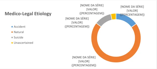
Figure 3 – Distribution of cases by medico-legal etiologies (INMLCF and PAHNT) ...29
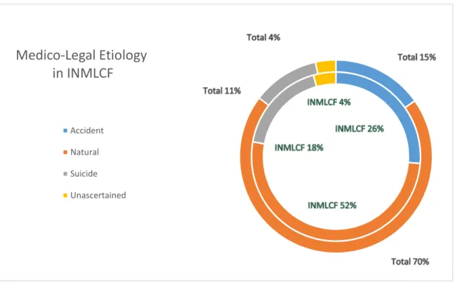
Figure 4 – Distribution of cases by medico-legal etiologies in INMLCF ...33
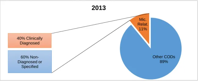
Figure 5 – Proportions of Infectious Diseases and Diagnosis in INMLCF (2013) ...34
Figure 6 – Proportions of Infectious Diseases and Diagnosis in INMLCF (2014) ...34
Figure 7 – Infectious diseases in 2013 (INMLCF) ...35
Figure 8 – Infectious diseases in 2014 (INMLCF) ...35
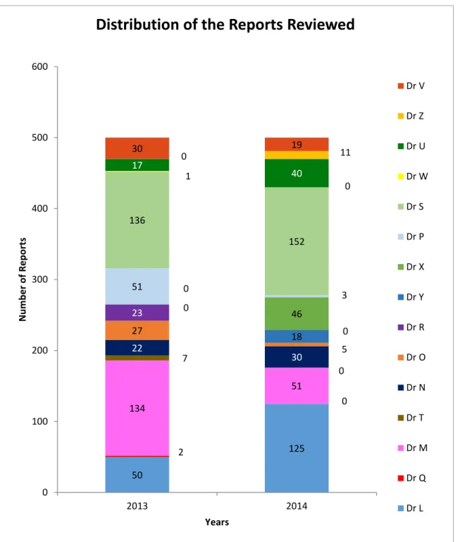
Figure 9 – Distribution of the Reports Reviewed in INMLCF ...36
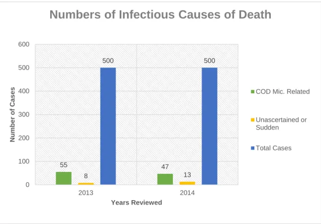
Figure 10 – Number of infectious or unascertained causes of death in INMLCF ...37
Figure 11 – Number of infectious or unascertained causes of death in PAHNT ...37
Figure 12 – Infectious causes of death in INMLCF ...38
Figure 13 – Infectious diseases in PAHNT ...38
Figure 14 – Distribution of cases by medico-legal etiologies in PAHNT ...40
Figure 15 - Proportions of infectious diseases and diagnosis in PAHNT (2013) ...42
Figure 16 - Proportions of infectious diseases and diagnosis in PAHNT (2014) ...43
Figure 17 - Infectious diseases in 2013 (PAHNT) ...44
Figure 18 – Infectious diseases in 2014 (PAHNT) ...45
Page | XII
Tables Index
Table 1 – Number of causes of death related to infectious diseases and unascertained or
sudden deaths (INMLCF and PAHNT) ...29
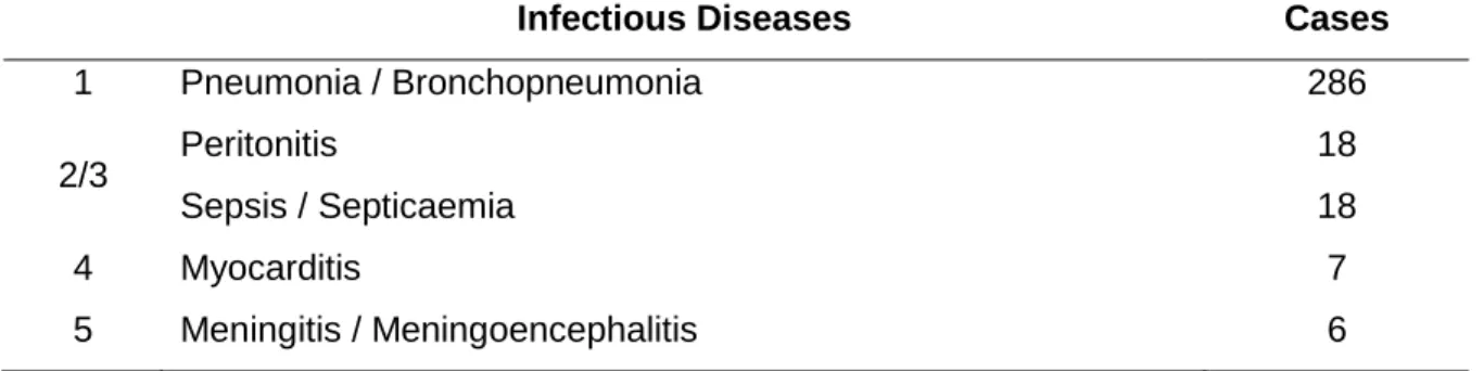
Table 2 – List of the 5 main infectious diseases as causes of death in INMLCF and PAHNT ...30
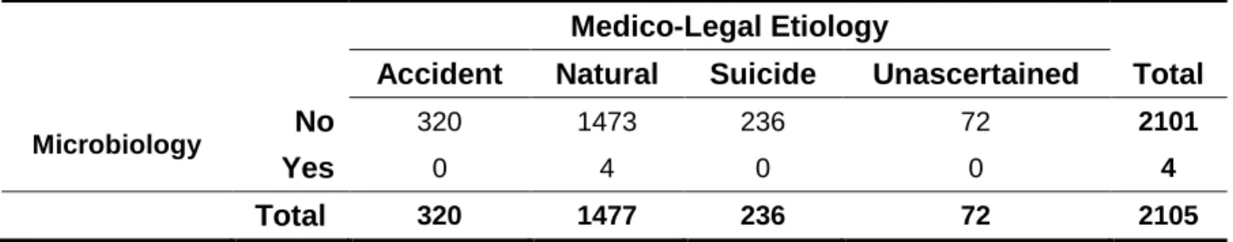
Table 3 – Distribution of the cases with microbiological examination by the medico-legal etiologies in INMLCF and PAHNT ...31
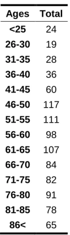
Table 4 – Distribution of the cases by age ranges in INMLCF ...32
Table 5 – Number of infectious or unascertained causes of death in INMLCF ...33
Table 6 – Number of infectious diseases, clinical diagnosis and microbiological tests performed in INMLCF ...34
Table 7 – List of Infectious Causes of Death in INMLCF ...35
Table 8 – Distribution of cases by age ranges in PAHNT ...39
Table 9 – Number of infectious or unascertained causes of death in PAHNT ...40
Table 10 – Number of infectious diseases, clinical diagnosis and microbiological tests performed in PAHNT...41
Table 11 – Number of cases with microbiological testing in each medico-legal etiology in PAHNT ...41
Page | XIII
List of Abbreviations
A.C. – “After Christ”
AIDS – Acquired Immune Deficiency Syndrome B.C. – “Before Christ”
COD – Cause of Death CSF – Cerebro-Spinal Fluid DL – Decreto-lei (Act)
e.g. – “exempli gratia” “for example” HIV – Human Immunodeficiency Virus HM – Her Majesty
ICBAS – Instituto de Ciências Biomédicas de Abel Salazar (Institute of Biomedical Sciences Abel Salazar)
IML – Instituto de Medicina Legal (Legal Medicine Institute)
INML – Instituto Nacional de Medicina Legal (Legal Medicine National Institute)
INMLCF – Instituto Nacional de Medicina Legal e Ciências Forenses (Legal Medicine and Forensic Sciences National Institute)
NHS – National Health Service
PAHNT – Pennine Acute Hospitals NHS Trust PCR – Polymerase Chain Reaction
ROH – Royal Oldham Hospital SCD – Sudden Cardiac Death
SIDS – Sudden Infant Death Syndrome
SPSS – Statistical Package for the Social Sciences SUDI – Sudden Unexpected Death in Infancy UK – United Kingdom
USA – United States of America WHO – World Health Organization
Page | XIV This page was intentionally left in blank.
Page | 1
1. Background
“The progress of science is strewn, like an ancient desert trail, with the bleached skeleton of discarded theories which once seemed to possess eternal life.”
– Arthur Koestler (1905/1983)
1.1
Motivation
In legal medicine, infectious diseases are very present but in different perspectives. On one hand, there are infectious diseases as a cause of death. For this, some countries, such as England, have well-defined protocols regarding autopsies with suspicion of infectious diseases, according to The Health and Safety Executive (2003), The Health Protection Agency North West (2004) and The Royal College of Pathologists (2002). However, not all institutions seem to accept or be prepared to perform this kind of autopsy (Lucas 2011).
On the other hand, as far as we could find out and understand, it seems there is no specific protocol in the Portuguese legal medicine for diagnosis of infectious diseases at autopsy (Instituto Nacional de Medicina Legal e Ciências Forenses 2015). This can lead that in some cases, certain procedures might be neglected, impairing the correct
diagnosis of the cause of death (Baptista 2014 and Bonds et al. 2003).
Furthermore, neglecting certain procedures can also lead to a hazard to the integrity of the medical examiner that carries out the autopsy or to other professionals who contact with the corpses, as defended by Nolte (2004) and Wilson (2006). Therefore we have infectious diseases as a recurrent danger in the day-to-day life of Legal Medicine professionals.
In another point of view there are the bureaucratic procedures, such as the repatriation of bodies, which require an expert to certify the absence of infectious diseases like in the act
Page | 2 of The Portuguese Council of Ministers (1998). So we observe as well infectious diseases on a body as a potential threat to public health.
It may then be stated that the correct diagnosis of infectious diseases is important not only in life but also post-mortem, particularly in an autopsy.
The main goals of this study are: 1) compare and evaluate the consequences of the existence or non-existence of national / international protocols for the diagnosis of infectious diseases in a medico-legal autopsy; 2) determine the frequency of infectious diseases as a cause of death, as well as, the importance of microbiology in medico-legal autopsies.
Later, if appropriate, it may be proposed an example of a procedural protocol for an autopsy with suspected infectious disease.
1.2
Structure of the Thesis
This thesis is structured in 7 chapters.
Chapter 1 is about the motivation, background and structure of this thesis.
Chapter 2 presents a resumed bibliographic review about the main subjects that based this thesis: the legal medicine, the medico-legal autopsy and the infectious diseases. Chapter 3 clarifies the methodology chosen and designates the sample used.
Chapter 4 condenses the results obtained in this study and some main observations. Chapter 5 discuss the results associating them with the review of the literature and its previous results.
Chapter 6 displays the main conclusions of this work.
Chapter 7 enunciates the limitations of the research and offers some suggestions for future works.
Page | 3
2. Bibliographic Revision
Legal Medicine
“Taceant colloquia. Effugiat risus. Hic locus est ubi mors gaudet succurrere vitae. – Let conversation cease. Let laughter flee. This is the place where death delights to help the living.”
– Giovanni Morgagni (1682/1771)
– Latin motto posted on the autopsy room of Dr Milton Helpern, the third medical examiner of New York City
2.1
The Legal Medicine in the History, Definition and Importance
Searching throughout the history, it is impossible to indicate a date for the origin of the legal medicine. As the first application of medical principles is attributed to the first cry of pain ever existent that made another human experiment something to ease the pain, the first application of medical knowledge for legal purposes might have happened in the early days of the humanity (Smith 1951).
The oldest record of legislation about the practice of medicine is The Code of Hammurabi from the year 2200 B.C. that included punishments for medical malpractice (Wecht 2005).
Some authors defend that the first medico-legal expert was Imhotep 3000 B.C. a grand vizier, chief justice, and physician to King Zozer, and also architect of the first great
Pyramid of Sakkara. However, because Imhotep became a God, all his actions have been mistaken with mystical or magical weakening Egyptian medicine (Smith 1951).
In China, on the first half of the thirteenth century there was compiled a volume with instructions to follow while investigating deaths and especially those with suspicious or obvious criminal circumstances (Smith 1951).
Page | 4 The legal medicine as we know it, a distinct and individualized subject, was probably born in Germany in 1553 with the “Constitutio Criminalis Carolina” promoted by the Emperor Carlos V. This document assigns the obligation of doctors being called to give their opinion in cases of murder, wounds, poisoning, hanging, drowning, infanticide and abortion (Baptista 2014).
This obligation gave birth to innumerous publications on the field, including one of the most important books on the matter “Quaestiones Medico-Legales” by Paulus Zacchias, published somewhere between 1621 and 1658.
There is a great difficulty in defining legal medicine because of its multiplicity of purposes and applications to the Law. It’s even frequent calling legal medicine and forensic
medicine synonyms despite the etiological differences. In fact, forensic medicine is just a part of legal medicine that deals with all the questions in Court (Pinto da Costa 2009). In the end we can define legal medicine as a subsidiary science of Law that applies medical knowledge to juridical matters in Civil Law, Criminal Law, Labour Law, Administrative Law or Family and Juvenile Law (Baptista 2014). This multiplicity of applications is what makes legal medicine such a wide and rich field for investigation and development of scientific knowledge.
Actually there are two great types of Juridical and Medico-Legal Organizations with authority to request an autopsy in order to determine a cause of death (Baptista 2014):
1) The countries following the Anglo-Saxon Code like England, Wales, Ireland and some parts of The United States of America (USA) among others politically influenced by the previous. In these countries the main character is the Coroner.
2) The countries following the Napoleonic Code like most of the Continental European countries and others politically similar to them. Notice that the Scottish system has more in common with this group than with the Coroner system.
Page | 5 The Portuguese medico-legal system started in the XIX century with the creation of three establishments distributed by the three main regions: North (Oporto), South (Lisbon) and Centre (Coimbra). Each of them had its own independent Medico-Legal Counsel and morgue in articulation with medical schools and local hospitals (Baptista 2014). In 1918, these structures were named “Institutos de Medicina Legal” (IML) – Legal Medicine Institutes, gaining some resemblance with the current organization, but there were some issues due to their different methodologies (Baptista 2014).
Later in 2001, these three institutes were extinguished giving place to one unified organization with three delegations, the “Instituto Nacional de Medicina Legal” (INML) – Legal Medicine National Institute. This new organization with administrative and financial independency operated through the values of pursuing the public interest, exemption, fairness, accuracy and quality.
With the evolution of the scientific knowledge and the necessity of hosting other sciences non-related to medicine to better serve the public interest the INML suffered another reformation to the current organization – the “Instituto Nacional de Medicina Legal e
Ciências Forenses” (INMLCF) – Legal Medicine and Forensic Sciences National Institute
– which is responsible for all the expert activity in the forensic medicine field. It is divided in three delegations: North based in Oporto, South based in Lisbon and Centre based in Coimbra that is also the head office; and twenty seven minor offices spread throughout all the Portuguese territory (Ministério da Justiça 2014).
INMLCF services are divided in four departments:
Forensic Clinic and Forensic Pathology Service: responsible for all the medico-legal post-mortems, body or human remains identification, embalming and study of anatomic parts, as well as the realization of exams in living people for describing and evaluating any injury to the psycho-physic integrity in criminal law, work law and civil law. Forensic Psychiatry, Forensic Psychology, Forensic Entomology, Forensic Anthropology and Dental Medicine are also included in these unities. This service is present in all the delegations and it supervises all the offices (Instituto Nacional de Medicina Legal e Ciências Forenses 2015).
Page | 6 Forensic Chemistry and Toxicology Service: laboratory responsible for the
chemistry and toxicology testing in the ambit of the activities of the Institute as well as other cases requested by the Court. It is located on the South Delegation although it may have some working unities in other delegations (Instituto Nacional de Medicina Legal e Ciências Forenses 2015).
Genetic and Forensic Biology Service: laboratory responsible for genetic
identification exams like paternity investigation, individual identification and biologic criminalistics, among others involved on the Institute activities or requested by the Court. It is located on the Centre Delegation although it might have some working unities in other delegations (Instituto Nacional de Medicina Legal e Ciências Forenses 2015).
Forensic Technologies and Criminalistics Service: responsible for the search, registration, collection and treatment of samples, as well as other exams not
mentioned or fitted in the other services like Ballistics, Physics, Writing and Document Analysis. It is located on the North Delegation (Instituto Nacional de Medicina Legal e Ciências Forenses 2015).
2.3
English Legal Medicine and the Coroner’s System
The Coroner’s System is a distinctive system applied to most of the countries with English influence since the Victorian time. It is focussed on the Coroner figure, from the Latin
corona or the Anglo-French coroune which means “crown”, whose main task was “keep
the pleas of the Crown”, this is to protect the financial interests of the Crown (Coroners' Society of England and Wales 2014).
Some say they existed since the Dark Ages but only in 1194 the Coroner’s Office was officially created by King Richard I of England in “Articles of Eyre”. At that time the
Coroner had a large variety of duties from tax gathering to investigating any other aspects of medieval life that could assist the Crown. Sudden deaths were always important and remained one of the duties of a Coroner till our days, although the reasons for its investigation are not the same (Coroners' Society of England and Wales 2014).
With the Norman Conquest, to detain local communities of keeping killing more Normans, every sudden death was investigated and every deceased would be presumed to be
Page | 7 and the penalty was known as the Murdrum from which the term “murder” has derived.
The changes to the Coroner’s System continued over the centuries with the Births and
Deaths Registration Act in 1836 as one of the differences (Coroners' Society of England
and Wales 2014).
The Coroners Act of 1887 refuted the fiscal tasks as the main duty of a Coroner, changing it to the investigation of sudden, violent or unnatural deaths for better serve the community and its concerns (Coroners' Society of England and Wales 2014).
In the Coroners Act of 1926, the investigation of homicides was transferred to the police services leaving to the Coroner the purpose of investigating all the other cases of sudden or violent deaths, like the system we know now, but still have the power to judge a person for murder or infanticide cases. It was also added that the Coroner would need to have knowledge in Medicine or Law (Baptista 2014).
Further adjustments took place to the laws that rule the Coroners and its system as a result of the needs and evolution of the society as well as the evolution of the English judicial and political system. During this path to the Coroners System as we know it, some countries adopted and adapted it to themselves while others gave it up, adopting the Medical Examiner System instead.
Nowadays in England, Wales and some other countries the coroner is mostly either a doctor or lawyer or both responsible for investigating the deaths when the body is within the Coroners Legislation territory and the death is: a) suspicious of violent or unnatural causes; b) the cause of death is unascertained; c) within custody (Baptista 2014).
Page | 8
The Medico-Legal Autopsy
“Vous auriez pendant vingt ans pris du matin au soir des notes au lit des malades sur les affections du coeur, du poumon (...) que tout ne sera pour vous que confusion dans les symptômes (...) Ouvrez quelques cadavres: vous verrez aussitôt disparaître l'obscurité que la seule observation n'avait pu dissiper.” – Marie François Xavier Bichat (1771/1802)
2.4
The History of the Autopsy
It is believed that it was the curiosity of a scientist or the helping instinct of a physician that lead to the first autopsy in the ancient ages.
The word autopsy, from Greek “autopsía”, means seeing for one’s self or making a personal inspection. However, its usage is only applied to the pathologic meaning i.e. opening a dead body for observing its organs and determining the cause of death according to King et al. (1973).
This practice was performed in animals in 3500 B.C. during the animistic period, not for the anatomic study but for offering rituals where a diviner would be able to predict the future by observing the liver of the sacrificed animal, considered to be the seat of the soul as said by Dada et al. (1996) and King et al. (1973).
With naturalism, Hippocrates influence and the birth of Medicine, the path to the autopsy was shortened but still very long. In Egypt with the ritual of embalming the bodies of the deceased, they had to remove the internal organs so it might be considered the first approach to an autopsy. During the third century B.C. in Alexandria dissection was allowed to be performed to study the normal and the changes caused by disease (King et
Page | 9 suspected about the wine being too strong. So after observing some changes in a pig’s liver drowned on the wine, he opened one of his fellow’s body and observed the same changes in his liver. This English chronicle brought to believe that the autopsy practice was already known (King et al. 1973).
Singer reported that autopsies were being done in Italy around 1266 and 1275, and the first ones were medico-legal to help the law solve some cases. It was also mentioned that by the approach of the fourteenth century the medical study increased the importance of anatomical dissections (King et al. 1973).
Till the nineteenth century, the autopsy was the main methodology of medical
investigation but with the advances in microscopy and Virchow’s contributes to Medicine, the naked-eye examination began to occupy the second place in investigation
methodology even though its practice hasn’t diminished. By the end of the century, the technique applied to the dissections and the methods of examination also improved, reducing the duration of the performance of an autopsy to less than 3 hours, which was Prost’s estimated minimum time for a post-mortem examination in 1802 (King et al. 1973). After the second great world war, the autopsy practice decreased, being even described as “routine work” or “burden”. In fact, with the big advances of other fields of science the autopsy stood important but only as a starting point for investigations and not as the final resource in searching the answers for the pathologist’s questions (King et al. 1973). With that the intrusive autopsy is still used but some other methods like Imaging or the so called Virtopsy are improving in order to replace possibly at least half of the autopsies performed today.
2.5
Types of Autopsy
There are two types of autopsies that differ mostly by its main objectives:
The anatomo-clinical: it is used to investigate more about the disease and the means to the cure. It is performed by request of the family or the hospital itself, in the hospital facilities by anatomopathologists and it needs the authorization of the deceased closest relatives (Baptista 2014 and Pinto da Costa 2009). This autopsy can never occur in cases of death or suspicion of death by violent causes (Santos 2008).
Page | 10 The medico-legal: it is used to investigate the cause of death and all its circumstances, in cases of suspicion of violent or criminal death of which its cause is unascertained. It intends to answer questions like: – What happened? – Who? – When? – Where? – Why? – How? – giving special attention to the external observations as opposed to the clinical autopsy. It doesn’t need authorization of the relatives and it can be requested or dispensed by the Coroner, a Judge, the Public Ministry, a Magistrate, a Tax Attorney or even the Police, depending on the geographic location. For example, in Portugal it takes place in the INMLCF and like many other European countries it is performed by expert forensic pathologists. It uses not only the external and internal examination of the body but also related information, examination of the scene where the body was found and eventual complementary laboratory test results, being all equally important (Baptista 2014 and Pinto da Costa 2009).
In some places the medico-legal autopsy can be divided in two categories:
The criminal deaths like infanticide, murder or suspiciously litigant. These autopsies are performed by forensic pathologists (Baptista 2014).
The non-criminal like suicide, accidental or sudden deaths presumed as natural. In European countries like Portugal these autopsies are performed by forensic pathologists even without criminal suspicion, but in countries like England they are performed by hospital anatomopathologists not necessarily with any forensic specialization (Baptista 2014).
2.6
Medico-Legal Investigation & Medico-Legal Autopsy Objectives
The medico-legal autopsy should be performed whenever there is a violent death (accident, suicide, homicide) or whenever the cause of death is unascertained and its circumstances raise suspicion on the interference of an external agent (Santos 2008). In cases like these it is necessary a medico-legal investigation including a medico-legal autopsy. The main objectives of a medico-legal investigation are (Santos 2008):
Deceased identification, Death mechanism, Cause of death,
Page | 11 autopsy alone can’t give the answers to all of them (Santos 2008).
The identification of the body reveals to be quite difficult when there’s an advanced stage of decomposition, when the deceased isn’t wanted (neither by family nor the police) and when there’s no information related to the case (Santos 2008).
Sometimes it’s impossible to determine the cause and mechanism of death even after a medico-legal autopsy. Studies reveal that the number of deaths by unascertained causes differs from center to center but it fluctuates between 4-10% (Santos 2008).
In other cases, even though the cause of death is determinate, the differential diagnosis is indeterminate because of lack of data to assure with certainty if it was an accident, a suicide or a homicide (Santos 2008).
This can happen if there is lack of information by the police, clinical records, family, etc.; by an inadequate investigation of the scene and the death circumstances; or by a poorly conducted or performed autopsy by inexperienced professional (Santos 2008).
Page | 12
The Infectious Diseases and Microbiology
“False facts are highly injurious to the progress of science, for they often endure long; but false views, if supported by some evidence, do little harm, for every one takes a salutary pleasure in proving their falseness.”
– Charles Darwin (1809/1882)
2.7
The Infectious Diseases and Microbiology in the History
Since the antiquity that we observe manifestations of infectious diseases and microbe proliferations like bacterial and algal slimes (Wainwright et al. 1992).
Microbes can invade the human body by cavities, wounds and mucosa, using the human cell’s energy to replicate and spread. Infectious diseases are diseases caused by
pathogens that can be transmitted human to human – directly or indirectly – by air or body fluids, by food and water or by insects and ticks, according to The Science Museum of Minnesota (2014) and The World Health Organization.
Today it is one of the leading causes of death all over the world and the third in the USA, next to cancer and cardiovascular diseases (Science Museum of Minnesota 2014).
However, there are at least five infectious diseases that were intimately connected to the changes in history and that caused the biggest and deadliest outbreaks all over the world.
2.7.1 The Oldest Disease: Tuberculosis
The oldest disease recorded is the tuberculosis with its earliest evidence dated 2400 B.C. consisting in fragments of spinal columns from Egyptian mummies. After that tuberculosis kept appearing and killing large numbers of people as Hippocrates records it in 460 B.C. as the wasting disease. The disease kept infecting people all over the world with many long outbreaks during centuries (Science Museum of Minnesota 2014).
Page | 13 Emily Bronte died from tuberculosis and some fictional characters in works like
Shakespeare’s Hamlet or Giacomo Puccini’s La Boheme, succumbed to the disease too (Science Museum of Minnesota 2014).
In 1819, Rene Laennec detected the tubercle – the mucus created in lungs of tuberculosis patients – and invented the stethoscope to ear the internal sounds of the lungs in order to help diagnose tuberculosis (Science Museum of Minnesota 2014).
Herman Brehmer, a tuberculosis sufferer, went to the Himalayas under doctor’s orders to live in a healthier climate and when he returned cured in 1859 he funded the first
sanatorium to treat other patients with fresh air (Science Museum of Minnesota 2014). In 1882, Robert Koch discovered the bacteria causing tuberculosis and defended that the disease spreads by sneezing and coughing, making great contributions to the emerging field of bacteriology as with his germ theory he gave the basis for understanding more about infectious diseases (Science Museum of Minnesota 2014).
During the 1900s there were anti-spitting campaigns as a way to stop the spreading of the disease. In 1926, three scientists created a vaccine to prevent tuberculosis, the BCG Vaccine – named after its creators Boquet, Calmette and Guerin – now used in every continent as a part of a national compulsory vaccination plan. In the USA this vaccine is not compulsory due to widespread public controversy as a consequence of the risk of infection (Science Museum of Minnesota 2014).
In 1943, Selman Walksman discovers the streptomycin, the first antibiotic against tuberculosis, for which he received the Nobel Prize years after his discovery (Science Museum of Minnesota 2014).
In 1989, officials of the World Health Organization (WHO) predicted the eradication of tuberculosis by 2010; however, later in 1990 tuberculosis re-emerged in a multi-drug resistant (MDR) form and affected a great number of people weakened with Acquired Immune Deficiency Syndrome (AIDS). It was declared as a global emergency (Nelson, et al., 2014) (Science Museum of Minnesota 2014).
In the 2000s, it was detected a case of extensively drug resistant tuberculosis (XDR TB) that didn’t respond to any known drug until today (Nelson et al. 2014 and Science Museum of Minnesota 2014).
Page | 14 2.7.2 Smallpox
The smallpox is dated since 1150 B.C. with its pustules appearing on the mummy of the Egyptian Pharaoh Ramses V (Brachman 2003). Later in 430 B.C. Thucydides in Athens reports that the survivors of the disease are immune to later infection, according to Nelson
et al. (2014) and The Science Museum of Minnesota (2014).
In 910 A.C. the Persian physician Rhazes is the first to differentiate measles from
smallpox, proposing that smallpox is contagious from person to person and that survivors acquire immunity (Nelson et al. 2014 and Science Museum of Minnesota 2014).
In 1718, a British ambassador’s wife, after surviving smallpox in Turkey, described a process called “variolation” that consisted in placing dried scabs of the disease under the skin causing a mild case of smallpox but giving lifelong immunity and after testing it in orphans and prisoners, the king promoted the procedure (Nelson et al. 2014 and Science Museum of Minnesota 2014).
In 1792, Edward Jenner inoculated a boy with fluid from a blister of cowpox of a milkmaid and created the first vaccine – the word “vaccine” from Latin means “from a cow” – leading to the first laws of vaccination in northern Europe in the nineteenth century (Nelson et al. 2014 and Science Museum of Minnesota 2014).
However, with the community’s movements protesting against the vaccination till the end of the century, the British Parliament created a conscience clause in 1907 for the parents objecting the procedure (Science Museum of Minnesota 2014).
By the end of the 40s, England was declared smallpox-free, and after the latest outbreak the USA promoted a campaign of massive vaccination. In 1966, the World Health
Organization (WHO) declared a program towards the eradication of the smallpox in ten years. The last reported case of smallpox was in a three-year-old girl that survived in Bangladesh in 1975 and in 1979 the WHO declared the world free of smallpox (Science Museum of Minnesota 2014).
Nowadays there are two reservoirs of the virus, one in the USA and another in Russia, but the discussion of if these should be destroyed or kept continues until today with two main premises: 1) if one of these reservoirs falls at the wrong hands it can be used for
bioterrorism and cause a devastating outbreak; 2) if a new kind of smallpox appears these reservoirs can be useful to create a new cure (Science Museum of Minnesota 2014).
Page | 15 In India, there are reports dating back to 450 B.C. and written in Sanskrit of a lethal illness that drains the water of its victims, which is believed to be the cholera. The first modern records of the disease are from the Portuguese Garcia de Orta who in 1563 was working in India on his book “Colóquios dos simples e drogas he cousas medicinais da Índia”, in English, Colloquy of the simple, and drugs and medical things of India (Science Museum of Minnesota 2014).
Around the nineteenth century, Cholera spread from India to western and central Europe, Canada and USA and some believed it to be a plot to kill the poor (Science Museum of Minnesota 2014).
In 1854, John Snow created the first map of an outbreak in a London district and found out that most of the ill people were using the same well water contaminated by an overflowing cesspool, leading to the practice of filtering and treating municipal water (Nelson et al. 2014 and Science Museum of Minnesota 2014).
In the same year Filippo Pacini describes the bacteria of cholera as a commalike shape during an outbreak in Florence, and in 1883 Robert Koch confirms Pacini’s findings (Science Museum of Minnesota 2014).
Cholera re-emerged in 1992 with the same toxin but with a different structure that makes it harder to detect and control. Today it affects mostly countries with less financial resources and hygiene habits. Regions prone to natural disasters have also a high probability of developing the disease (Science Museum of Minnesota 2014).
2.7.4 The Plague
The earliest evidence of the plague was reported in China and India back in 400 B.C. and then in the Roman Empire in 250-266 A.C and became known as The Plague of Justinian. In 540 the bubonic plague killed 5.000 to 10.000 people a day in the Byzantine Empire, especially in Constantinople – now Istanbul, Turkey – and kept its outbreaks in all Europe for centuries with many names like Black Death in 1347 (Nelson et al. 2014 and Science Museum of Minnesota 2014).
In 1400, when the rates of the killing were high in Europe, the legend of The Three Living
Page | 16 saying “Such as I was you are, and such as I am you will be”, became popular (Science Museum of Minnesota 2014).
Around 1700 A.C. Europe was not only being devastated by the smallpox and
tuberculosis, named as “the great white death”, but also trying to prevent the plague to enter European territory by commercial routes (Science Museum of Minnesota 2014). The last serious European outbreak occurred in Marseille, France, in 1720 but it remained virulent in Asia. In 1855-1900 the plague spread from the Hong Kong’s port to every continent and some islands like Hawaii and Philippines, becoming the last major plague epidemic (Science Museum of Minnesota 2014).
In 1894, Alexandre Yersin and Kitasato Shibasaburo identified the bacteria causing the plague, later named Yersinia Pestis (Nelson et al. 2014 and Science Museum of Minnesota 2014).
In the 1900s, scientists found that rat fleas could spread the disease to humans; so the community started campaigns to kill the rats in an attempt to stop the spread of the disease (Nelson et al. 2014 and Science Museum of Minnesota 2014).
In 1938, doctors discovered that sulfa drugs were effective against the plague, but later the discovery of streptomycin revealed a super effective treatment (Science Museum of Minnesota 2014).
There have been some sporadic outbreaks of plague over the 80s-90s in Africa, Asia and South America. Globally the WHO reports 1.000 to 3.000 cases of plague per year and in some less developed countries the disease is still a problem with rodents carrying the bacteria that causes it (Science Museum of Minnesota 2014).
2.7.5 The HIV/AIDS
The earliest evidence of the virus is dated from 1930s when the Simian Immunodeficiency Virus (SIV) mutated to a form that could infect humans – HIV. Hunters while handling with bushmeat became infected by contacting with the human form infected blood (Science Museum of Minnesota 2014).
Page | 17 the locals because later some unknown Haitians went to the USA and infected some Americans (Science Museum of Minnesota 2014).
In 1979, symptoms of pneumonia associated with skin lesions – Kaposi’s Sarcoma – detected in young, homosexual men left the Centers for Disease Control curious and later the infection was named Acquired Immune Deficiency Syndrome (AIDS) (Nelson et al. 2014 and Science Museum of Minnesota 2014).
Somewhere around 1981-1983, two scientists identified separately a virus they thought to be the cause of AIDS but later their discoveries were found to be the same virus which was named HIV (Brachman 2003 and Science Museum of Minnesota 2014).
The AIDS memorial quilt – the NAMES Project – was created in 1987 to honour those who have died with AIDS and it consisted of about 40.000 panels 3 x 6 feet long, displayed in several communities in the USA (Science Museum of Minnesota 2014).
During the 90s, red ribbons became the symbol of the AIDS awareness and many public figures and athletes such as Freddy Mercury, Rock Hudson and Magic Johnson either died of AIDS or assumed publicly their status of HIV-positive. There’s still no cure to the disease (Nelson et al. 2014 and Science Museum of Minnesota 2014).
2.7.6 Microbiology
Although the presence of microbe-related diseases and phenomena existed ever since the very beginning of life on Earth, microbiology remained with very little practical knowledge until the second millennium A.C. Even so, it was only after the invention of microscopes that microbiology was disseminated to truly rise as a science (Wainwright et
al. 1992).
The microscope was invented in the late 1600s but the first microscopes didn’t have enough magnification to allow the observation of bacteria by the human eye (Wainwright
et al. 1992).
A microscopist named Van Leeuwenhoek was the first to make a microscope that could show bacteria in rainwater, well water, seawater and other preparations (Brachman 2003) that he ended up drawing and publishing. His sketches now are clear evidence that he observed various types of bacteria and protozoa. The botanist Pietro Antonio Micheli
Page | 18 described over 900 species of microbes during the eighteenth century but still there were no big advances for microbiology itself (Nelson et al. 2014 and Wainwright et al. 1992).
It was only when Louis Pasteur refuted the theory of spontaneous generation by
conducting a series of experiments to sterilize air, water and a bit of meat broth that some scientists became convinced to try the same experiments and new ones to completely refute the theory of their most ferocious opponent H. Charlton Bastian. From this discussion, one of the Pasteur’s followers, John Tyndall showed, with a process of tyndallisation (heating then leave to cool repeatedly and alternately) to kill heat-stable forms of bacteria, that microbes don’t appear spontaneously (Nelson et al. 2014 and Wainwright et al. 1992).
After this, microbiology needed two main things to completely advance: 1) improvements on microscopes and tools to help the visualization of the microorganisms and 2) methods of growing microorganisms (Wainwright et al. 1992).
Robert Hooke, in the sixteenth century, invented the first compound microscope which consists in the combination of two or more magnifying systems or lenses. However, his microscopes suffered from chromatic aberrations that made the observation of bacteria very difficult. Only in the nineteenth century was this problem fixed with the invention of achromatic lenses by Professor Amici (Wainwright et al. 1992).
The next major advance was the creation of staining methods. The first staining procedure was used in 1849 by Ferdinand Cohn, a botanist, using vegetable dyes like carmine and haematoxylin. Robert Koch used methylene blue in 1877 to stain bacteria and gave a big step to the process of creating permanent preparations. Later in 1884, Hans Christian Gram developed his famous coloration method, allowing the classification of bacteria as Gram-positive if they retained the violet coloration or Gram-negative if they didn’t. This was based on the biochemical and morphological properties of these microorganisms (Wainwright et al. 1992).
Rapid advances in the identification and differentiation of microorganisms were made during these decades but the microscope advances were very little. Only in 1919, with the creation of the ultraviolet microscope, was it possible to look at elementary virus. Then appeared the electron microscope in 1934 achieving unbelievable magnifications comparing to either one of the previous types and in 1965 the scanning electron microscope was invented (Wainwright et al. 1992).
Page | 19 medium to cultivate bacteria was created by Pasteur in 1860. Before that meat broth was used as a medium for bacteria cultures and it persisted in some branches of microbiology, e.g. bacteriology, until nowadays. Mycologists also tend to prefer undefined medium (Wainwright et al. 1992).
All these media were in liquid form until the introduction of gelatine and then agar in 1882 what made solid medium possible. Later the invention of media with silica gel promoted even more advances for microbiologic studies. One of Kohn’s assistants, Petri, introduced the Petri dish revolutionizing the experiments with such a simple modification in 1887 (Wainwright et al. 1992).
More advances were made during the nineteenth century like the selective medium and the anaerobic jar but none of them was as important as an effective procedure of sterilization – the autoclaves, large pressure cookers – created by Pasteur’s colleague, Chamberland, in 1884. After this, new methods of sterilization allowed the conversion of some tools to plastic copies that eased the work of microbiologists (Wainwright et al. 1992).
Microbiology had also an important role on the creation of the first vaccines and antibiotic treatments, like the penicillin by Alexander Fleming in the 1930s, for lethal diseases such as the ones described previously and also chemotherapy (Brachman 2003, Nelson et al. 2014 and Wainwright et al. 1992).
2.8
Microbiology in Post-mortem Practices
The use of tissue samples from autopsies for microbiological examination is very
controversial, because of the post-mortem changes and the possibility of contamination, in addition to the difficulty of interpretation of the results by specialists and the lack of
consensus (Price et al. 2011). However, for some authors it has an important role in the specific post-mortem diagnosis of an infectious disease (Finkbeiner et al. 2009 and Riedel 2014).
Tsokos et al. (2001), support that the collection of specimens from at least two different sites for sampling should be autopsy standard procedure, although multiple cultures from
Page | 20 a large variety of sampling sites could raise the probability of identifying the etiologic agent of an ante-mortem infection. They also refer that the best samples are usually spleen and heart blood in opposition to the lung that frequently gives unreliable results due to false positives.
According to Oliva et al. (2011), in cases of Sudden Cardiac Death (SCD) not only microbiologic samples like blood, organs with septic lesions, cerebro-spinal fluid (CSF) and any other recovered fluids should be collected for cultures, but also complementary techniques like PCR should be used to exclude or identify cases of HIV, hepatitis B and C and for detection of viral proteins.
Moore et al. (2015) tried to improve the value of Microbiology sampling in autopsy using swabs instead of tissue collection and they came to the conclusion that in coroner’s cases, even though results of swabbing were supportive rather than tissue collection, the omission of virology findings as contributors to death leads to an under-estimation of virus’s burden on mortality.
When observing cases of Sudden Unexpected Death in Infancy (SUDI), Weber et al. (2010) recommend routine microbiology sampling, regardless the post-mortem interval (PMI) or the difficulties on the interpretation of the positive results. Carmichael et al. (1996) defend that microbiological tests contributed to the diagnostic of Sudden Infant Death Syndrome (SIDS) by revealing no infectious disease in most cases, but in those where an infection was diagnosed, microbiology was crucial to correctly identify the cause of death (COD).
There are some authors that seem to believe that with careful technique the artefacts in post-mortem microbiology tests can be significantly reduced and that it should be performed regardless the age of the deceased (Morris et al. 2006).
Others even made a possible optimized protocol/guideline regarding the best samples to collect in each case by suspicion or by age-subgroup and they recommend the inquiry of the cost-benefit for every case in forensic or clinical pathology (Fernández-Rodríguez et
Page | 21 Can et al. (2014) define thanatomicrobiome as microorganisms present in a healthy human body prior to death and in their study they try to find out what happens to these microorganisms. Thanato-microbiology is a new branch of microbiology which studies the microorganism colonies and tries to distinguish which colonies are originally from post-mortem phenomena, using it to estimate time since death (Metcalf et al. 2013) and which ones colonized the tissue while in life and that might be the probable cause of death (Can
et al. 2014). This area of microbiology isn’t established in the UK yet, because it’s very
recent and still in development, but it can be potentially helpful in the future. For this reason it is imperative to discuss the contribute that thanato-microbiology could bring to infectious diseases diagnosis in Legal Medicine.
2.10
Infectious Diseases, Autopsy and Hazards
Infections caused by organisms in dead bodies are unlikely in healthy people with intact skin, but there are other ways they can be spread. The needlestick injuries with a sharp instrument or fragment of bone, aerosols from fissures in the body or wounds or oral/anal orifices that carry intestinal pathogens onto the eyes, mouth or nose cavities are some examples of hazards when dealing with dead bodies according to The Health Protection Agency North West (2004).
The Health and Safety at Work etc. Act from 1974 was the first attempt of regulation, by the British Government, about the safety of workers both in industry and other
occupations (National Archives 1974).
Comparing with other forensic specialties, the autopsy-related infectious risk is one of the highest, even after standard disinfection (Hostiuc et al. 2011). However, by using the appropriate protective clothing and some standard precautions it can be avoided (Health Protection Agency North West 2004).
Embalming is another hazardous process because blood has to be drained before injecting the solution with formaldehyde. In the UK, approximately 70% of the bodies are
Page | 22 at least partially embalmed. So in cases with blood-borne infections like hepatitis B or C, HIV and septicaemias, it is not advised to embalm the body without special training and it must be prohibited in bodies with one of the infections from the Group A, considered of very high risk (Health Protection Agency North West 2004).
In cases like HIV, the possibility of contamination while performing an autopsy leads to discrimination problems for refusal of performing a standard post-mortem. However, the solution for these cases is on wearing standard protection clothing like a full-body water resistant disposable gown, a mask combined with an eye protection or a visor and appropriate gloves to protect against possible cuts (Rutherford 2013).
Page | 23
3. Methodology
“We must revisit the idea that science is a methodology and not an ontology.”
– Deepak Chopra (1947)
3.1
General Characteristics of the Study
In this chapter it is described how this investigation is structured and all the advantages and disadvantages of the methods applied to collect the information about the subject being studied.
One part of this problematic is the research and analysis of the protocols for autopsies with suspicion of infectious disease. As far as it was possible to determine during this investigation, Portugal doesn’t have any protocols that include specific procedures for this. To evaluate how this may affect the results of an autopsy as well as the frequency of hazardous behaviours during the procedure, it was proposed to observe some autopsies at both countries and make a comparison. Also, if needed it would be suggested an example of a protocol based on those available at PAHNT’s Mortuary based at Royal Oldham Hospital in Oldham, Manchester, UK, as well as those found on the literature. The Royal Oldham Hospital (ROH) serves a population of about 400.000 and it has some of the main services of Pennine Acute Hospitals NHS Trust (PAHNT) serving the North-East Sector of Greater Manchester, UK (Pennine Acute Hospitals NHS Trust 2015).
The other component of this study intends to summarize the statistics of both countries post-mortems practices by doing a retrospective study that took place at the Forensic Pathology Department of the North Delegation of INMLCF, Porto, Portugal and the Pathology and Microbiology Services from PAHNT based at ROH in Oldham, UK.
The investigation consisted in the retrospective analysis and comparison of the medico-legal autopsy reports from the years 2013 and 2014 in both institutions, and intends to
Page | 24 give a contribution to diminish the risk of contaminations at the autopsy rooms, especially under an epidemic outbreak. The reports were obtained from the files of each respective institution for review.
Only cases of adults (ages of 18 years or above) with or without internal examination were considered in this study, because perinatal and paediatric autopsies are not performed in ROH as well as the multiplicity of the experts opinion about microbiologic sampling in these. Cases of bone analysis were excluded for not having tissue or fluids for
microbiological examination. Cases of suspicion of homicide were also excluded, because in the UK they are investigated with other protocols, by specialized forensic professionals and by the police.
The data collected from the reports were the following: age, gender, cause of death, microbiology related (yes/no) and if affirmative was it clinically diagnosed (yes/no), microbiology performed (yes/no) and death circumstances (natural, accident, suicide or indeterminate). Any other observation relevant for the case was also noted.
The cases with causes of death microbiologically related were separated in different categories, depending on the organs affected and similarities between diseases. Microbiology examination was not performed in this study.
3.2
Sampling
This study was based on the analysis of 1105 valid reports from the Pathology
Department of PAHNT based at ROH, North Manchester, UK (555 reports from 2013 and 550 from 2014) and 1000 from the Forensic Pathology Department at the North
Delegation of INMLCF, Porto, Portugal, distributed equally between 2013 and 2014 (500 reports each).
Page | 25 3.2.1 Sampling from INMLCF
From the Pathology Department of INMLCF in Porto, all cases were analysed by medico-legal/forensic experts, because they are all cases of violent death or unascertained causes.
643 cases were reviewed from 2013 and 704 from 2014, from which 143 and 204 cases were considered invalid, for being respectively exhumations or bone analysis, for the autopsy being dispensed, for homicide suspicion and for being of individuals under the age of 18, unborn foetus, or stillbirth children.
500 valid cases were reviewed for each year 2013 and 2014, including the medico-legal etiologies of natural, accidental, suicidal or indeterminate death.
3.2.2 Sampling from PAHNT
From the Pathology Department of ROH, PAHNT, Manchester all the cases were of the responsibility of a Coroner and were analysed by specialized anatomopathologist doctors. Were reviewed 558 cases from 2013 and 553 cases from 2014, from which 3 cases were considered invalid in each year, for being of individuals under the age of 18, leaving 1105 valid reports from the same etiologies described on the previous institution.
There were not found cases of homicide, exhumations or babies because they are performed by forensic professionals or in other facilities.
Page | 26
3.3
Processing and Statistic Analysis of Data
The results were statistically analysed, on Statistical Package for the Social Sciences Software (SPSS) version 23.0® for Microsoft Windows® and Microsoft Excel 2013®, using the variables above described for evaluation.
The observations were summarized in descriptive statistics, based on absolute or relative frequency tables and cumulative frequency tables as well as central tendency and
variability measures, allowing the crossover of the multiple variables investigated and obtaining a great variety of results answering the objectives proposed previously and other questions. However, only the most relevant results for the better comprehension of this investigation’s conclusions will be displayed in this work.
Page | 27
4. Results
“Say not always what you know, but always know what you say.”
– Claudius (10BC/54AD)
4.1
Observations/General Results of the Study
In this study a total of 2458 reports were reviewed under the responsibility of the
Pathology Departments from North Delegation of INMLCF and the Pathology Department of PAHNT based at ROH.
From these, 353 were considered invalid for the study for not respecting the criteria described in the methodology of this investigation, for example: 1) exhumations or bone analysis for biological investigation; 2) autopsy dispensation determined by the legal authority; 3) homicide suspicion 4) embalming cases with no autopsy performed 5) children (under 18 years old), unborn foetus or embryos or stillbirth children.
The distribution of ages as it’s shown in Figure 1 revealed that the most predominant range is between 76 and 80 years old, being 65 the average age verified as a whole (range from 18 to 98 years old). However, as the results show that most of the individuals in the INMLCF cases died much younger than the ones from PAHNT’s files.
Page | 28 Figure 1 – Distribution of the cases by age ranges (INMLCF and PAHNT)
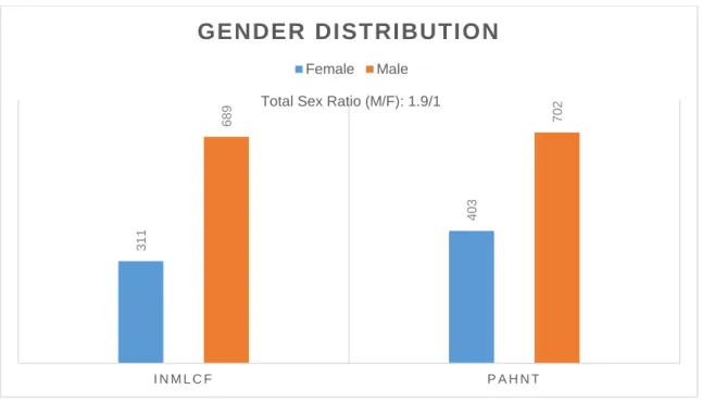
Regarding the gender of the deceased as shown in Figure 2, both institutions revealed a male prevalence, with a proportion M/F of approximately 1.9/1.
Figure 2 – Distribution of cases by gender (INMLCF and PAHNT) 0 50 100 150 200 250 300 N u m b e r o f C a se s Age
Age Distribution
Age Distribution
Age Distribution
Age Distribution
INMLCF PAHNT Total
3 1 1 4 0 3 6 8 9 7 0 2 I N M L C F P A H N T