Universidade de Aveiro 2016
Departamento de Biologia
Tatiana Moura Branco
Combining Antibiotics and Photodynamic Therapy to
inactivate Staphylococcus aureus on skin
Combinação entre Antibióticos e Terapia Fotodinâmica
para inativar Staphylococcus aureus na pele
DECLARAÇÃO
Declaro que este relatório é integralmente da minha autoria, estando
devidamente referenciadas as fontes e obras consultadas, bem como
identificadas de modo claro as citações dessas obras. Não contém, por isso,
qualquer tipo de plágio quer de textos publicados, qualquer que seja o meio dessa
publicação, incluindo meios eletrónicos, quer de trabalhos académicos.
Universidade de Aveiro 2016
Departamento de Biologia
Tatiana Moura Branco
Combining Antibiotics and Photodynamic Therapy to
inactivate Staphylococcus aureus on skin
Combinação entre Antibióticos e Terapia Fotodinâmica
para inativar Staphylococcus aureus na pele
Dissertação apresentada à Universidade de Aveiro para cumprimento dos requisitos necessários à obtenção do grau de Mestre em Microbiologia, realizada sob a orientação científica da Doutora Maria Adelaide de Pinho Almeida, Professora Auxiliar com Agregação do Departamento de Biologia da Universidade de Aveiro e co-orientação da Professora Doutora Maria do Amparo Ferreira Faustino, Professora Auxiliar do Departamento de Química da Universidade de Aveiro.
This work was supported by funding FEDER through COMPETE – Programa Operacional Factores de Competitividade, by National funding through Fundação para a Ciência e Tecnologia (FCT), and Marine Studies (CESAM).
CESAM
Centro de Estudos do Ambiente e do Mar www.cesam.ua.pt
O júri
Presidente
Vogais
Doutora Isabel da Silva Henriques
Investigadora Auxiliar do Centro de Estudos do Ambiente e do Mar da Universidade de Aveiro
Doutora Anabela de Oliveira Pereira
Técnica Superior do Centro de Estudos do Ambiente e do Mar da Universidade de Aveiro
Prof. Doutora Maria Adelaide de Pinho Almeida
Professora Auxiliar com Agregação do Departamento de Biologia da Universidade de Aveiro
Agradecimentos À Professora Doutora Adelaide Almeida, orientadora da tese, pela sua disponibilidade e constante colaboração, pelo incentivo e dedicação a este trabalho.
À Professora Doutora Maria do Amparo Ferreira Faustino do Grupo de Química Orgânica do Departamento de Química pela cedência das porfirinas.
À Dona Helena pelo apoio técnico disponibilizado.
Quero agradecer a toda a gente que diretamente ou indiretamente
contribuiu para este trabalho. Aos meus colegas do laboratório de Microbiologia Aplicada.
Ao talho BioBom que me forneceu gratuitamente a pele de suíno necessário para o desenvolvimento deste trabalho.
Palavras-chave Porfirinas, Terapia Fotodinâmica Antimicrobiana, Staphylococcus aureus, Antibióticos, Pele.
Resumo Staphylococcus aureus é uma bactéria de Gram-positivo que está
comumentepresente em infeções da pele podendo espalhar-se através da corrente sanguínea e afetar outros órgãos. Para o tratamento destas infeções são normalmente utilizados os antibióticos, no entanto, os microrganismos têm a capacidade de adquirir resistência aos agentes antimicrobianos. A terapia fotodinâmica antimicrobiana está a ser estudada ativamente como alternativa ao tratamento de infeções localizadas. Este estudo foi orientado para avaliar a atividade antibacteriana da terapia fotodinâmica no tratamento de infeções por S. aureus na superfície da pele. Foi também avaliado o efeito sinérgico da terapia fotodinâmica antimicrobiana e de antibióticos (ampicilina, cloranfenicol, canamicina, penicilina G e tetraciclina) na inativação de S. aureus. Para este fim, foi utilizado uma porfirina tetra catiónica (Tetra-Py+-Me) que foi testada in vitro para inativar a bactéria numa solução tampão e ex vivo em pele de porco artificialmente contaminada com S. aureus. Os resultados mostraram inactivação eficaz de S. aureus (redução de 8 log) em tampão fosfato salino utilizando a porfirina Tetra-Py+-Me numa concentração de 5.0 µMapós 180 minutos de irradiação com luz branca (com incidência de 40 W.m-2) Na combinação de ampicilina (MIC 0,25 µg mL-1) em concentrações de 0,5 µg mL-1 e 1 µg mL-1 com a porfirina Tetra-Py+-Me a 5.0 µM (em tampão fosfato salino) foi observado
um decréscimo mais rápido (8 log) na inativação total do número de bactérias após 30 e 60 min de irradiação, respetivamente. Para os outros antibióticos não foi observado qualquer aumento na inactivação bacteriana. Nos ensaios ex vivo houve uma redução de ~4 log após tratamento com a porfirina Tetra-Py+-Me a 50 µM após 180 minutos de
irradiação. A eficiência da inativação na pele nas mesmas condições, mas na presença de 5 μg mL-1 de ampicilina foi significativamente diferente da obtida com PS na ausência do antibiótico com uma inativação de ~5,6 log. Os resultados deste estudo mostraram que a DPT é uma abordagem eficaz para controlar infeções por S. aureus na pele, inativando as bactérias até ao limite de deteção após três ciclos de tratamento. Além disso, a combinação de aPDT com antibióticos pode aumentar a eficácia da inactivação bacteriana, permitindo a redução do tempo de tratamento para um quarto.
Keywords Porphyrins, Antimicrobial Photodynamic Therapy, Staphylococcus aureus, Antibiotics, Skin.
Abstract Staphylococcus aureus is a Gram-positive bacterium common in skin
infections, but this bacterium can spread through the bloodstream and infect distant organs. To the treatment of this infections antibiotics are usually used, however, microorganisms have acquired the capacity to develop resistance against antimicrobial agents. Antimicrobial photodynamic therapy (aPDT) is being actively studied as a possible alternative to antibiotics to treat localized infections. This study was conducted to evaluate the antibacterial activity of aPDT for treatment of S. aureus infections on skin. The synergistic effect of aPDT and antibiotics (ampicillin, chloramphenicol, kanamycin, penicillin G and tetracycline) to inactivate S. aureus was also evaluated. To this purpose, a tetracationic porphyrin, the 5,10,15,20-tetrakis(1-methylpiridinium-4-yl)porphyrin tetra-iodide (Tetra-Py+-Me) was used to inactivate S. aureus in vitro using a buffer solution (PBS) and ex vivo, on pork skin artificially contamined with S. aureus. The results show an efficient inactivation of S. aureus in PBS using 5.0 μM of Tetra-Py+-Me during 180 min in the presence of a
white light at an irradiance of 40 W m-2 (reduction of 8 log). When aPDT was done in the presence of ampicillin at 0.5 and 1.0 μg mL-1 (MIC dose 0.25 μg mL) in PBS a faster decrease (8 log) in total bacterial number was observed at 60 and 30 min, respectively. For the other antibiotics no increase in bacterial inactivation was observed. In ex vivo experiments a reduction of ~4 log of S. aureus after treatment with 50 µM of Tetra-Py+
-Me under after 180 min. The efficiency of inactivation in the skin in the same conditions but in the presence of 5 μg mL-1 of ampicillin at 50 µM was significantly different of that obtained with PS in the absence of antibiotic with an inactivation of ~5.6 log. The results of this study showed that aPDT is an effective approache to control S. aureus infection in skin, inactivating the bacteria to the detection limit after three cycles of treatment. Moreover, the combination of aPDT with antibiotics can increase the efficacy of bacterial inactivation, allowing the reduction the treatment time for a quarter.
TABLE OF CONTENTS
LIST OF FIGURES ... i
LIST OF TABLES ... ii
LIST OF ACRONYMS AND ABBREVIATIONS ... iii
THESIS OUTLINE ... 1
CHAPTER 1 ... 3
1. Photodynamic Therapy ... 3
1.1. Briefly History ... 3
1.2. Principle and mechanisms of Photodynamic Therapy ... 3
1.3. Light Source ... 5
1.4. Photosensitizers (PS) ... 6
1.5. Antimicrobial Photodynamic Therapy (aPDT) ... 11
1.5.1. Advantages and disadvantages of antimicrobial photodynamic therapy ... 13
1.5.2. Antimicrobial Photodynamic Therapy of Staphylococcus aureus ... 13
2. Antibiotics ... 16
2.1. Penicillin’s - β-lactam antibiotics ... 17
2.1.1. Natural penicillin’s ... 17 2.1.2. Aminopenicillins ... 17 2.2. Chloramphenicol ... 18 2.3. Aminoglycosides ... 18 2.4. Tetracycline ... 19 2.5. Microbial resistance ... 21 CHAPTER 2 ... 24
Combining Antibiotics and Photodynamic Therapy to Inactivate Staphylococcus aureus on skin ... 24
1. Introduction ... 24
2. Methods ... 27
2.1. Photosensitizer ... 27
2.2. Bacterial strains and growth conditions ... 27
2.3. Irradiation conditions ... 27
2.4. Photoinactivation assays in PBS ... 27
2.5. Photoinactivation assays in PBS combined with antibiotics ... 28
2.6. Photoinactivation assays in skin model (ex vivo) ... 29
2.7. Photoinactivation assays combined with antibiotic in the skin model (ex vivo) ... 30
2.8. Statistical Analysis ... 30
3. Results ... 31
4. Discussion ... 40
5. Future perspectives ... 46
i
LIST OF FIGURES
Figure 1 – Scheme of photosensitization [14]. ... 4
Figure 2- Example of structure of one cationic porphyrin derivative [58] ... 11
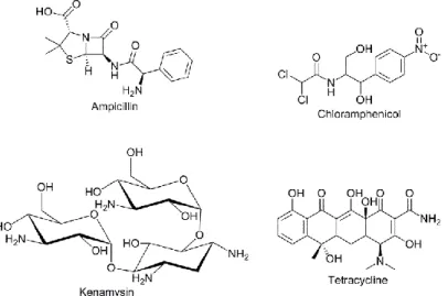
Figure 3 – Structures of some antibiotics. ... 16
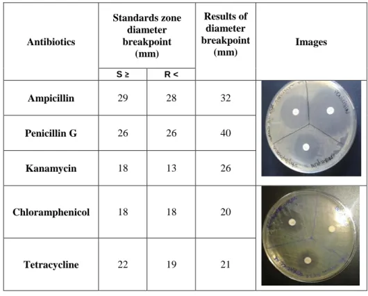
Figure 4 – Drug-resistance profile to antibiotics for the studied strain. ... 31
Figure 5 - Photoinactivation of S. aureus (ATCC 6538) in PBS, after 30, 60, 90 and 180 min, incubated with Tetra-Py+-Me at 5.0 µM and irradiated with white light at an irradiance of 40 W m-2. ... 32
Figure 6 - Photoinactivation of S. aureus in PBS, after 30, 60, 90 and 180 min of irradiation at 40 W m-2, at 5.0 µM of Tetra-Py+-Me and ampicillin concentrations of 0.25 μg mL-1 (A), 0.5 μg mL-1 (B), 1.0 μg mL-1 (C) ... 33
Figure 7 - Photoinactivation of S. aureus in PBS, after 30, 60, 90 and 180 min of irradiation at 40 W m-2 with a concentration 5.0 µM of Tetra-Py+-Me and chloramphenicol concentrations of 8.0 μg mL-1 (A), Kanamycin 2.0 μg mL-1 (B), Penicillin G 0.125 μg mL-1 (C) and Tetracycline 1.0 μg mL-1 (D) ... 34
Figure 8 – (A) Photoinactivation of S. aureus on porcine skin, after 60, 90 and 180 min of irradiation at 40 W m-2. (B) Photoinactivation of S. aureus in porcine skin, after 60, 90, 120 and 180 min of irradiation at 150 W m-2. ... 36
Figure 9 - Photoinactivation of S. aureus on porcine skin, after 60, 90, 120 and 180 min of irradiation at 150 W m-2 with a concentration 50 µM of Tetra-Py+-Me and ampicillin in concentrations of 5 μg mL-1 ... 37
Figure 10 - Photoinactivation of S. aureus on porcine skin, after 60, 90, 120 and 180 min of irradiation at 150 W m-2 with a concentration 40 µM of Tetra-Py+-Me and ampicillin in concentrations of 1.0 μg mL-1 (A), Ampicillin 5 μg mL-1 (B) ... 38
Figure 11- Photoinactivation of S. aureus in porcine skin, after three cycles successive of aPDT of 180 min with an irradiation at 150 W m-2 with a concentration 50 µM of Tetra-Py+ -Me ... 39
ii
LIST OF TABLES
Table 1 - Photosensitizer wavelength absorbance maxima in PBS (adapted from
Wainwright 1998) [36]. ... 6
Table 2 - Classification of photosensitizers as porphyrin-like or nonporphyrin-like
molecules (adapted from O’Connor et al, 2009) [41]. ... 7
Table 3 – Same advantages and disadvantages of aPDT [16,20,41,48,68]. ... 13 Table 4 – Summary of some studies demonstrating the effect of different PDT conditions
on S. aureus survival in vitro, ex vivo and in vivo. ... 15
Table 5 – Some of antibiotics used to treat bacterial infections by Staphylococcus aureus
(EUCAST- European Committee on Antimicrobial Susceptibility Testing 2016) [99]. .... 20
iii
LIST OF ACRONYMS AND ABBREVIATIONS
1O2 Singlet oxygen
3O2 Molecular oxygen in the ground state
AMP Ampicillin
ANOVA Analysis of variance
aPDT Antimicrobial Photodynamic Therapy
BHI Brain-Heart Infusion
BPA BD Baird-Parker Agar
BPA BD Baird-Parker Agar
CFU Colony-forming Unit
CLOR Chloramphenicol
EUCAST European Committee on Antimicrobial Susceptibility Testing
FDA Food and Drug Administration
Gram (–) Gram-negative Gram (+) Gram-positive HpD Hematoporphyrin derivative J Joule KAN Kanamycin log Logarithm
MIC Minimum Inhibitory Concentration
O2 Molecular oxygen ºC Degree Celsius PBS Phosphate-buffered Saline PDI Photoinactivation PDT Photodynamic Therapy PEN G Penicillin G PRT Photoradiation Therapy PS Photosensitizer
rpm Revolutions per minute
TETRAC Tetracycline
iv
TSA Trypticase Soy Agar
1
THESIS OUTLINE
The objective of this study was to evaluate the antibacterial activity of aPDT for the treatment of Staphylococcus aureus infections on skin and also to assess if aPDT efficacy increase in the presence of antibiotics.
For this, the photodynamic effect on S. aureus using a tetracationic porphyrin as photosensitizer (PS) Tetra-Py+-Me at different concentration (5.0 µM, 25 µM, 40 µM, 50
µM) and two artificial white light of irradiances of 40 W m-2 and 150 W m-2) was evaluated. The photodynamic inactivation was evaluated in vitro and ex vivo. The assay in ex vivo was produced on skin of porcine, which has served as a model for human tissuet. The efficiency of photodynamic therapy combinated with different antibiotics (ampicillin, chloramphenicol, kanamycin, penicillin G and tetracycline) was also tested in the same conditions.
Chapter 1 consists on a brief background, focusing on the photodynamic therapy, the antimicrobial photodynamic therapy, the mechanisms of photodynamic inactivation, types of antibiotics used for treatment of infections by S. aureus and some mechanisms of resistance of antibiotics.
Chapter 2 describes the experimental work, the results obtained and their discussion as well as the main conclusions.
3
CHAPTER 1
1. Photodynamic Therapy
1.1. Briefly History
The benefits, for restoration of health, of exposure to sunlight was known in centuries [1,2] and it association with photosensitizers has been used for centuries from the ancient civilizations of Egypt, Greece and India. In fact, they used the photodynamic action by the ingestion of plants containing for instance psoralens which in combination with the exposure to sunlight treated skin disorders such as vitiligo, psoriasis and rickets [3,4].
With the understanding of the photodynamic mechanism, von Tappeiner and colleagues performed the first PDT trial in patients with skin carcinoma using eosin as photosensitizer (PS), applied topically together with the application of sunlight [3,5].
The success of this and other tests conducted, in 1993 in Canada, the semi-purified preparation of HpD known as the Photofrin® (porfimer sodium), to be the first regulatory approval PS for the treatment of bladder cancer. This was later, in 1995, approved by the Food and Drug Administration (FDA) for the treatment of esophageal cancer, and later to be used in several countries in Europe. Today, there are already several PS approved in several countries for applications in PDT [1,5,6]. The PDT, appears in the past years as a viable alternative, cost efficient and with promising applications in several areas, namely, as an antimicrobial approach, in clinical field, food industry and environmental control [7,8].
1.2. Principle and mechanisms of Photodynamic Therapy
Photodynamic therapy is a therapeutic modality requiring the combined action of three main components in promoting cytotoxicity: a PS, a visible light source (a coherent laser light or a non-coherent one, for instance, an LED or Xenon lamp), and the molecular oxygen usually present in the biological target at an affordable concentration [9]. PDT is based on the visible light activation of a PS molecule in the presence of molecular oxygen which results in generation of reactive oxygen species (ROS) [10–12].
4
Reactive oxygen species (ROS) is a collective term used for oxygen derived free radicals (superoxide, hydroxyl radical, nitric oxide) and non-radical oxygen derivatives of high reactivity (singlet oxygen, hydrogen peroxide, peroxynitrite, hypochlorite) [13,14]. The mechanisms of photosensitization involve generation of singlet oxygen (1O2) via energy
transfer from the triplet excited state of PS to the molecular oxygen which is always abundant in a biological environment (Type II) or via electron or hydrogen transfer from the triplet excited state of PS to surrounding molecules (Type I) [15–18]. The photophysical process is illustrated in Figure 1, using the energy levels or Jablonski diagram, illustrated the states according to their energy as well as spin multiplicity [16].
Figure 1 – Scheme of photosensitization [14].
One essential property of a good PS is a high intersystem crossing (ISC) yield, a high probability of transition from S1 to an excited triplet state T1 [17]. The excited triplet state is
the main mediator of the photodynamic reactions. In the T1 state, the photosensitizer can transfer energy to molecular oxygen (3O2), exciting it to its highly reactive singlet state (1O2).
During the energy transfer process the PS is simultaneously brought back to its singlet ground state (S0) where it can, in principle, take part in further sensitisation cycles [16,17].
The basic protocol of PDT involves PS administration followed by a wait time of varying duration to allow for the accumulation of the PS in the cells/tissue, after which the target tissue is irradiated with light [11,19]. The irradiation activates the PS in singlet ground state (S0) to transit from a low power state into a singlet excited state (S1). In this case, there
5
are a transit of electrons to a different orbital, exciting the PS to the form of an unstable molecule with a short half-life (first excited singlet-state, S1) [17,20]. In order to return to its
stable ground state, the PS emits fluorescence or phosphorescence [21]. Fluorescence emission does not alter the electron spin but phosphorescence changes [10]. In other hand, the unstable molecule can transit to triplet state (T1) and transfers energy with the production
of ROS as it was mentioned before.
In aPDT, the high levels of ROS, surrounding pathogenic microorganisms lead to cellular damage, specifically oxidate lipids, peptides or nucleic acids [10,22]. Those can elicit either cell survival or dead depending on severity and duration of exposure [10,23,24]. These radicals reduces the probability of the selection of resistant strains, which is the main problem faced by the current conventional antibacterial therapies [25–29] .
1.3. Light Source
The efficiency of PDT requires an adequate light that activates the used PS. This light must be monochrome and centered on PS absorption band used [30]. The first light sources to be used were conventional bulbs with no coherent and polychromatic light [31]. Since the emergence of the first laser equipment and LEDs, those light sources have been more used in PDT due to its properties: high concentration of energy, low energy dispersion, coherence and monochromaticity, possibility of lighting a medium composed of different materials and only interaction with a particular component (selectivity) [32]. The wavelength of light used for PDT is typically in the wavelength range between 600–800 nm, the so called “therapeutic window” which corresponds to a depth of light penetration 0.5 to 1.5 cm [17].
The dosimetry of light is a major factor in the effectiveness of treatment and depends on the area and depth of the lesion [33]. This parameter is usually expressed by the light irradiance, which is defined in terms of energy incident on a given area of lesion per unit time (W.m-2, W=J.s-1) [34].
The clinical efficacy of PDT dosimetry will also depends on illuminated system used: the total dose and irradiation time delivery mode (continuous or fractionated irradiation) [35]. The use of optical fibers allowed the light to be directed easily to deliver irradiation to desired regions without the requirement of a straight light path [17].
6 1.4. Photosensitizers (PS)
PS are usually aromatic molecules that can be a natural or a synthetic nature which may absorb light of certain energy and may undergo an electronic transition to the singlet excited state (electron spins paired) after interaction with an appropriate light [35]. Different PSs have different wavelength range of maximum efficiency, as it is demonstrated in Table 1.
Table 1 - Photosensitizer wavelength absorbance maxima in PBS (adapted from Wainwright 1998) [36].
Photosensitizer Type Wavelength range absorbance
maximum in buffer solution (nm)
Psoralen 300-380 Porphyrin 400-450 Acridine 400-500 Phenazine 500-550 Cyanine 500-600 Perylenequinonoid 600-650 Phenothiazinium 620-660 Phthalocyanine 660-700
Depending on its molecular structure and environment, the molecule may then lose its energy by electronic or physical processes, thus returning to the ground state, or it may undergo a transition to the triplet excited state (electron spins unpaired) which may then react further by one or both of two pathways already known as the type I and type II photoprocesses [1,36]. This gives rise to activated species which are very reactive towards the chemical environment thus producing molecular damages on important biological targets [36–38]. Typically, these compounds do not persist in the environment for long periods of time [39,40].
A photosensitizing agent with potentially optimal properties should be endowed with specific features, in addition to the expected photophysical characteristics such as a high quantum yield for the generation of both the long-lived triplet state and the cytotoxic singlet oxygen species [32].
7
Such features include [41–44]:
a good absorption capacity at the wavelength of the spectral region where the light source is emitted and a good efficiency to generate ROS;
broad spectrum of action, since one PS can act on bacteria, fungi, yeasts and parasitic protozoa;
efficacy independent of the antibiotic resistance pattern of the given microbial strain;
possibility to develop photodynamic protocols which lead to an extensive reduction in pathogen population with very limited damage to the host tissue; Cell inactivation provide a mechanism that makes minimum the risk of
developing resistant strains and mutagenic processes;
availability of formulations allowing a ready and specific delivery of the PS to the infected area;
adequate solubility in body fluids, which affects the transport and the retention time;
can be used in low concentrations and necessity to use of low cost light sources for activation of the photosensitizing agent;
Be a low energetic toxic compounds and not suffer degradation by light. In summary, the photodynamic activity of the PS to induce cell damage or death is determined by an overall lipophilicity and ionization of the photoreactive PS; quantum yield of the triplet state formation; redox potentials of the excited states (singlet and triplet) of the PS, if the reaction follows the type I pathway or the type II; the molecular extinction coefficient e and the quantum yield of the singlet oxygen generation, if the reaction occurs by the type II pathway [45].
A large number of different PS with photodynamic activity are now available and they are generally classified as porphyrins or non-porphyrins as described in Table 2.
7
Table 2 - Classification of photosensitizers as porphyrin-like or nonporphyrin-like molecules (adapted from O’Connor et al, 2009) [41].
Compound Type Trade name
Photosensitizers Porphyrin-like
Hematoporphyrin Hematoporphyrin derivative (Photofrin®) 1 st G ener a tio n P ho to sens it izer s Metalloporphyrins Lutrin® 2 nd G ener a tio n P ho to sens it izer s Porphycenes Pheophorbides Tookad® Purpurins Clorins NPe6 Foscan® Phthalocyanines Photosens® Photosensitizers Non-porphyrin-like Psoralens Anthracyclines Chalcogenopyrylium dyes
ADPMs ADPM derivatives
Cyanines Merocyanine 540
Cationic cyanines
Phenothiazinium dyes Methylene Blue
Toluidine Bue Nile Blue
Pro-drug 5-ALA Levulan®
8
Porphyrin-derived PSs are further classified as first, second or third generation PSs. Porphyrins known from cancer photodynamic therapy were also used to kill bacteria [39].
Porphyrins are present in nature and they are essential in biochemical processes such as in oxygen transport and photosynthesis. Molecules, such as haemoglobin, myoglobin, cytochromes, chlorophylls and vitamin B, are part of the porphyrinic compounds [46]. Porphyrins are a class of aromatic heterocyclic compounds PS comprise of four pyrrole type subunits linked together by four methine bridges that forming a tetrapyrrole ring structure. Tetrapyrroles are naturally occurring pigments and one benefit of these compounds is that they don’t react with other compounds and they are quenching naturally in a normal environment [20]. Accordingly, the tetrapyrrole macrocycle nucleous structure is named porphin and its derivatives are named porphyrins. Porphyrins can be transformed into cationic entities through the insertion of positively charged substituents in the peripheral positions of the tetrapyrrolic macrocycle that affect the kinetics and extent of binding with microbial cells [41].
A porphyrin skeleton is essentially hydrophobic, so this factor affecting the preferential accumulation in cellular hydrophobic loci since such molecules must be able to get into cells by crossing lipid membranes [20,47]. Structure activity relationship studies suggest that amphiphilic derivatives (as tetracationic porphyrins) exhibit the greatest affinity for Gram (+) [26,38,48]. Some studies revealed that tetracationic porphyrins are efficient PS against both Gram (+) and Gram (-) bacteria on visible light [35,49].
First generation PSs are based on naturally porphyrins, such as hematoporphyrin (Hp) that is an endogenous porphyrin and is formed from acid hydrolysis of hemoglobin. Its derivative, hematoporphyrin derivative (HpD), gave rise to the first PS to be approved for use in PDT for treatment of cancer marketed by the name of Photofrin® [32,33].
This PS remains the most widely used and studied, but have several drawbacks, such as contamination with impurities, relatively low absorbance at 630 nm, where tissue penetration of light is not optimal, and prolonged skin photosensitivity lasting up to 6–8 weeks [41].
A number of second generation PSs have been developed to prevent certain problems associated with the first generation molecules such as prolonged skin photosensitization and suboptimal tissue penetration [37]. These PS are chemically pure compared with first
9
generation compounds, absorb light at a longer wavelength and cause significantly less skin photosensitization post-treatment [41].
Among the second generation of PS are the chlorins, the phthalocyanines, the metalloporphyrins and others identified in the Table 2. Chlorins are reduced porphyrins characterized by a strong enhancement of the far-red absorption band compared to metal-free porphyrins. The meso-tetraarylchlorin derivative 5,10,15,20-tetrakis(3-hydroxyphenyl)chlorin (mTHPC) is a hydrophilic chlorin derivative, which has the trade name Foscan® that was approved in 2001 in the European Union (EU) for the palliative treatment of head and neck cancer [1,42].
Phthalocyanines has been tested as alternatives for porphyrin and chlorin derivatives. Aluminum phthalocyanine sulfonate (AlPcS, Photosens®) is used routinely in Russia for PDT [41].
Still within the second generation are endogenous PS that are found in the body, like the protoporphyrin IX (PpIX) [31]. PpIX is an intermediate of heme synthesis. In the biosynthetic pathway of heme, the 5-aminolevulinic acid (ALA), is the biosynthetic precursor of all porphyrins in nature [50]. ALA is not a PS itself, but induces in vivo biosynthesis of endogenous porphyrins. ALA is normally formed from the reactants glycine and succinyl-coenzyme A in the mitochondria and is then converted into the PS-PpIX [51]. Normally, the synthesis of endogenous porphyrins is limited by the presence of free heme, but exogenous ALA bypasses this negative feedback control. The trade name of ALA is Levulan® and this compound was approved in 1999 for the treatment of actinic keratoses on the face and scalp. Studies have shown that its penetrates the skin when topically applied, however, its penetration into and nodular tumors is quite limited [50,51].
In an attempt to solve the problem of ALA hydrophilicity and hence their difficult to cell penetration, have been developed alkyl esters of ALA, the metilaminolevulinato (MAL), which presents as a trade name Metvix®. It was approved in the EU in 2001 for the topical treatment of actinic keratosis and Bowen's disease. MAL has a higher lipophilicity and are able to penetrate the cell more easily [52,53].
The third generation emerged when bound the second generation PSs with antibodies and liposomes for selective accumulation within tissue/cells/microorganisms and currently represent an active research area in the field [54].
10
Although the majority of PSs at the preclinical stage are porphyrin derivatives, a diverse number of non-porphyrin PSs also exist. First of all, the synthetic non-porphyrin compounds have demonstrated photosensitizing ability, like the phenothiazine dyes: methylene blue and toluidine blue [43,55]. Another group of dyes belongs to the naturally occurring PS. Psoralens (furanocoumarins) and perylenequinonoids are two examples of natural products which originally act in plants as chemical defence substances against microbial or eukaryotic organisms [45]. Significant effort is now being employed in the synthesis of pure chemical derivatives with improved activity and minimal side effects [43].
It would be desirable to have an effective PS for microbial inactivation without the need of additional chemicals, which ensures that it will bind preferentially to microbial cells instead of mammalian cells [32,52]. An important step forward in this direction was prompted by the discovery that PS that are positively charged at physiological pH values such as phenothiazines, phthalocyanines and porphyrins can directly promote the photoinactivation of both Gram (+) and Gram (-) bacteria [43,44]. Another important characteristic during the design of a PS for PDT is their water solubility where must have hydrophilic and hydrophobic characteristics, because they must be administrated in a solution, but on the other hand they must be able to cross the bacterial cell wall [56]. On the other hand, must have a positive charge being extremely important in gram-negative bacteria inactivation since their membrane structure excludes many anionic and uncharged lipophilic molecules that would lead to phototoxicity [14,35].
While phenothiazine derivatives are naturally cationic, owing to the involvement of one amino group in the π electron cloud resonance, porphyrins and phthalocyanines can be transformed into cationic entities trough the insertion of positively charged substituents in the peripheral positions of the tetrapyrrolic macrocycle (meso positions) and, respectively, tetraazaisoindole macrocycle, which may largely affect the kinetics and extent of binding with microbial [25,35,57].
The PS more frequently tested in microbial photoinactivation are based mainly in meso-substituted tetracationic porphyrins (Figure 2). The popularity of this type of PS results from their easy synthesis and potentiality toward further elaboration [43]. In fact, the synthetic approaches usually involve the condensation of pyrrole with adequate aldehydes, which are available in a wide range, providing porphyrins with different aryl or heteroaryl substituents at the meso-positions. Further manipulations of those substituents can give
11
access to a high number of porphyrins that can be designed for the desired application [40,43].
Figure 2- Example of structure of one cationic porphyrin derivative [58]
Some bacteria are also known to produce endogenous porphyrins [56]. A few studies revealed that bacteria that produce reasonable amounts of endogenous porphyrins can be efficiently degraded by photosensitization, since there is no need of break through cell barriers [59].
Therefore, porphyrins and their derivatives have been reported as one of the most promising compounds used in aPDT [46,58,60]. Porphyrins are currently used in PDT due to their unique physico-chemical properties, particularly in the treatment of superficial cancer; topical treatment of dermatological problems, such as psoriasis, acne, and Bowen’s disease; gastrointestinal cancer; age related macular degeneration; cutaneous leishmaniasis; and viral infections, such as papillomatosis [42,61,62] and in the photoinactivation of microorganisms [23,43].
1.5. Antimicrobial Photodynamic Therapy (aPDT)
Antimicrobial Photodynamic Therapy (aPDT) has emerged as a potential alternative to antibiotics to treat microbial infections [28].Membrane permeability barriers, differences
12
in antioxidant enzymes or DNA repair mechanisms and the size of the microbial cell are among the factors that affect the photoinactivation process. These factors are described as:
Bacterial target: Microbial cells display a large variety of size, sub-cellular architecture and biochemical composition [63]. The susceptibility to photoinactivation processes can be significantly different for the various microorganisms [64]. Gram-positive bacteria have an outer wall, which is separated from the plasma membrane by a periplasmic space. Once that in their wall can be diffused macromolecules with molecular weight up to 60000 Da, and the most common PS have in generally a molecular weight of 1500 Da, these can cross the outer wall and localize in the immediate surroundings of the photosensitive endocellular sites [35]. So, the Gram (+) bacteria are relatively easy to kill by PDT, while Gram (-) bacteria show significant resistance [64]. On the contrary, the outer wall of Gram(-) bacteria possesses an additional structural element, which is external to the peptidoglycan network consisting of a glycocalyx, lipolysaccharide, outer membrane lipid bilayer, periplasm, peptidoglycan cell wall, and plasma membrane lipid bilayer [64]. This barrier keeps out most PS therefore specific methods have to be adopted to ensure that the PS can penetrate the bacterium [48].
Physiological state: When the cells are in the logarithmic phase of growth are more susceptible to photodynamic inactivation than the corresponding cells in the stationary phase [63].
Cell density: The cell density influence the competition for binding with the available PS, as well as for reaction with photogenerated cytotoxic species [65].
In general, the positively charged PS bound to the negatively charged surface of bacteria (Gram + and Gram −) can cause damage to bacteria by two mechanisms: DNA damage and damage to the cytoplasmic membrane, allowing inactivation of membrane transport systems and enzymes or leakage of cellular contents [66,67].
13
1.5.1. Advantages and disadvantages of antimicrobial photodynamic therapy
Antimicrobial photodynamic therapy as other type of therapies includes advantages and disadvantages. Below (Table 3) are described the most significant advantages/disadvantages of the treatment by aPDT.
Table 3 – Same advantages and disadvantages of aPDT [16,20,41,48,68].
Advantages Disadvantages
Non-invasive;
Selective: targeted to select part of organism/tissues (does not influence on whole organism);
Few side effects;
Can be combined with other conventional treatments; Equally effective at killing both
multi-drug resistant microbes as well as native bacterial strains;
The effect of aPDT on
microorganisms is much more rapid as compared to that of other
antimicrobial agentes;
Does not damage the tissues, in particular tissue elements such as collagen and elastin in skin;
Not generating specific mechanisms of resistance.
Difficulty to obtain the ideal PS with all of the adequate characteristics; The lack of highly effective
antimicrobial clinically approved PSs; New PSs have not yet to be subjected
to the rigorous and costly toxicological and safety studies necessary for approval for human use; Treatment may be associated with
pain;
Cutaneous photosensitivity is common;
Occasional blistering may occur and hyperpigmentation can occur at treated sites;
Located: PDT can only treat areas where light can reach.
PDT can’t be used in people who have certain blood diseases, such as any of the porphyrias or people who are allergic to porphyrins.
1.5.2. Antimicrobial Photodynamic Therapy of Staphylococcus aureus
Staphylococcus aureus is an important human pathogen and a common etiological factor of health care associated as well as community acquired infections [69]. The ability of S. aureus to develop multidrug resistance is well documented, so it is crucial to find
14
alternative antimicrobial treatment to which this bacterium [70,71]. A potential alternative can be the aPDT to inactivate microbial cells [19,21,72,73].
S. aureus is a versatile opportunistic pathogen that is responsible for a wide variety of conditions, ranging from superficial skin infections to severe, invasive diseases [74,75]. S. aureus skin infections are very contagious, including the following [75–77]:
Folliculitis is the least serious. A hair root (follicle) is infected, causing a slightly
painful, tiny pimple at the base of a hair.
Impetigo consists of shallow, fluid-filled blisters that rupture, leaving
honey-colored crusts. Impetigo may itch or hurt.
Abscesses (boils or furuncles) are warm, painful collections of pus just below
the skin.
Cellulitis is infection of skin and the tissue just under it. Cellulitis spreads, causing
pain and redness.
Toxic epidermal necrolysis and, in newborns, scalded skin syndrome are
serious infections. Both lead to large-scale peeling of skin.
Necrotizing fasciitis is a severe, rare, potentially lethal soft tissue infection that
develops in the scrotum and perineum, the abdominal wall, or the extremities.
The use of antibiotics is yet the first treatment option to treat and sometimes to eradicate diseases caused by S. aureus [28,78]. An example of treatment could be the use of penicillin regardless of strain, now many of its strains are resistant to beta-lactams, macrolides, and even vancomycin, the ‘‘drug of last resort’’[70,79]. Methicillin-resistant (MRSA) and vancomycin resistant S. aureus (VRSA) are collectively recognized as a very serious health threat [8,80].
In vitro studies have shown that S. aureus is sensitive to porphyrins, phthalocyanines and others PS. Some of these studies are summarized in Table 4.
15
Table 4 – Summary of some studies demonstrating the effect of different PDT conditions on S. aureus survival in vitro, ex vivo and in vivo.
Article Title PS Total light dose Results
In vitro
Bartolomeu, 2016 [81]
Effect of Photodynamic Therapy on the Virulence Factors of Staphylococcus aureus.
5,10,15,20-tetrakis(1-methylpyridinium-4-yl)porphyrin tetra-iodide (Tetra-Py+-Me) - 5.0 µM
40 W/m2 Reduction of 5 log CFU mL -1
Almeida, 2014 [82]
Photodynamic inactivation of multidrug-resistant bacteria in hospital wastewaters: influence of residual.
5,10,15,20-tetrakis(1-methylpyridinium-4-yl)porphyrin tetra-iodide (Tetra-Py+-Me) - 5.0 µM
40 W/m2 reduction of 4 – 5 log CFU
mL-1
Mai, 2016
[29]
The Antibacterial Effect of Sinoporphyrin Sodium Photodynamic Therapy on Staphylococcus aureus Planktonic and Biofilm Cultures.
Sinoporphyrin sodium (DVDMS) –
2 µM
10 J/cm2 ≥ 90% of the bacteria were
eradicated Sinoporphyrin sodium (DVDMS) –
5µM
100 J/cm2 Reduction of 4 log CFU mL -1
Hsieh, 2014
[83]
5-Aminolevulinic acid induced photodynamic inactivation on Staphylococcus aureus and Pseudomonas aeruginosa
5-aminolevulinic acid (ALA) – 1 mM
162 J/cm2 90% of the bacteria were
eradicated
Ex vivo
Lambrechts, 2005 [11]
Photodynamic therapy for Staphylococcus aureus infected burn wounds in mice.
meso-mono-phenyl-tri(N-methyl-4-pyridyl)-porphyrin (PTMPP)
(Tri-Py+-Me-Ph)- 500 µM
210 J/cm2 98% of the bacteria were
eradicated
Maisch, 2007 [84]
Determination of the antibacterial efficacy of a new porphyrin-based photosensitizer against MRSA ex vivo
XF-73 (exeporfinium chloride) – 10
µM
210 J/cm2 Reduction of ≈ 3.5 log CFU
mL-1 In vivo
Zolfaghari, 2009 [85]
In vivo killing of Staphylococcus aureus
using a light-activated antimicrobial agent Methylene Blue (MB) – 100 µg/mL 360 J/cm
2 Reduction of ≈ 1.5 log CFU
mL-1
Grinholc, 2015 [86]
Antimicrobial photodynamic therapy with fulleropyrrolidine: photoinactivation mechanism of Staphylococcus aureus, in vitro and in vivo studies
N- mthylpyrrolidimium fullerene
iodide - 30 µM 500 W/m
2 Reduction of 2 log CFU
16
Because the delivery of visible light to living tissue is almost by definition a localized process, PDT for infections is likely to be applied exclusively to localized disease, as opposed to systemic infections such as bacteremia [11].
2. Antibiotics
Antibiotics are derived from three sources: moulds or fungi; bacteria; or synthetic or semi-synthetic compounds [87]. They can be used either internally or topically, and their function is to either inhibit the growth of pathogens or to kill them [88]. Antibiotics represents one of the most revolutionary progresses made in scientific medicine to destroy selectively microorganisms, resulting in the treatment and sometimes complete eradication of earlier incurable diseases [28,89].
It might have been supposed that at the beginning of the twenty first century, microbiologically-based diseases would have been reduced to a level that no longer had a serious impact on human health [88]. There are various antibiotics available and they come in various different brand names [87]. Antibiotics are usually grouped together based on how they work [71]. Each type of antibiotic only works against certain types of bacterium [28]. This is why different antibiotics are used to treat different types of infection [89]. Some antibiotics was referred and illustrated in the figure 3.
17 2.1. Penicillin’s - β-lactam antibiotics
The β-lactam antibiotics include a number of drugs with a chemical structure and a common mechanism of action. In fact, this class of antibiotics has a β-lactam ring which may be fused or linked with cyclic non-cyclic radical structures [70,71]. These antibiotics inhibit the peptidoglycan synthesis of the bacterial wall [78].The integrity of the β-lactam ring is essential for exercise the antibiotic activity. Some bacteria produce enzymes, the β-lactamases, that opening the β-lactam ring and completely nullifies the antibiotic action [90]. The penicillin’s can be classified according to their antibacterial activity [70]: Natural penicillin’s; aminopenicillins and others.
2.1.1. Natural penicillin’s
The benzylpenicillin (penicillin G) are a natural penicillin that currently are use in clinical practice [78,91]. Currently, a growing emergency producing bacterial strains that produces β-lactamases (resistant to penicillins), led to the need for administered this antibiotic with the combination with inactivators of β-lactamases [71]. The action of benzylpenicillin on aerobic gram-positive cocci as Staphylococcus spp. are efficient when this specie is sensitive, because do not produce beta-lactamase enzymes [70,78]. However, the majority of clinical Staphylococcal infections are caused by strains producing β-lactamases, so that benzylpenicillin has no further interest in the treatment of such infections [70]. Nevertheless, there are still situations in which the benzylpenicillin continues to be used as a drug of choice [70].
2.1.2. Aminopenicillins
The aminopenicillins including amoxicillin, ampicillin and bacampicillin [71]. These aminopenicillins have an additional hydrophilic groups and are particularly effective against gram-negative bacteria that not produce β-lactamases [92]. However, your spectrum of action may include some gram-positive bacteria. The differences from penicillin are the presence of an amino group. That amino group helps the drug penetrate the outer membrane
18
of gram (-) bacterium [92]. Ampicillin acts as a competitive inhibitor of the enzyme transpeptidase, which is needed by bacteria to make their cell walls. It inhibits the third and final stage of bacterial cell wall synthesis in binary fission, which ultimately leads to cell lysis [70].
2.2. Chloramphenicol
Chloramphenicol is an antibiotic produced synthetically, which acts by inhibition of bacterial protein synthesis. Chloramphenicol binds reversibly to the 50S ribosomal subunit and preventing binding of end of the tRNA, and therefore bacterial protein synthesis. Chloramphenicol has a very broad spectrum of action and it is mainly bacteriostatic, acts against most bacteria except the Mycobacteriaciae, Treponema and Actinomyces [93]. Furthermore, this antibiotic is ineffective against viruses, fungi and protozoa.
2.3. Aminoglycosides
Aminoglycoside antibiotics comprising a set formed of two or more amino sugars joined by a glycosidic linkage to a hexose aminated. Most of aminoglycosides have a natural origin. However, amikacin and gentamicin are two exceptions to the extent they are respectively semisynthetic derivative of sisomicin and kanamycin [94]. The kanamycin was formerly used in the treatment of severe infections caused by Gram-negative, but currently this antibiotic has low clinical use. Aminoglycosides act by interfering with protein synthesis and have a bactericidal effect (and not bacteriostatic effect, as all the other inhibitors of protein synthesis) [95]. The antibacterial spectrum of these antibiotics is quite extensive, including gram-negative bacteria, gram-positive and bacteria do not stain the Gram (such as the Koch's bacillus). In turn, the obligatory and facultative anaerobes are resistant to the action of aminoglycosides [94].
S. aureus and epidermidis are sensitive to aminoglycosides, whether or not produce β-lactamase. However, Staphylococcus quickly develop resistance against aminoglycosides.
19
Aminoglycosides are generally used in combination with other classes of antibiotics which have a synergistic effect between these antibiotics, especially the β-lactam [95].
2.4. Tetracycline
Tetracyclines act by blocking bacterial protein synthesis, bind to the 30S ribosome subunit, preventing the aminoacyl tRNA binding to ribosomal receptor, and thus the growth of the peptide chain [96]. The effect of tetracyclines on the bacteria is reversible, such that, upon interruption of their use, the non-return eliminated microorganisms to multiply [97].
Tetracyclines are antibiotics with broad-spectrum action, with bacteriostatic action against various gram-positive and gram-negative and bacteria that not stain the Gram (including mycoplasmas, chlamydiae, rickettsiae) and some protozoans [97,98].
20
Table 5 – Some of antibiotics used to treat bacterial infections by Staphylococcus aureus (EUCAST- European Committee on Antimicrobial Susceptibility Testing
2016) [99].
Class/Antibiotics Mechanism of
action Effect on bacteria Spectrum
Penicillins
Penicillin G
Interfer with bacterial
cell wall synthesis Bactericidal
Active against non β-lactamase–producing gram (+) cocci (Pneumococci, Staphylococci, Streptococci), few gram (-) cocci (meningococci andgonococci), gram (+) bacilli (Bacillus anthracis, Bacillus
erfringens, Bacillus diphtheriae) and anaerobes (Clostridum perfringens, C. tetani).
Aminopenicillins
Ampicillin
Inhibitor of cell wall
synthesis Bactericidal
Aminopenicillins are similar to penicillin G in the activity against Gram (+) organisms but are slightly weaker than the latter. Aminopenicillins are more active against enterococci and Listeria monocytogenes compared to penicillin G. Gram (-) spectrum incudes Haemophilus influenza, Salmonella, Shigella, Escherichia coli, Proteus mirabilis, N. gonorrhoeae, N. meningitidis.
Chloramphenicol
Chloramphenicol
Inhibition of bacterial
protein synthesis Bacteriostatic
Among the bacteria more sensitive are H. influenzae, N. meningitidis, N.
gonorrhoeae, Salmonella typhi, Brucella, Bordetella pertussis, Streptococcus pyogenes, Streptococcus pneumoniae and anaerobic bacteria (Clostridium
and Bacteroides fragilis), Staphylococcus aureus and some
Enterobacteriaceae (such as E. coli, Proteus mirabilis, Shigella,
Pseudomonas pseudomallei, Klebsiella, and some strains of Enterobacter
and Serratia). Aminoglycosides
Kanamycin
Inhibition of bacterial
protein synthesis Bactericidal
Aminoglycosides have high activity against aerobic gram (-) bacilli (E. coli,
Proteus, Klebsiella, Enterobacter, Salmonella, Shigella and Pseudomonas aeruginosa), Staphylococcus aureus and epidermidis.
Tetracycline
Tetracycline
Inhibition of bacterial
protein synthesis Bacteriostatic
Action against various gram (+) and gram (-) such as Staphylococci,
Enterococci, Pneumococci, Gonococci, some Enterobacteriaceae (such as Salmonella and Shigella), bacteria that not stain the Gram (including
21 2.5. Microbial resistance
The rapidly increasing emergence of antibiotic resistance amongst pathogenic bacteria assume increasing economic and social impact due to the high morbidity and mortality induced by the proliferation of antimicrobial resistance [78,80]. Bacteria replicate very rapidly and a mutation that helps a microbe to survive in the presence of an antibiotic drug will quickly become predominant throughout the microbial population [28,100]. The inappropriate prescription of antibiotics and the failure of some patients to complete their treatment regimen also exacerbate the problem [101].
The intended modes of action of antibiotics may be counter-acted by bacterial organisms via several different means [102]. This may involve preventing antibiotic access into the bacterial cell or perhaps removal or even degradation of the active component of the antimicrobial agent [78,103]. In fact, several different mechanisms may work together to confer resistance to a single antimicrobial agent [88]. Some of this mechanisms are described below:
Intrinsic Resistance
Intrinsic resistance is the innate ability of a bacterial species to resist activity of a particular antimicrobial agent through its inherent structural or functional characteristics, which allow tolerance of a particular drug or antimicrobial class [88,104].
Acquired Resistance
Acquired resistance is said to occur when a particular microorganism obtains the ability to resist the activity of a particular antimicrobial agent to which it was previously susceptible [89,105]. This can result from the mutation of genes involved in normal physiological processes and cellular structures, from the acquisition of foreign resistance genes through a small circular DNA (plasmids) or from a combination of these two mechanisms [103,106].
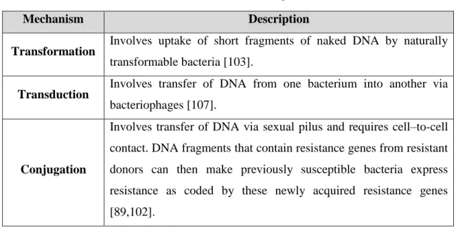
Acquired resistance results from successful gene change and/or exchange that may involve: mutation or horizontal gene transfer via transformation, transduction or conjugation [89,102] described in Table 6.
22
Table 6 – Mechanisms that envolve the acquired resistance.
Mechanism Description
Transformation Involves uptake of short fragments of naked DNA by naturally
transformable bacteria [103].
Transduction Involves transfer of DNA from one bacterium into another via
bacteriophages [107].
Conjugation
Involves transfer of DNA via sexual pilus and requires cell–to-cell contact. DNA fragments that contain resistance genes from resistant donors can then make previously susceptible bacteria express resistance as coded by these newly acquired resistance genes [89,102].
Mutation
A mutation is a spontaneous change in the DNA sequence within the gene that may lead to a change in the trait which it codes for [104,106]. Any change in a single base pair may lead to a corresponding change in one or more of the amino acids for which it codes, which can then change the enzyme or cell structure that consequently changes the affinity or effective activity of the targeted antimicrobials [102,105].
24
CHAPTER 2
Combining Antibiotics and Photodynamic Therapy to Inactivate
Staphylococcus aureus on skin
1. Introduction
Staphylococcus aureus belongs to the natural microflora, resides on the surface of the skin and on mucous membranes of warm-blooded animals (present in the nose of about 30% of healthy adults and on the skin of about 20%, and at higher percentages in patients admitted to hospitals and people who work there), but it may become pathogenic in conditions such as breakage of the skin barrier or decreased immunity [74,108–110]. Is a common Gram-positive bacteria that is responsible for a wide variety of disorders, causing wound infections and colonize the mucous of nasal cavity and normal skin of healthy population [110,111].
Its pathogenesis is dependent on the secretion of an array of virulence factors, the surface exposure of multiple cell wall anchored proteins [74] and extracellular components that are expressed during the different stages of infection: colonization, avoidance or invasion of the host immune defense, growth and cellular division culminating in bacterial dissemination, causing toxic effects to the host [112,113].
S. aureus infections are common in skin, but the bacterium can spread through the bloodstream and infect distant organs. As the bacterium tends to infect the skin, often causing abscesses [79], but as the bacterium can travel through the bloodstream, causing bacteremia and infect almost any site in the body, particularly heart valves (endocarditis) and bones (osteomyelitis) [76,114]. The bacterium also tend to accumulate on medical devices in the body, such as heart pacemakers, and catheters inserted through the skin into blood vessels [76].
These infections are usually treated with antibiotics that are chosen based on whether they are likely to be effective against the strain causing the infection. The penicillin, aminopenicillins, cephalosporin, aminoglycoside, tetracyclines and chloramphenicol are effective antibiotics to combat infection by S. aureus. However, both community-associated and hospital-acquired infections with S. aureus have increased in the past three decades, and
25
the rise in incidence has been accompanied by a rise in antibiotic-resistant strains [24,101], particularly methicillin-resistant S aureus (MRSA) and vancomycin-resistant strains (VRSA) [115,116] which are collectively recognized as a very serious health threat [8,80]. The resistance to penicillin emerged in the mid-1940s, only a few years after the introduction of this antibiotic in the clinical practice [101]. Later, in 1959, the semi-synthetic antibiotic methicillin was introduced for the treatment of infections caused by penicillin-resistant S. aureus [80]. Yet, in 1961 the first cases of methicillin- resistant S. aureus (MRSA) isolates [24] were reported and currently, only few compounds are still effective in the treatment of MRSA infections [101]. Vancomycin, the ‘‘drug of last resort’’has been the most reliable therapeutic agent against infections caused by meticillin-resistant S. aureus (MRSA). However, in 1996 the first MRSA to acquire resistance to vancomycin, was isolated from a Japanese patient. Subsequent isolation of several vancomycin resistant S. aureus (VRSA) strains from USA, France, Korea, South Africa, and Brazil has confirmed that emergence of vancomycin resistance in S. aureus is a global issue [70,79]. This resistance comes from the bacterial gene mutation and horizontal transfer of resistance genes from external sources [117,118].
Although the intensive overuse of antimicrobial drugs began to exert new survival pressure on relevant microorganisms, antibacterial resistance was hardly acknowledged in the past as novel antibiotics became steadily available and were readily modified and improved for clinical use [24,101,117]. The development of novel but still conventional antibiotics, is not likely to solve the problem as it is probably only a matter of time until they will also ineffective. Bacterial resistance is undoubtedly recognized as a major medical challenge in most healthcare systems. Consequently, the traditional treatment of bacterial infections with antibiotics can be difficult, emphasizing the importance and urgency to develop new alternative treatments to treat bacterial infections. In this sense, new alternative therapies such as antimicrobial photodynamic therapy (aPDT) have been proposed.
aPDT is being actively studied as a possible alternative to antibiotic treatment as in localized infections [60,64,119]. aPDT involves the combination of light, oxygen and PS that trust be able to produce ROS upon irradiation with light. Thus, when the dye absorbs a photon, an electron is promoted from its ground state to an electronically excited state that returns the energy. Although originally employed in the treatment of cancer during the last decade, an increasing number of studies on PDT application have been published about
26
microbiocidal effect in addition to better access to sites that are inaccessible to conventional therapy [120]. This technology has already shown to be effective against Gram-positive and Gram-negative bacteria, viruses, fungi, and parasites [64,119,121]. The major advantage of this technology over antibiotics is the multi-target action [122] and no emergence of resistances [28,81,123].
Although it is well known that the use of large amounts of antibiotics in clinical practice is undesirable, since they give rise to the selection of antibiotic-resistant strains, little effort has been made to employ aPDT in order to increase the efficacy of such antibiotics, as well as to employ antibiotics to potentiate the efficacy of aPDT. Xing et al. (2011) tested a vancomycin-porphyrin conjugate to inactivate vancomycin-resistant enterococci, showing strong PDI activity of the conjugate against vancomycin-sensitive and resistant strains, when compared to vancomycin and porphyrin alone [124]. The combination of vancomycin with PDI was also useful in the disruption of S. aureus biofilms. Pre-treating the biofilms with PDI and then apply vancomycin at concentrations bellow the biofilm inhibitory concentration, causes a disintegration of the biofilm matrix and allows the killing of bacteria almost entirely [27]. Malik and Nitzan (1995) tested also the combination of different natural porphyrin derivatives and antibiotics (methicillin, ampicillin, polymyxin B nonapeptide, tetracycline) to inactivate both multi-resistant Gram-positive (S. aureus) and Gram-negative bacteria (E. coli) [125]. The photoinactivation of four multidrug resistant bacteria by a porphyrin derivative in the presence of ampicillin and chloramphenicol was also evaluated and the results showed that in the presence of porphyrins and antibiotics the bacterial photoinactivation was higher than when the porphyrin derivatives were used alone [49].
The aim of this study was to evaluate the antimicrobial applicability of aPDT to treat skin infections by S. aureus. Besides, the synergism of the combination of this therapy with conventional antibiotics was also studied.
27
2. Methods
2.1. Photosensitizer
The PS applied in this study was a tetracationic porphyrin, the 5,10,15,20-tetrakis(1-methylpiridinium-4-yl)porphyrin tetra-iodide (Tetra-Py+-Me) and was synthetized according to previously described procedure [126]. A stock solution of this porphyrin, at 500 µM, was prepared in dimethyl sulfoxide and stored in the dark. Before each assay the porphyrin solution was sonicated during 30 min at room temperature (ultrasonic bath 0.6L | Nahita).
2.2. Bacterial strains and growth conditions
The strain of S. aureus (ATCC 6538) was inoculated on solid medium BD Baird-Parker Agar (BPA)(Liofilchem) at 37 ºC during 24 h and posteriorly kept a 4 ºC (Work Sample). The bacterium was inoculated whenever necessary in liquid medium Brain-Heart Infusion (BHI) (Liofilchem) and grown aerobically at 37 ºC for 24 h under stirring (130 rpm). For each assay, an aliquot of this culture (300 μL) was transferred twice into a new fresh BHI medium (subcultured in 30 mL) and grew overnight at 37 °C under stirring.
2.3. Irradiation conditions
Following the dark incubation period (10 min for in vitro and 30 min for ex vivo), samples were exposed, in parallel, to white light: PAR radiation (13 OSRAM 21 lamps of 18 W each, 380-700 nm) with an irradiance of 40 W m-2 or Lumacare system (an interchangeable fiber optic probe - 400–800 nm - coupled to a 250 W quartz/halogen lamp (LC-122; LumaCare, Newport Beach, CA, USA) with an irradiance of 150 W m-2, at 25 °C for 180 - 270 min.
2.4. Photoinactivation assays in PBS
Overnightbacterial cultures, were tenfold diluted in phosphate-buffered saline (PBS) to a final concentration of ~107-8 CFU mL-1 (colony-forming unit per mililitre). The diluted
bacterial suspensions were distributed in 50 mL beakers (final volume of 10 mL per beaker) and incubated in the dark for 10 min at room temperature under 100 rpm stirring with 5.0 μM of porphyrin (Tetra-Py+-Me), to promote the PS binding to cells. Then the beakers are
28
irradiated by white light with an irradiance of 40 W m-2. Bacterial suspensions were irradiated up to 270 min (total light dose of 64.8 J cm-2) and sub-samples of 100 µL were collected at the beginning of the irradiation (time 0) and after 15, 30, 60, 90, 180 and 270 min of exposure to light. After each photosensitization period, the cells were serially diluted in PBS, pour-plated in solid medium trypticase soy agar (TSA) (Liofilchem) and incubated at 37 °C for 24 h, for viability monitoring. The cell viability was determined by counting the CFU mL-1 on the most appropriate dilution. Control samples were included in all PDI experiments: light control (LC) consisted of a bacterial suspension that was exposed to light; and dark control (DC) consisted of a bacterial suspension incubated with PS at the maximum concentration under the same conditions as the samples, but protected from light. Three independent experiments were conducted.
2.5. Photoinactivation assays in PBS combined with antibiotics
In these assays, the photoinactivation of S. aureus with the PS at 5.0 µM in the presence of five antibiotics: ampicillin (Applichen Panreae), chloramphenicol (Applichen Panreae), Kanamycin (Applichen Panreae), Penicillin G (Sigma Life-Science) and Tetracycline (Sigma Life-Science) was evaluated. All the antibiotics were tested at the minimum inhibitory concentrations (MIC) according EUCAST- European Committee on Antimicrobial Susceptibility Testing (2016) and accordingly to the Clinical and Laboratory Standards Institute [CLSI] (2013) [99,127]. The MIC concentrations were, 0.25 μg mL-1, 8 μg mL-1, 2 μg mL-1, 0.125 μg mL-1 and 1.0 μg mL-1, respectively, for ampicillin,
chloramphenicol, kanamycin, penicillin G and tetracycline. For ampicillin two more concentrations were tested 0.5 μg mL-1 and 1.0 μg mL-1.
For each assay four goblets of 50 mL were prepared: one with bacteria diluted 1:10 in PBS and PS (Sample); other with bacteria diluted 1:10 in PBS and PS, plus antibiotic (PS + (abbreviation of the used antibiotic)); other with bacteria diluted 1:10 in PBS and antibiotic (Antibiotic Control - AC); other with bacteria diluted 1:10 in PBS and PS, which was kept in dark conditions, wrapped in aluminium foils (DC) and other with bacteria diluted 1:10 in PBS (LC). The quantity of antibiotic was the same used in the Sample and in the Antibiotic Control. The goblets were kept under irradiation 180 min and aliquots were taken at 0, 15, 30, 60, 90 and 180 min. The aliquots were diluted in PBS and plated in TSA by
![Figure 1 – Scheme of photosensitization [14].](https://thumb-eu.123doks.com/thumbv2/123dok_br/15933925.1095398/17.892.221.691.457.720/figure-scheme-of-photosensitization.webp)
![Table 1 - Photosensitizer wavelength absorbance maxima in PBS (adapted from Wainwright 1998) [36]](https://thumb-eu.123doks.com/thumbv2/123dok_br/15933925.1095398/19.892.165.775.386.662/table-photosensitizer-wavelength-absorbance-maxima-pbs-adapted-wainwright.webp)
![Table 2 - Classification of photosensitizers as porphyrin-like or nonporphyrin-like molecules (adapted from O’Connor et al, 2009) [41]](https://thumb-eu.123doks.com/thumbv2/123dok_br/15933925.1095398/21.1262.150.1163.143.752/table-classification-photosensitizers-porphyrin-nonporphyrin-molecules-adapted-connor.webp)
![Figure 2- Example of structure of one cationic porphyrin derivative [58]](https://thumb-eu.123doks.com/thumbv2/123dok_br/15933925.1095398/25.892.348.599.214.464/figure-example-structure-cationic-porphyrin-derivative.webp)
![Table 3 – Same advantages and disadvantages of aPDT [16,20,41,48,68] .](https://thumb-eu.123doks.com/thumbv2/123dok_br/15933925.1095398/27.892.129.784.337.848/table-same-advantages-and-disadvantages-of-apdt.webp)

![Table 5 – Some of antibiotics used to treat bacterial infections by Staphylococcus aureus (EUCAST- European Committee on Antimicrobial Susceptibility Testing 2016) [99]](https://thumb-eu.123doks.com/thumbv2/123dok_br/15933925.1095398/34.1262.122.1133.207.743/antibiotics-bacterial-infections-staphylococcus-european-committee-antimicrobial-susceptibility.webp)