CASE REPORT
Right cardiac intracavitary metastases from
a primary intracranial myxo
fibrosarcoma
Diogo Alpuim Costa,
1,2,3Pedro Barata,
3,4Emanuel Gouveia,
1Manuela Mafra
5,61Department of Medical
Oncology, Instituto Português de Oncologia de Lisboa Francisco Gentil, EPE, Lisbon, Portugal
2
Underwater and Hyperbaric Medicine Centre, Portuguese Navy, Lisbon, Portugal
3Nova Medical School, Lisbon,
Portugal
4Department of Medical
Oncology, Centro Hospitalar de Lisboa Central, Lisbon, Portugal
5Pathology Department, Centro
Hospitalar de Lisboa Central, Lisbon, Portugal
6
Instituto Português de Oncologia de Lisboa Francisco Gentil, EPE, Lisbon, Portugal Correspondence to Dr Diogo Alpuim Costa, [email protected] Accepted 23 February 2016
To cite: Costa DA, Barata P, Gouveia E, et al. BMJ Case Rep Published online: [ please include Day Month Year] doi:10.1136/bcr-2015-214052
SUMMARY
Primary intracranial myxofibrosarcoma is exceedingly rare, with less than 10 cases published. We present a case of a 23-year-old man with previous history of a primary low grade myxofibrosarcoma of the left parietal-occipital convexity resected in March 1999. He
subsequently underwent several interventions for multiple local recurrent disease until March 2004. At that time, complete remission was documented. About 8 years later, in February 2012, the patient was admitted to the emergency room with refractory acute pulmonary oedema. On work up, sustained
monomorphic ventricular tachycardia and hyperechoic myocardial mass with invasion of the right ventricular cavity were detected. Electrical cardioversion was unsuccessful and irreversible cardiac arrest followed. The autopsy confirmed multiple bilateral lung metastases, malignant pulmonary embolism and myocardial invasion by the primary tumour, with intracavitary cardiac thrombosis and absence of intracranial disease. To the best of our knowledge, this is thefirst report of extracranial metastases of this neoplasm.
BACKGROUND
Cardiac neoplasms are rare, and mostly metastatic in origin. Cardiac metastases are found at autopsy in 6–20% of patients with malignant neoplasms.1
More than one-third (36%) of cardiac metastases originate from lung cancer. Leukaemia, lymphoma and Kaposi sarcoma account for 20% of cardiac metastases, breast carcinoma for 7% and oesopha-geal carcinoma for 6%.2Cardiac metastases are the immediate cause of death in as many as one-third of the cases in which they occur.3 Usually, the related symptoms are insidious in young and middle-aged patients.4 Symptoms may be specific, such as pericardial effusion with tamponade, arrhythmias, valvular dysfunction, intracardiac blood flow abnormalities, congestive heart failure, dyspnoea, chest pain, syncope, haemoptysis and sudden cardiac death. Patients may also present with other symptoms related with the primary tumour.5 Myxofibrosarcoma (MFS) is a tumour
comprising malignant spindle cells in a myxoid stroma.6 Clinically, MFS tends to occur as masses
in the proximal limb girdles of elderly patients where they may involve skeletal muscle, fascia, sub-cutaneous adipose and through their propensity for infiltrative growth, the dermis.7 Sarcomas
metasta-sise mainly to the lungs, but they may also involve the liver, lymph nodes, bone and brain. In the lit-erature, there are only three reports of intracerebral metastases from MFS.8 We report a different
pattern of a primary intracranial MFS with extra-cranial metastases. The rarity of these tumours leads to a paucity of data and experience regarding their management, and there is no consensus on the best treatment strategy.4 5 To our knowledge,
this is thefirst report of a primary intracranial MFS with cardiac metastases ( particularly on the right ventricle) in a patient with long survival after the initial diagnosis. The purpose of this report is to present this unusual case.
CASE PRESENTATION
In February 2012, a 23-year-old man was admitted to the emergency room with a severe unspecified chest pain, lasting more than 24 h, inconstant, without relief symptoms, associated with shortness of breath, cold sweats and dizziness. At the time, he was under antibiotics for a lower respiratory infec-tion. A month before, a right inferior pulmonary lobectomy had been performed on suspicion of a late lung relapse of a primary central nervous system (CNS) tumour, which was not confirmed histologically.
Thirteen years prior, at the age of 10 years, and with an unremarkable medical and family history, he had undergone a complete excision of an extra-axial lesion in the left parietal-occipital convexity.
At the time, histological examination revealed a low-grade MFS, characterised by fibrous and myxoid areas with low mitotic index. Neither haem-orrhage nor necrosis was present. The neoplasm expressed immunoreactivity for vimentin and CD34 (focal); epithelial membrane antigen (EMA), S100, smooth muscle actin (SMA) and desmin were nega-tive (figure 1A and B). Pathology was reviewed by experts in CNS and Soft Tissues tumours at the Mayo Clinic’s Pathology Department in Rochester, USA, confirming the diagnosis.
The patient had remained asymptomatic until February 2000, when local relapse was detected (figure 2). A second surgery was then carried out, with similar histological diagnosis, followed by adju-vant external-beam radiotherapy (39.6 Gy in 32 fractions). Shortly after, brain magnetic resonance imaging (MRI) documented total remission. About 1 year later, a second small local recurrence was detected. In June 2002, a third surgery was per-formed with partial removal, since the parasagittal lesion was not amenable to resection. Again, the histological diagnosis was identical. Unfortunately, the patient developed rapid symptomatic disease progression, with throbbing headaches, a small visual field defect and nausea. At that time, the patient presented a poor response to symptomatic
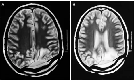
treatment, including corticosteroids. In October 2002, he was submitted to a fourth surgery with subtotal resection of the lesion, with the same histology. Thereafter, he was treated in another institution in Spain, where fractionated external beam stereotactic radiotherapy (SRT) (57.6 Gy in 32 fractions) was per-formed, which was completed in February 2003. A tumour regression of about 80% was documented, with a residual hetero-geneous lesion involving the posterior portion of the superior sagittal sinus and parietal-occipital left side. In December 2003, the patient underwent surgery for the fifth time, after new disease progression at the same location. Again, the resection was incomplete, with persistent disease adherent to the venous drain-age system. At that time, the patient was 15 years old, and the tumour board decided to perform a second SRT scheme (45 Gy in 25 fractions), which was completed in March 2004. The response evaluation by brain MRI (figure 3A and B), with single-voxel spectroscopy sequence included, confirmed complete remission (figure 3A and B). Despite this optimal response, rele-vant neurological sequelae related to the treatment were docu-mented: the visual field defect, which worsened on discontinuation of corticosteroids, focal epilepsy and mild right hemiparesis with crural predominance.
About 8 years later, in December 2011, the patient reported of persistent dry cough and low-grade fever. He reported neither dyspnoea, pleuritic pain, tiredness, profuse sweating, haemopty-sis, anorexia nor weight loss. There were no obvious infection locations other than the respiratory tract. The chest X-ray
showed lower right lobe opacity with pleural effusion (figure 4). Laboratory findings included: haemoglobin 14.1 g/dL, platelet count 362×109/L, total leukocyte count 11.1×10⁹/L, neutro-phils 63.8×10⁹ (57.5%), lymphocytes 17.8×10⁹ (16%), erythrocyte sedimentation rate 35.0, C reactive protein 6.7 mg/ dL and lactate dehydrogenase 264 UI/L. Culture tests were nega-tive. Symptoms were initially interpreted as a lower respiratory infection and the patient was treated with antibiotics. Brain MRI excluded recurrence. Two weeks later and despite some clinical improvement, a bronchoscopy was conducted to clarify the clin-ical picture. This examination showed partial occlusion of the right bronchus and extrinsic compression of the basal pyramid. The possibility of a late distant recurrence was admitted and the patient was submitted to right inferior lung lobectomy. However, no evidence of tumour tissue was observed and only a necrotising inflammatory granulomatous process was identified (negative microbiological tests, including Koch’s bacillus (BK) test and Löwenstein culture).
One month later, he presented with a similar clinical presenta-tion, with cough, low-grade fever and pleuritic pain on the right side. Thoracic computerised tomography (CT) scan revealed moderate right pleural effusion, septate pneumothorax related to previous lung surgery, as well as multiple suspicious nodules in both lungs (figure 5). He was admitted to our Institution, diagnosed as having a lower respiratory infection, and treated with antibiotics and supportive medication. However, the patient did not improve and a few days later, he was transferred to the emergency department. At the time, he was tachycardic (105 bpm), his blood pressure was 150/90 mm Hg and a para-doxical pulse was present. He had mild jugular distension. Chest X-ray revealed a right pleural effusion with an ipsilateral confluent heterogeneous opacity as well as other bilateral nodules. Electrocardiogram (ECG) and echocardiogram revealed a sustained monomorphic ventricular tachycardia with left bundle branch block, hyperechoic myocardial mass with inva-sion of the right ventricular cavity and a mild pericardial effu-sion (figure 6andvideo 1). Troponine T was slightly above the upper limit: 0.057 (<0.5 ng/mL); D-dimers increased: 932.0 (<230 ng/mL); and arterial blood gases were: pH 7.44, pCO2
28.4 mm Hg, pO2 102.3 mm Hg, HCO3 18.8, peripheral
Figure 1 (A) H&E staining, magnification ×40. (B) Vimentin staining confirming mesenchymal origin of the tumour, magnification ×100. Histological examination revealedfibrous and myxoid areas with abrupt transition between both. Myxoid areas had loosely whorled growth pattern around blood vessels, variable cellularity and variable amount of myxoid matrix. They were composed of spindle cells with ovoid nuclei and little cytoplasm. Fibrous areas had low to moderate cellularity and bland spindle cells with minimal pleomorphism and no mitosis. The neoplasm expressed immunoreactivity for vimentin.
Figure 2 MRI T1 sagittal section showing a nodular heterogeneous lesion in a cortical-subcortical left occipital location.
saturation 97.9% (FiO2=21%). Owing to the rapid
deterior-ation of the clinical condition with refractory acute pulmonary oedema, the patient was transferred to the cardiology intensive care unit and emergency manoeuvres were initiated. Electrical cardioversion was attempted with 200, 300 and 360 J. Unfortunately, the situation evolved into an irreversible cardiac arrest.
OUTCOME AND FOLLOW-UP
The patient died 2 h after admission, with an irreversible cardiac arrest, on 24 February 2012. The autopsy confirmed multiple bilateral lung metastases, bilateral pulmonary tumour embolism, pericardium and myocardial invasion by primary tumour, with intracavitary cardiac thrombosis and no evidence of intracranial recurrence.
DISCUSSION
MFS,first described by Angervall et al,6is a variant of a group of malignant fibrous histiocytomas (MFH), a highly heteroge-neous group of soft tissue neoplasms.9 In fact, because ultra-structural studies have shown afibroblastic differentiation, today
it is considered a distinct tumour, with myxoid features, myo-genic differentiation with immunoreactivity to SMA and a better prognosis than other cellular subtypes of MFH.10 11MFS
is the second most frequent subtype, representing 20% of all cases. In our case, the neoplasm histology showed fibrous and myxoid areas with abrupt transition between both. The myxoid areas had a loosely whorled growth pattern around blood vessels, variable cellularity and a variable amount of myxoid matrix. They were composed of spindle cells with ovoid nuclei and little cytoplasm. Variable nuclear pleomorphism and mitotic figures were present in the nodules; the more proliferative showed 8 mitosis/10 HPF. Fibrous areas had low to moderate cellularity and bland spindle cells, with minimal pleomorphism and no mitosis. Neither haemorrhage nor necrosis was present. The neoplasm expressed immunoreactivity for vimentin and CD34 (focal); EMA, S100, SMA and desmin were negative. Recurrences showed more myxoid areas, more cellularity and more cellular pleomorphism. Differential diagnosis between low-grade MFS and low-grade fibromyxoid sarcoma was recently aided by the absence of the chromosomal translocation, t(7;16)(q34;p11) with fusion gene FUS-CREB3L2 (BBF2H7), searched for but not found in this tumour.12
An intracranial location is extremely rare and, to the best of our knowledge, only four other cases have been published. One of the cases reported was of a 19-year-old woman, in whom the was located in the temporal bone, with endocranial extension, and resembling a meningioma on imaging.13 Surgery and
Figure 3 (A and B): MRI T2 axial section revealing complete disease remission. The single-voxel spectroscopy sequence showing, bilaterally, signal changes in white matter of the parietal-occipital lobe due to iatrogenic causes.
Figure 4 Plain chest radiography showing lower right lobe opacity
adjuvant chemotherapy was performed. At 18 months of follow-up the patient was doing well with no evidence of disease.
Another case was that of a 21-year-old woman with an exten-sive frontoparietal-occipital lesion. After afirst gross total resec-tion, the patient had two recurrences within a time frame of 2.5 years, with increasing tumour extent and histological grade, permitting only decompression during the third surgery.14One other case occurred in a 9-year-old boy. Nine months after surgery, local recurrence was noted and surgery performed. With 6 months of follow-up, the patient was well and with no recurrence.10
A case of MFS secondary to radiation was also documented in a 28-year-old man, 17 years after the treatment of basal ganglia germinoma.15Long-term follow-up details are unknown for this patient.
The differential diagnosis of intracardiac mass lesions includes benign and malignant tumours, such as a thrombus, vegetation and foreign bodies. However, intracardiac metastases, even though rare, should also be included in the differential diagno-sis, as well as infectious and non-bacterial thrombotic or maran-tic endocarditis.16
Reflecting the age distribution of malignant diseases, cardiac metastases predominantly occur in patients in the sixth and seventh decade of life. There is no sex preference and they
mostly appear in disseminated disease, as solitary metastases are very rare.17Metastases may reach the heart via the lymphatic or
the haematogenous route, or by transvenous extension. Lymphatic spread usually originates from pericardial metastases while myocardial metastases usually arise from a haematogenous route. In our case, at the time of necropsy, extensive multiple and bilateral pulmonary metastases were documented as well as multiple instances of mediastinic nodal involvement. It is rea-sonable to assume that tumour cells from the right cardiac chambers were being released from the heart and spread into both lungs, over time.
On the other hand, there was evidence of invasion of the par-ietal pericardium of the right ventricle by the tumour itself. Metastases to the epicardium are most commonly the result of direct invasion by an intrathoracic or mediastinal tumour, retro-grade lymphatic spread through tracheal or bronchomediastinal lymphatic channels or secondary involvement of the pericar-dium through spread from myocardial or epicardial metasta-ses.16 Furthermore, the hypothesis that tumour cells travelled from the intracranial venous system to the right cardiac cham-bers, where they would deposit for several years until clinical evidence of cardiac involvement, seems to be remote.
Cardiac tumours are generally asymptomatic, with most being detected following diagnostic investigation of an unrelated problem or in staging of a recently diagnosed primary tumour. Obviously, the symptoms differ greatly according to the most heavily involved site.18When they grow, the most common clin-ical manifestations are the result of large pericardial effusion, heart failure, obstruction of cardiac chambers or embolic phe-nomena. Cardiac tumours can also lead to rhythm disturbances, especially in atrioventricular conduction, as well as non-specific changes on the ECG, particularly ST-segment and T-wave altera-tions. Although less common, invasion of the myocardium can cause sustained ST-segment elevation, mimicking acute trans-mural myocardial infarction.19 Interestingly, there is no strong correlation between the extent of cardiac metastases and the clinical picture.20 Similar to most patients, our patient showed symptoms of congestive heart failure and embolisation.21
Autopsy confirmed multiple bilateral lung metastases, malignant pulmonary embolism and myocardial invasion by the primary tumour, with intracavitary cardiac thrombosis. Generally, the clinical pattern tends to be proportional to the degree of myo-cardial infiltration, or related to the wall infiltration site. Indeed, tumours spreading more extensively to the pericardium or cardiac chambers may give a dramatic presentation, causing medical emergencies. Whenever the right heart is involved, the interventricular septum is likely to be involved in the process, and the conduction system may be compromised.20 In our
patient, the cardiac invasion led to a sustained monomorphic ventricular tachycardia with left bundle branch block, as docu-mented when the patient was admitted to the emergency room.
The evidence of cardiac metastasis has clear prognostic impli-cations. It depends on several factors, such as the primitive tumour, the extent of the disease and the potential curative management of cardiac involvement.17 20Owing to the rarity of the case, no specific reports are available for patients with cardiac metastases from intracranial MFS. According to some reports of other primitive tumours, an average survival period after the diagnosis of cardiac metastases has been approximately 5.5 months.22 Furthermore, it is very important to know that,
in patients with cancer with clinical symptoms of heart involve-ment, metastatic cardiac deposits must always be considered in the differential diagnosis. In fact, clinicians should be alert to the possibility of cardiac metastases in a patient with, or even Figure 6 Right ventricle inflow view: intracardiac mass in the free
wall of the right ventricle, near the posterior leaflet of the tricuspid valve.
Video 1 Emergency department echocardiogram, right ventricle inflow view.
without, clinical symptoms. To note, cardiac involvement does not only have prognostic implications: even when curative therapy is not available, palliative measures may improve the quality of life of affected patients. However, one must keep in mind that, in some cases, there is no time to offer any kind of palliative treatment and cardiac involvement is not noticed until after death.17
The case presented is about cardiac metastases from MFS, a rare entity that has seldom been reported in the literature. Unfortunately, the diagnosis of cardiac metastases was only sus-pected when cardiac symptoms led the patient to the emergency department with rapid clinical deterioration. Definitive diagno-sis is frequently confirmed only after death, as happened in this case.
Learning points
▸ The rare occurrence of primary intracranial
myxofibrosarcoma (MFS) leads to a paucity of data and experience regarding its management.
▸ We report the first case of primary intracranial MFS with extracranial metastases and, particularly, in the right ventricle, an even rarer phenomenon. This required an independent external confirmation of the histological diagnosis in another specialised centre.
▸ The process of spreading of the metastases to the heart remains unclear: whether by direct invasion, retrograde lymphatic spread of lung or mediastinal metastases, by secondary involvement of the pericardium through myocardial or epicardial metastases or, the most remote hypothesis, related to cardiac deposition of the tumour cells migrating from the intracranial venous system.
▸ Despite the long disease-free survival obtained with multiple treatments, one cannot neglect the neurological sequelae that impacted on the patient’s quality of life.
Acknowledgements The authors are most grateful to the deceased and honoured colleague from Mayo Clinic’s Pathology Department, Dr Bernd Scheithauer, for revising the pathology, and to Dr Duarte Salgado, the neurologist who closely followed the evolution of the patient. The authors also thank ITClinical for the linguistic revision; Samanta McMurray for photography and digital work; and Dr Pedro Ferreira for cardiac imaging and video; and,finally, a special thanks to the patient’s parents, Teresa Santos and Francisco Patrício, for their indispensable collaboration.
Contributors DAC, PB, EG and MM equally participated in writing and reviewing the case report.
Competing interests None declared. Patient consent Obtained.
Provenance and peer review Not commissioned; externally peer reviewed. REFERENCES
1 Pinho T, Rodrigues-Pereira P, Araújo V, et al. Cardiac metastasis of melanoma as first manifestation of disease. Rev Port Cardiol 2009;5:633–9.
2 Chiles C, Woodard PK, Gutierrez FR, et al. Metastatic involvement of the heart and pericardium: CT and MR Imaging.Radiographics2001;21:439–49.
3 Hallahan DE, Vogelzang NJ, Borow KM, et al. Cardiac metastases from soft-tissue sarcomas. J Clin Oncol 1986;4:1662–9.
4 Agaimy A, Rösch J, Weyand M, et al. Primary and metastatic cardiac sarcomas: a 12-year experience at a German heart center. Int J Clin Exp Pathol 2012;5:928–38. 5 Takenaka S, Hashimoto N, Araki N, et al. Eleven cases of cardiac metastases from
soft-tissue sarcomas.Jpn J Clin Oncol2011;41:514–18.
6 Angervall L, Kindblom LG, Merck C. Myxofibrosarcoma. A study of 30 cases. Acta Pathol Microbiol Scand A 1977;85A:127–40.
7 Fanburg-Smith JC, Spiro IJ, Katapuram SV, et al. Infiltrative subcutaneous malignant fibrous histiocytoma: a comparative study with deep malignant fibrous histiocytoma and an observation of biologic behavior.Ann Diagn Pathol1999;3:1.
8 Wernhart S, Woernle CM, Neidert MC, et al. A deeply seated brain metastasis from a primary myxofibrosarcoma: case report.Clin Neurol Neurosurg2013;115:2296–8.
9 Castronovo C, Arrese JE, Quatresooz P, et al. Myxofibrosarcoma: a diagnostic pitfall.
Rare Tumors2013;5:e60.
10 Buccoliero AM, Castiglione F, Garbini F, et al. Primary cerebral myxofibrosarcoma: clinical, morphologic, immunohistochemical, molecular, and ultrastructural study of an infrequent tumor in an extraordinary localization.J Pediatr Hematol Oncol
2011;33:e279–83.
11 Al-Agha OM, Igbokwe AA. Malignantfibrous histiocytoma: between the past and the present.Arch Pathol Lab Med2008;132:1030–5.
12 Storlazzi CT, Mertens F, Nascimento A, et al. Fusion of the FUS and BBF2H7 genes in low gradefibromyxioid sarcoma.Hum Mol Genet2003;12:2349–58.
13 Nakayama K, Nemoto Y, Inoue Y, et al. Malignantfibrous histiocytoma of the temporal bone with endocranial extension. AJNR Am J Neuroradiol 1997;18:331–4. 14 Majumdar K, Mandal S, Saran RK, et al. Recurrent intracranial myxofibrosarcoma
presenting as an extensive fronto-parieto-occipital SOL: an unusual sarcoma of meningeal origin.Clin Neurol Neurosurg2013;115:354–8.
15 Kuo JR, Chio CC, Wang CC, et al. Radiation-induced intra- and extra-cranial high-grade myxofibrosarcoma with tumor bleeding.J Clin Neurosci
2008;15:1151–4.
16 Bussani R, De-Giorgio F, Abbate A, et al. Cardiac metastases.J Clin Pathol
2007;60:27–34.
17 Reynen K, Kockeritz U, Strasser RH. Metastases to the heart.Ann Oncol
2004;15:375–81.
18 Vaitkus PT, Herrmann HC, LeWinter MM. Treatment of malignant pericardial effusion.JAMA1994;272:59–64.
19 Goldberg AD, Blankstein R, Padera RF. Tumors metastatic to the heart.Circulation
2013;128:1790–4.
20 Katalinic D, Stern-Padovan R, Ivanac I, et al. Symptomatic cardiac metastases of breast cancer 27 years after mastectomy: a case report with literature review— pathophysiology of molecular mechanisms and metastatic pathways, clinical aspects, diagnostic procedures and treatment modalities.World J Surg Oncol2013;11:14. 21 Perchinsky MJ, Lichtenstein SV, Tyers GF. Primary cardiac tumors: forty years’
experience with 71 patients.Cancer1997;79:1809–15.
22 Ohnishi M, Niwayama H, Miyazawa Y, et al. Echocardiography in patients with malignant metastatic neoplasms of the heart and great vessels. J Cardiol 1990;20:377–84.
Copyright 2016 BMJ Publishing Group. All rights reserved. For permission to reuse any of this content visit http://group.bmj.com/group/rights-licensing/permissions.
BMJ Case Report Fellows may re-use this article for personal use and teaching without any further permission. Become a Fellow of BMJ Case Reports today and you can:
▸ Submit as many cases as you like
▸ Enjoy fast sympathetic peer review and rapid publication of accepted articles ▸ Access all the published articles
▸ Re-use any of the published material for personal use and teaching without further permission For information on Institutional Fellowships contact [email protected]