C A S O C L Í N I C O
T H O R A C I C O U T L E T S Y N D R O M E ( T O S ) M I M I C K I N G
T A K A Y A S U
’
S A R T E R I T I S–
C A S E R E P O R TEdgard Torres dos Reis Neto,
*Mário Luis Cardoso Pucinelli,
**Alexandre Wagner Silva de Souza,
**Emília Inoue Sato
***na primeira costela e fractura de clavícula. Os au-tores descrevem o caso de uma mulher jovem com dor no antebraço esquerdo acompanhada de clau-dicação, perda de peso, mialgias e lesões isquêmi-cas nos dedos, com pulso radial não palpável e pressão arterial não mensurável no membro supe-rior esquerdo, que inicialmente havia sido diag-nosticada como Arterite de Takayasu. A radiografia de tórax revelou presença de costela cervical bila-teral e os métodos de imagens dinâmicos (ultra-so-nografia com Doppler e angiografia) mostraram compressão bilateral da artéria subclávia, confir-mando o diagnóstico de SDT.
Palavras-Chave: Síndrome do Desfiladeiro
Torá-cico; Síndrome da Costela Cervical; Arterite de Ta-kayasu.
Introduction
Thoracic outlet syndrome (TOS) is defined as a set of symptoms that may occur due to compression of the brachial plexus and subclavian vessels in the thoracic outlet region, between the neck and the axilla. Bone or soft tissues anomalies can be respon-sible for TOS, including prolonged transverse pro-cess of the seventh cervical vertebra, cervical rib, anomalous first rib, first rib or clavicle fracture’s and fibromuscle abnormalities.1,2
In the literature there are some TOS classifica-tions according to the compressed structures or the etiology of the lesion.
Huang et al. classified TOS in three groups: com-pression of the brachial plexus (neurogenic TOS), compression of the subclavian vessels (vascular TOS) and another nonspecific form (pain and sen-sitive symptoms, without either objective signs of neurological compression or alterations in the neu-rophysiological tests).3
Colli et al. classified TOS into five groups. They
*Pos-graduated student **Assistant
***Professor of Rheumatology
Rheumatology Division, Paulista Medical School – Universidade Federal de São Paulo, São Paulo, Brasil.
Abstract
Thoracic outlet syndrome (TOS) is defined as a set of symptoms caused by the compression of the bra-chial plexus and subclavian vessels in the thoracic outlet region. Anomalies in musculoskeletal struc-tures may be responsible for TOS, including prolon-ged transverse process of the seventh cervical ver-tebra, cervical rib, and first anomalous rib and cla-vicle fractures. The authors describe a case of a young woman with pain in the left forearm, accom-panied by intermittent claudication, weigh loss, myalgias and ischemic lesions in the fingers, with no pulses and no measurable blood pressure in the left arm, who was initially diagnosed as Takayasu arteritis. The chest radiography showed accessory cervical ribs and the dynamic vascular image tests (Doppler ultra-sound and angiography) showed bi-lateral compression of the subclavian artery, con-firming the diagnosis of TOS
Keywords: Thoracic Outlet Syndrome; Cervical Rib
Syndrome; Takayasu Arteritis.
Resumo
O Síndrome do desfiladeiro torácico (SDT) é defi-nido como o conjunto de sintomas que pode ser causado por compressão do plexo braquial e dos vasos subclávios na região do desfiladeiro torácico. Alterações da morfologia dos elementos músculo--esqueléticos na região podem ser responsáveis pelo SDT, incluindo apófise transversa longa da sé-tima vértebra cervical, costela cervical, anomalias
E D G A R D T O R R E S D O S R E I S N E T O E C O L.
subdivided the neurogenic TOS in two groups: one with classic signs and symptoms of neurogenic compression accompanied by specific eletroneuro-myography (ENMG) findings and other with clini-cal and ENMG nonspecific findings. The vascular TOS are also subdivided in: arterial or venous sub-clavian compression and the fifth group comprises patients with signs and symptoms of post-trauma-tic neurovascular compression.4
Sanders et al. classified TOS in three groups: neurogenic TOS, arterial TOS and venous TOS.5
Concerning the frequency of different forms of TOS, Huang et al. considered the nonspecific type as the most common and the vascular as the rarer form.3
Neurological symptoms occur over 90% of all TOS cases3,5,6and the vascular form corresponds
appro-ximately to 5% (the arterial TOS represents less than 1%).5,6 The neurogenic and vascular forms may
coexist and the distinction between these forms is not easy.3
There is no gold standard test for TOS diagno-sis.2,7Detailed anamnesis and physical
examina-tion are the most important tools for the TOS diag-nosis. Provocative tests, such as Adson´s test, are nonspecific, and can be positive in asymptomatic individuals. The electro-diagnostic and image stu-dies are not always useful for the diagnosis. Since there is no specific confirmatory test, TOS inciden-ce is variable, ranging from 3 to 80 cases/1,000 in-habitants. It is more frequent in women with age ranging from 20 to 50 years3possibly because
ge-nerally women present weakened muscular struc-tures which makes their scapulas lower, predispo-sing the compression of the structures involved in TOS.1
The differential diagnosis is wide and includes cervical disc lesions, osteophytes, Pancoast tumor, nerve sheath tumor, ulnar and median nerve en-trapment, brachial plexitis, spinal cord tumor, shoulder’s diseases, fibromyalgia, multiple sclero-sis, Raynaud phenomenon, acute coronary artery disease, venous thrombosis, micro-embolism, Takayasu arteritis, vasospastic disorders, complex regional pain syndrome, brachial plexus injuries and myofascial syndrome.1,3,6
There is no consensus for the best treatment of TOS and it depends on the etiology. The surgical treatment is usually indicated in cases of true neu-rogenic or vascular TOS and for patients with nons-pecific form refractory to the conservative treat-ment. The best surgical approach in those patients is not defined yet.3
Takayasu’s arteritis (TA) is a chronic inflamma-tory disease that affects primarily large vessels like aorta and its branches.8The inflammation leads to
stenosis and occlusion of the involved arteries, aneurysms formation or both.9It may cause the
decrease or the absence of arterial pulses, cerebral ischemia, acute myocardial infarction, aortic in-sufficiency, cardiac congestive failure, limb claudi-cation, hypertension, aneurysm and blindness.8,10
TA affects mainly women in the second and third decades of life8and can cause premature death.9
Case Report
A 23 y.o. mulatto woman was admitted at our uni-versity hospital complaining about burning pain and intermittent claudication in the left forearm for 5 months. She had noticed her skin cold and pale on the left forearm and hand associated with a painful lesion on the second digital pulp in the last two months. The pain and intermittent clau-dication became worse and were associated with new necrotic lesion on the third finger of the left hand. She was being treated with cilostazol, ace-tylsalicylic acid and pentoxifylline without impro-vement. The patient also reported headache, diz-ziness, fever and a weight loss of 5 Kg. She referred a traumatic left clavicle fracture when she was child and had a history of heavy smoking, alcoholic ha-bits and drug abuse with cannabis and cocaine. The patient had been attended in another hospi-tal where it was prescribed 60 mg/day of predni-sone due to the suspicion of TA.
When the patient was attended at our service, there was no left brachial and radial arterial pulse or limb edema. Blood pressure was 140 × 90 mmHg in the right arm, not measurable in the left arm and 160 × 90 mmHg in both lower limbs. A bruit over the left subclavian artery, pale skin in the left hand, mild hypotrophy in thenar and hypothenar regions and diminished temperature in the left fo-rearm and hand were observed. She also presen-ted necrotic lesions on pulp of the second and third fingers and on subungueal region in the fingers of the left hand (Figure 1). Hypoesthesia and pares-thesias over the thenar region and palmar aspect of the first, second and third fingers of the left hand became worse with hyper-abduction and exten-sion of the left shoulder. The Adson’s test, Roos’ test and Wright test were positive bilaterally. No episodes of fever were detected during the whole
T H O R A C I C O U T L E T S Y N D R O M E M I M I C K I N G TA K AYA S U’S A R T E R I T I S
inpatient period.
Laboratory tests: hemoglobin level was 11,6g/ /dL, white-cell count was 19,700/mm (1% bands, 78% segmented, 17% lymphocytes, 2% eosi-nophils, 2% monocytes)and erythrocyte sedimen-tation rate: 10 mm per hour. Serum creatinine, electrolytes, transaminases and the urinalysis were normal. Serologic tests for viral hepatitis B and C, HIV and syphilis and the anticardiolipin antibody test were all negative. Blood and urine cultures we-re also negatives.
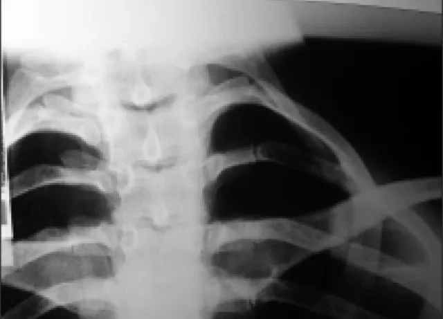
The chest radiography showed bilateral acces-sory cervical ribs (Figure 2) and the Doppler ul-trasound of the left upper limb exhibited reduced flow in subclavian artery due to costoclavicular en-trapment at provocative test. The distal artery was fulfilled by collateral circulation and the subcla-vian and axillary arteries presented partial throm-bosis, with a total thrombosis of the brachial artery.
The exam in the right upper limb also showed re-duced subclavian artery flow at hyperabduction without thrombosis. The digital arteriography con-firmed the alterations observed at Doppler ul-trasound (Figure 3) and the echodopplercardio-gram was normal.
The ENMG showed altered sensory and motor fibers conduction of all nerves in the left upper limb with no abnormality in the right one.
The patient was submitted to bilateral cervical rib excision and anticoagulation with improve-ment of all symptoms.
Discussion
We describe the case of a young woman with neu-rological and vascular symptoms mainly in the left arm who was erroneously diagnosed as TA. The pa-tient fulfilled 5 of 6 American College of Rheuma-tology TA classification criteria11(age at disease
on-set < 40 years, claudication of extremities, decrea-sed brachial artery pulse, bruit over subclavian ar-tery, difference of blood pressure between arms > 10 mmHg). The complaints of fever and weight loss also had suggested systemic disease, however, no episode of fever was observed during the hos-pitalization. The leucocytosis was attributed to the prednisone use and the weight loss was attributed to the ischemic pain and her appetite normalized and she recovered weight after the pain treatment. No signs of infection were observed either at physi-cal exams or at laboratory tests.
The left subclavian artery is described as one of the most frequently affected vessels in TA,
va-Figure 1. Left hand. 1A. Ischemic lesions under nails. 1B. Ischemic lesions on pulp of second and third fingers.
Figure 2. Radiography of the thorax showing bilateral
cervical ribs.
A
E D G A R D T O R R E S D O S R E I S N E T O E C O L.
rying from 46 to 67.8% in different parts of the world.9,12-15However, digital ischemic lesions
usual-ly are not part of the clinical spectrum of TA. This finding was the first clinical sign to indicate other diagnostic possibility in our case. The vascular form of TOS is less frequently found than the neu-rological form, and in most of cases requires sur-gical treatment. We should be alert for this diagno-sis in a young individual without high risk for athe-rosclerosis who presents ischemic manifestations in upper limbs.16
Arterial form of TOS usually presents as cold ex-tremity, weakness, intermittent claudication, dif-fuse pain and decreased amplitude of the arterial pulse. Venous involvement usually presents with venous thrombosis, superficial vessels distension, edema and pain.6
Massionneure et al. classified arterial
manifesta-tions of TOS in 3 types: partial or complete throm-bosis, post-stenotic dilatation and post-stenotic aneurysm of subclavian artery. These lesions may cause distal embolization, claudication, vasomo-tor phenomena and digital gangrene. Retrograde embolization from subclavian to vertebral or caro-tid arteries are rarely described.17Thrombosis of
the subclavian vein occurs most often in men with strenuous job and presents as edema and cyano-sis of the upper limb or distended superficial veins of the shoulders or chest.3
Our patient presented partial thrombosis of the subclavian and axillary arteries and a total throm-bosis of the left brachial artery, without aneurisms. The distal ischemic lesions could be due to embo-lism of proximal thrombus or insufficient collate-ral vessel for irrigation of distal regions.
There is no specific diagnostic method for none
Figure 3. Angiography on neutral position (3A) and hyper-abduction of the right arm (3B).Angiography of the left arm
T H O R A C I C O U T L E T S Y N D R O M E M I M I C K I N G TA K AYA S U’S A R T E R I T I S
of TOS forms. On physical examination, various tests had been described, such as Adson’s test and hyper-abduction, but their sensibility and specifi-city are not still defined.18 Costo-clavicle or
Hals-ted’s test, Roos’ test and Wright test are others ma-neuvers that can be used to evaluate TOS patients. However, false-positive or false-negative results can be found.7
Cervical ribs are described in 10% of TOS pa-tients and in 0,01 to 0,5% in the general population in which most of them are asymptomatic.3 Cervical
ribs are more common in women than men and bi-lateral in more than 50%.19The presence of
cervi-cal rib is not diagnostic for TOS and its absence does not invalidate the diagnosis.1It is unknown
why only few patients with cervical rib develop TOS. The format and the consistency of fibrous band that leagues this rib to the first rib seem to be one of the factors involved in the development of symptoms. Nevertheless, it seems to be related to some sports and jobs that demand a prolonged arm hyper-abduction.6
In comparison to other image methods, Dop-pler ultra-sound and angiography in neutral and hyper-abduction of the arm are useful for the diag-nosis of the vascular form of TOS.16 Doppler
ul-trasound has limitations in obese individuals and in areas in which bone structures overlie vessels. In these cases conventional or magnetic resonan-ce (MR) angiography are useful20specially to
monstrate fibrous bands and brachial plexus de-flection in patients without cervical rib.7
Nervous conduction studies and ENMG can help the diagnosis by revealing decreased senso-rial or motor action potential in ulnar and/or me-dian nerves. ENMG can also reveal abnormalities in the intrinsic muscles of the hands.3
The treatment of TOS is still controversial21and
can be conservative or surgical.
Conservative treatment includes behavioral changes by avoiding activities and positions that determine its appearance, beside rehabilitation with strengthening of pectoral musculature and postural positioning. The improvement after con-servative treatment varies from 50 to 90% and de-pends on its etiology.3
Surgical treatment involves surgical decompres-sion and, when necessary, vascular reconstruc-tion.3Cervical rib excision and/or first rib excision;
resection of cervical muscles, brachial plexus neu-rolysis and complementary vascular procedures are some of surgery procedures applied to TOS
pa-tients.16,18The presence of cervical rib per se is not
an indication for surgery, unless there is failure in conservative treatment or incapacitating symp-toms.19 Vascular reconstruction after
decompres-sion presents good results on a short and long term follow-up. Endovascular treatment with stents is also described, however long term studies showed high chance of re-stenosis. The treatment of ve-nous involvement include early use of thrombolytic agents followed by anticoagulation and late surgi-cal decompression, or thrombolytic therapy through catheter followed by early surgical decom-pression and anticoagulation followed by balloon angioplasty in cases of stenosis. These options had better results when compared to anticoagulation alone.16
We described a young woman with neurogenic and vascular TOS characterized by intermittent claudication, bruit over subclavian arteries, absen-ce of arterial pulse and non measurable blood pres-sure in the left arm. All of these features could be suggestive of TA. However, TA is a chronic arteritis and rarely causes ischemic cutaneous lesions as presented by our patient. This case emphasizes the importance of a detailed clinical examination and the differential diagnosis between these pathologi-es which have different treatment and prognosis. We should pay attention to suspicious cases of TOS, because some of its manifestations may be irrever-sible in case of late diagnosis.
Correspondence to Emilia I Sato
UNIFESP – Disciplina de Reumatologia
Rua Botucatu, 740, CEP 04023 900 – São Paulo-SP, Brazil E-mail: eisato@unifesp.br
References
1. Rosa Filho BJ. Síndrome do desfiladeiro torácico. Available from: http://www.wgate.com.br/conteu-do/medicinaesaude/fisioterapia/desfiladeiro.htm. Acessed on Sep. 2006.
2. Charon J-PM, Milne W, Sheppard DG et al. Evaluation of MR angiographic technique in the assessment of thoracic outlet syndrome. Clin Radiol 2004;59:588--595.
3. Huang JH, Zager EL. Thoracic outlet syndrome. Neu-rosurgery 2004;55:897-902.
4. Colli BO, Carlotti CG, Assirati JA, Marques Jr. W. Neu-rogenic thoracic outlet syndromes: a comparison of true and nonspecific syndromes after surgical treat-ment. Surgical Neurology 2006;65:262-272.
5. Sanders RJ, Hammond SL, Rao NM. Diagnosis of tho-racic outlet syndrome. J Vasc Surg 2007;46:601-604. 6. Scola RH, Werneck LC, Iwamoto FM et al. Síndrome
E D G A R D T O R R E S D O S R E I S N E T O E C O L.
do desfiladeiro torácico tipo neurogênico verdadeiro. Arq Neuropsiquiatr 1999;57:659-665.
7. Cruz M, Matos AA, Saldanha T et al. Angiografia co-mo método de diagnóstico da síndrome do desfila-deiro torácico neurovascular. A propósito de um ca-so. Rev Bras Reumatol 2003;43:267-271.
8. Valente RM: Vasculitic syndromes. In: KELLEY, W. Textbook of rheumatology. Philadelphia, WB Saun-ders, 1997: 1079-1122.
9. Subramanyan R, Joy J, Balakrishnan KG: Natural his-tory of aortoarteritis (Takayasu´s disease). Circulati-on 1989;80:429-437.
10. Numano F, Kakato T: Takayasu arteritis – five doctors in the history of Takayasu arteritis. Int J Cardiol 1996;54:S1-S10.
11. Arend WP, Michel BA, Bloch DA et al. The American College of Rheumatology 1990 criteria for the classifi-cation of Takayasu´s arteritis. Arthritis Rheum 1990;33:1129-1134.
12. Sato EI, Sassaki Jr RH, Leão CS, Hata FS, Nunes DS, Santo BE. Arterite de Takayasu: estudo clínico e an-giográfico. Rev Bras Reumatol 1998;38:9-14.
13. Watzko WL, Chaves AMA, Rachid A, Rachid Filho A, Radominski SC. Takayasu´s arteritis: by the way of 24 case histories: revision. Rev Bras Reumatol 1989;29: 161-165.
14. Cañas CA, Jiménez CA, Ramirez LA et al. Takayasu ar-teritis in Colombia. Int J Cardiol 1998;66: S73-79. 15. Kerr GS, Hallaman CW, Giordano J et al. Takayasu
ar-teritis. Ann Intern Med 1994;120: 919-929.
16. Davidovic LB, Kostic DM, Jakovljevic NS et al. Vascu-lar outlet syndrome. World J Surg 2003;27:545-550. 17. Massionneure H, Planchon B, De Faucal P et al.
Vas-cular manifestations in TOS. Prospective study of 104 patients. J Mal Vasc 1991;16:220-225.
18. Paiva ES, Engelhorn AL, Mazer S. Síndrome do desfi-ladeiro torácico predispondo a trombose arterial e venosa em membro superior associada à síndrome antifosfolipídica. Rev Bras Reumatol 2001;41:315--318.
19. Sanders RJ, Hammond SL. Management of cervical ribs and anomalous first ribs causing neurogenic thoracic outlet syndrome. J Vasc Surg 2002;36:51-56. 20. Hagspiel KD, Spinosa DJ, Angle JF et al. Diagnosis of
vascular compression at the thoracic outlet using ga-dolinium-enhaced high-resolution ultrafast MR an-giography in abduction and adduction. Cardiovasc Intervent Radiol 2000;23:152-154.
21. Samarasam I, Sadhu D, Agarwal S et al. Surgical ma-nagement of thoracic outlet syndrome: a 10 year ex-perience. ANZ J Surg 2004;74:450-454.