UNIVERSIDADE DA BEIRA INTERIOR
Ciências
Mitochondrial genetic therapy in Parkinson’s
disease
Cloning mitochondrial gene ND1
Cátia Marlene do Carmo Baptista
Dissertação para obtenção do Grau de Mestre em
Biotecnologia
(2º ciclo de estudos)
Orientador: Prof.ª Doutora Diana Costa
Co-orientador: Prof.ª Doutora Fani Sousa
iii
Aos meus queridos pais.
À melhor irmã do mundo e afilhado lindo.
Às minhas grandes amigas Sandra, Ana e Vanessa.
v
Acknowledgements
First of all, a big and sincere thanks to my supervisor Profª Diana Costa, PhD and co-supervisor Profª Fani Sousa, PhD for all the support and understanding throughout the development of this master's thesis.
I would like to thank Ruben Salvado for all explanations and tips that he gave me in the initial phase of the thesis.
To all Endocrinology group thank you for everything. To Eduarda Coutinho for her support and assistance throughout the development of the thesis. To Maria Inês Alvelos for the advices. To Joana Raimundo companionship. To Catarina Gonçalves for the friendship and support in difficult times.
To Patricia Pereira and Augusto Pedro for the tips regarding the growth and productivity studies and the tips in relation to P.pastoris. To Joana Valente for all the tips given throughout the thesis and the explanation and help in the purification step. To Carolina Costa for the company and help during the preparation of nanoparticles.
Also, I would like to thank João Matos and Engª Ana Paula for assistance in SEM sessions and to Carlos Gaspar for assistance in Zeta Sizer measurements.
Lastly, I’m very thankful to everyone that in a way or another contributed to the success of this thesis.
Thank you all so much, Cátia Baptista
vii
Resumo
A mitocôndria é um organelo celular que tem o seu próprio genoma, de cadeia dupla e circular que codifica 13 proteínas envolvidas no transporte de eletrões e fosforilação oxidativa. Estão presentes entre 10 e 100 cópias por célula somática e, dentro das células, as cópias de ADN mitocondrial (mtDNA) podem ser idênticas em sequência (homoplasmia) ou mtDNA “wild-type” e mutado podem existir em diferentes proporções (heteroplasmia). A heteroplasmia ocorre principalmente porque o mtDNA é particularmente suscetível a mutações, devido à sua proximidade a locais de geração de espécies reactivas de oxigénio.
Alterações no mtDNA podem provocar disfunções no mecanismo de fosforilação oxidativa, onde as exigências de ATP não são atingidas. O fenótipo da doença mitocondrial depende da heteroplasmia do mtDNA, quando é atingido um valor de mtDNA mutado entre 60-90%.
A ideia de que a disfunção mitocondrial poderia influenciar a doença de Parkinson surgiu com descoberta do mecanismo de ação do MPTP no início de 1980, que afeta o fluxo de eletrões no Complexo I. Diferentes mutações que afectam os genes que codificam as proteínas deste complexo têm sido associadas com a doença de Parkinson, duas delas sendo identificadas no gene mitocondrial ND1. Este facto faz com que o desenvolvimento de um vector com o gene mitocondrial ND1 seja uma abordagem muito interessante para aplicação na terapia génica mitocondrial (MGT) para o potencial tratamento da doença de Parkinson.
No presente trabalho foi desenvolvido um sistema para transportar um vector, desenhado no laboratório, com possível aplicação em terapia génica mitocondrial. O vector foi construído usando o plasmídeo pCAG-GFP e o gene mitocondrial ND1. Posteriormente, nanopartículas de CaCO3 foram desenvolvidas para o seu transporte direccionado para a
mitocôndria. O desenvolvimento deste projecto foi dividido em três etapas principais: i) construção do vector baseado na clonagem do gene mitocodrial ND1 no plasmídeo pCAG-GFP; ii) estudo de diferentes hospedeiros recombinantes para a amplificação do vector pCAG-GFP-mtND1; e iii) desenvolvimento de uma formulação adequada ao direccionamento do vector pCAG-GFP-mtND1 de forma eficiente para a mitocôndria.
O vector pCAG-GFP-mtND1 foi inserido em diferentes estirpes de E. coli e os estudos de crescimento foram muito semelhantes entre as estirpes transformadas, tendo sido observadas apenas algumas diferenças nos rendimentos específicos. Os sistemas de entrega preparados, demonstraram ser biocompatíveis, com eficiências de encapsulação acima de 50%, tamanhos inferiores a 280 nm e potenciais zeta positivos tornando-os adequados para MGT.
Palavras-chave
ix
Abstract
Mitochondria have their own genome, a circular double-stranded genome that encodes 13 proteins involved in electron transport and oxidative phosphorylation (OXPHOS). They are present in 10–100 copies per somatic cell and within the cell mitochondrial DNA (mtDNA) copies could be identical in sequence (homoplasmy) or wild-type and mutant mtDNA could exist in different ratios (heteroplasmy). Heteroplasmy occurs mainly because mtDNA is particularly susceptible to mutations due its proximity to reactive oxygen species (ROS) generation sites.
MtDNA dysfunction is characterized by a defective OXPHOS where ATP demands are not reached. The phenotype of the mitochondrial disease depends on the mtDNA heteroplasmy, when a threshold in the region of 60–90% mutated mtDNAs is reached.
The idea that mitochondrial dysfunction could influence Parkinson disease (PD) arose with the discovery of the mechanism of action of MPTP in the early 1980s that affected electron flow in Complex I (CI). Different CI related mutations have been associated with PD and two of them were identified in mtND1 gene. This fact makes the development of a mtND1 construct a very interesting approach for mitochondrial gene therapy (MGT) purposes in PD treatment.
Throughout this year, it was developed a nanocarrier to transport a designed vector with possible application in MGT. The vector was constructed using pCAG-GFP plasmid and the mitochondrial gene ND1 and CaCO3 were developed for the directed transport of the plasmid
towards mitochondria.
This research development was divided into three main stages: i) construction of a vector using an OXPHOS CI gene, mtND1 and the pCAG-GFP plasmid; ii) study of different recombinant hosts to produce the pCAG-GFP-mtND1 vector; and iii) development of a formulation for the efficient pDNA delivery into mitochondria.
The mtND1 construct was successfully obtained and inserted into different E. coli strains. The growth profiles were very similar between the strains, being observed sligth differences in the pDNA specific yields. The nanocarries obtained were biocompatible, displaying encapsulation efficiencies (EE) above 50%, sizes below 280 nm and positive zeta potential, making them very suitable for MGT.
Keywords
xi
Index
Resumo ... vii Abstract ... ix Introduction ... 1 1.Mitochondria ... 21.1.Structure and function ... 2
1.2.Mitochondrial DNA (mtDNA) ... 3
1.2.1.MtDNA Replication ... 4
1.2.2.MtDNA mutations and repair mechanisms ... 5
1.2.3.Heteroplasmy and threshold effect ... 6
1.2.4.Inheritance of mtDNA ... 7
1.3.Role of mitochondria in the living cell ... 8
1.3.1.Electron transfer system and ATP synthesis ... 8
1.3.2.Production of Reactive oxygen species (ROS) ... 9
1.3.3.Ca2+ homeostasis ... 10
1.3.4.Programmed Cell Death (PCD) ... 11
1.3.5.Mitochondrial dynamics: Fission and Fusion ... 12
2.Mitochondrial diseases ... 13
2.1.Cytopathies and mutations ... 13
2.2.Aging ... 14
2.3.Neurodegenerative diseases: Parkinson (PD) ... 15
2.4.Diagnosis of mtDNA disease ... 16
xii
3.1.Construction/amplification of a recombinant human mtDNA construct for MGT ... 18
3.2.Nanotechnology ... 19
3.2.1.CaCO3 Nanoparticles ... 20
Aims of the Project ... 22
Materials and Methods ... 23
1.Materials ... 23 1.1.Reagents ... 23 1.2.Hosts ... 23 2. Methods ... 23 2.1.Vector construction ... 23 2.1.1.ND1 gene amplification ... 23 2.1.2.Binding Reaction ... 24 2.2.Enzymatic digestion ... 24 2.3.Sequencing ... 25 2.4.Cell transformation ... 26 2.5.Cell Banks ... 26
2.6.Plasmid DNA production studies ... 26
2.7.Growth of pCAG-mtND1 for nanoparticle synthesis ... 27
2.8.Plasmid Purification ... 27
2.8.1.Purification of the samples for yield studies ... 27
2.8.2.Purification of pCAG-GFP-mtND1 for nanoparticles production ... 27
2.9.Agarose gel electrophoresis ... 28
2.10.Nanoparticles studies ... 28
xiii
2.10.2.Particles Morphology ... 28
2.10.3.pDNA Encapsulation Efficiency ... 29
2.10.4.Nanoparticles Size and Zeta (ζ) Potential ... 29
2.10.5.Cell Citotoxicity ... 29
Results ... 31
1.ND1 gene amplification ... 31
2.Plasmid Amplification and purification ... 31
2.1.pCAG-GFP-ND1 Agarose gel electrophoresis ... 31
2.2.pCAG-GFP-ND1 sequencing ... 33
3.Host studies ... 34
3.1.P. pastoris X33 and S. cerevisiae NRRL Y-12632 ... 34
3.2.E. coli JM109, XL1B and DH5α ... 35
4.CaCO3 Nanoparticles synthesis ... 36
5.Encapsulation Efficiency ... 37
6.Nanoparticle Size and Zeta potencial (ζ) ... 38
7.Cell Citotocixity ... 41
Discussion ... 43
Conclusions and Future Perspectives ... 45
xv
Figures List
Figure 1- Eukaryotic cell with a detailed mitochondria diagram showing the inner and outer
membranes and the folded cristae ... 2
Figure 2 - Human mtDNA ... 3
Figure 3 – Proposed models of mammalian mtDNA replication.. ... 5
Figure 4 - Base excision repair pathways ... 6
Figure 5 - Manipulation of mtDNA heteroplasmy ... 7
Figure 6 - Mitochondrial genetic bottleneck theory ... 8
Figure 7 - Mitochondrial respiratory chain. ... 9
Figure 8 - Effects of reactive oxygen species ... 10
Figure 9 - Mitochondrial Ca2+ channels/transporters and Ca2+ ... 10
Figure 10 - Molecular pathways that lead to apoptosis.. ... 11
Figure 11 - Schematic representation of mitochondrial fusion and fission events ... 12
Figure 12 - Mitochondrial etiology of complex disease ... 13
Figure 13 - MtDNA mutations and corresponding diseases ... 14
Figure 14 - Algorithm for investigation of mitochondrial disease ... 17
Figure 15 - Delivery of therapeutic agents into Mitochondria ... 18
Figure 16 - Nanocarriers with potential for mitochondrial targeting ... 20
Figure 17 - Agarose gel electrophoresis of mtND1 gene amplification by PCR ... 31
Figure 18 - Colony PCR of E.coli DH5α colonies ... 32
Figure 19 - Enzymatic digestion of pCAG-GFP-mtND1 with SmaI and XbaI ... 32
Figure 20 - pCAG-GFP-mtND1 sequencing obtained from E.coli DH5α transformed colony .... 33
Figure 21 - Growth of P. pastoris X33 and S. cerevisiae NRRL Y-12632 on YPD plates complemented with 500 µg/ml ampicillin ... 34
xvi
Figure 22 - Growth curve of E.coli JM109 transformed with pCAG-GFP-mtND1 ... 35
Figure 23 - Growth curve of E.coli DH5α transformed with pCAG-GFP-mtND1 ... 36
Figure 24 - Growth curve of E.coli XL1B transformed with pCAG-GFP-mtND1... 36
Figure 25 - CaCO3 Nanoparticles synthesis ... 37
Figure 26- pCAG-GFP-mtND1/Rho123 CaCO3 nanoparticles ... 39
Figure 27 - pCAG-GFP-mtND1/Rho123/gelatin CaCO3 nanoparticles ... 39
Figure 28 - pCAG-GFP-mtND1/Rho123/cellulose CaCO3 nanoparticles ... 40
Figure 29 - Cytotoxicity profile of 5 µg pDNA and 10 µg pDNA of pCAG-GFP-mtND1/Rho123 CaCO3 nanoparticles on human fibroblast cells after 24, 48 and 72 hours incubation, measured by the MTT assay ... 42
xviii
Tables List
Table 1 - Genetic Code in Mitochondria. ... 4
Table 2 - Confirmed mtDNA variations associated with PD disease. ... 16
Table 3 - Thermal cycling program for mtND1 PCR amplification. ... 24
Table 4 - Conditions for enzyme digestion. ... 25
Table 5 - Thermal cycling program for DNA sequencing reaction. ... 25
Table 6 - Loading efficiency of pCAG-GFP-mtND1/Rho123, pCAG-GFP-mtND1/Rho123/cellulose and pCAG-GFP-mtND1/Rho123/gelatin CaCO3 nanoparticles with 5 µg and 10 µg pCAG-GFP-mtND1 loading amount. ... 38
Table 7 - Average size and zeta potential of pCAG-GFP-mtND1/Rho123, pCAG-GFP-mtND1/Rho123/cellulose and pCAG-GFP-mtND1/Rho123/gelatin CaCO3 nanoparticles with 5 µg and 10 µg pDNA loading amount. ... 41
xx
Acronyms List
AD – Alzheimer disease
ADP - adenosine diphosphate AIF - apoptosis-inducing factor ALS - Amyotrophic lateral sclerosis
Apaf -1 - Apoptotic protease activating factor 1 APE1 - AP endonuclease-1
ATP - adenosine-5′-triphosphate BER - Base excision repair Ca 2+ - Calcium
CaCl2 - Calcium Chloride
CaCO3 - Calcium Carbonate
CI – Complex I
DISC - death-inducing signaling complex DNA - Deoxyribonucleic acid
DNA SSB - DNA single strand break Dnm1l - dynamin-1-like protein
DOPE - Discrete Optimized Protein Energy
DQAsomes - DeQuAlinium-based liposome-like vesicles DR - death receptor
dRp - DNA single strand break flanked by 3’-hydroxyl and a 5’-deoxyribosephosphate
EDTA - Ethylenediamine tetraacetic acid
EE - Encapsulation Efficiency FADH2 - Flavin Adenine Dinucleotide
FEN-1 - Flap endonuclease-1 Fis1 - fission protein 1 H2O2 - hydrogen peroxide
HD – Huntington disease
xxi
Kb - Kilobase LB - Luria Broth LP - Liposomes
LP-BER - Long pathway BER Mfn - mitofusin
MGT - mitochondrial gene therapy Mn - manganese
MnSOD - superoxide dismutase
MOMP - mitochondrial outer membrane permeabilization mPTP - mitochondrial permeability transition pore mtDNA - mitochondrial DNA
MTT - 3-(4,5-dimethylthiazol-2-yl)-2,5-diphenyltetrazolium bromide NADH - nicotineamide-adenine-dinucleotide O2−• - superoxide anion OH - H-strand origin OH• - hydroxyl radical OL - L- strand origin OM - outer membrane
OXPHOS - Oxidative phosphorylation PCR - polymerase chain reaction PD – Parkinson Disease
Pi - inorganic phosphate PNA – peptide nucleic acid Pol β - polymerase β Q – Ubiquinone R8 - octaarginine
RES - reticuloendothetial system Rho123 - Rhodamine123
RITOLS - RNA incorporation throughout the lagging strand RNA - Ribonucleic acid
xxii
ROS - Reactive oxygen species rRNA - Ribosomal RNA
SDS - Sodium dodecyl sulfate SEM - Scanning Electron Microscopy
Smac - second mitochondria-derived activator of caspases SP-BER - Short pathway BER
TAE - Tris-Acetate-EDTA
TB - Terrific Broth TCA - tricarboxylic acid TFN - tumor necrosis factor TPP - triphenylphosphine tRNA - Transfer RNA
YPD - Yeast extract peptone dextrose ZFB – Zinc-finger binding proteins
1
Introduction
Human mitochondria are small cytoplasmic organelles, present in 10–100 copies per somatic cell (1). Mitochondria are different from other organelles with respect to their complex two membrane structure (2) and they contain a circular genome, mitochondrial DNA (mtDNA) that has been reduced during evolution through gene transfer to the nucleus (3). MtDNA encodes 13 polypeptides, 2 ribosomal RNAs and 22 RNA transcripts that contribute to 4 of the 5 respiratory chain complexes (1,4). By a process of oxidative phosphorylation (OXPHOS) mitochondria converts biochemical energy stored in food, through the oxidation of nutrients to produce adenosine-5′-triphosphate (ATP) (5).
MtDNA dysfunction is typically characterized by an inability to meet cellular ATP demand as a result of defective OXPHOS (4). Mitochondrial disease phenotype is dependent on mtDNA heteroplasmy, where disease manifests in cells carrying more than a certain threshold percentage of mutated mtDNA copies. Normally this threshold is in the region of 60–90% of mutated mtDNAs and changes between organs depending upon their energy requirements (1,6). It is described that 1 in 8000 individuals carry a mtDNA genetic disorder or are affected by a pathogenic mtDNA mutation (4).
Research undertaken to apply gene therapy for the treatment of mtDNA-related disorders is an emerging field known as mitochondrial gene therapy (MGT). Currently there are no recombinant human mtDNA constructs available for MGT (4) however cloning attempts of the human mtDNA were made in S.cerevisiae with success (1). Successful MGT has to follow three key conditions. First, the delivery of nucleic acids has to be made to the correct compartment, the mitochondria. Second, when applied to living cells, a beneficial effect on mitochondrial function should be obtained. Third, the modulation of mitochondrial function through MGT should occur in vivo and have a significant beneficial effect of the progress of the disease or disability (7).
Research in the field of nanotechnology has provided the development of systems that include nanovesicles and nanoparticles that have the potential of probing or manipulating mitochondrial function. Their affinity towards mitochondria comes from the presence of mitochondriotropic moieties, like rhodamine, methyl-TPP and dequalinium chloride, that reside in these novel carrier systems (DQAsomes, lipossomes, polymeric micelles, lipid based nanoparticles, quantum dots and MITO-porters) (8).
Nowadays, CaCO3 based nanoparticles are extensively studied because of their many
advantages such as biocompatibility, biodegradability, loading ability of different therapeutic agents, easy and cheap production and pH dependent dissolution. These nanoparticles are produced by co-precipitation of Ca2+ with nucleotides in the presence of CO
32−. This is an
attractive option since it is a very simple technique, promotes gene delivery with protection of the encapsulated DNA from degradation and the nanoparticles can be internalized into different histological cell types (9 - 12). Natural compounds such alginate and cellulose were used in the
2
formulation of the CaCO3 based nanoparticles to improve their stability and transfection
efficiency and Rhodamine was used to direct the nanoparticles towards mitochondria (11-13).
1. Mitochondria
1.1. Structure and function
Mitochondria are small (0.5-1 µm) tubular-shaped cytoplasmic organelles present in 10– 100 copies per somatic cell (1,14) Mitochondria are surrounded by two membranes, the outer membrane (OM), which is highly permeable, and the inner mitochondrial membrane (IMM) that restricts the entry of the polar molecules that lack their specific transporters (Figure 1). The inner membrane forms numerous folds (cristae), which extend into the mitochondrial matrix. Every one of these components have different roles, with the matrix and IMM representing the major working compartments of mitochondria (8,15).
Mitochondria plays a central role in growth, development and maintenance of vital processes. Their most crucial role is the generation of energy in the form of ATP through oxidative phosphorylation (OXPHOS) via the electron transport chain. Mitochondria are also involved in apoptosis, in dynamic movements (fusion/fission) required for correct respiratory activity and metabolic efficiency, regulation of intracellular calcium levels and generation of signaling molecules and toxic metabolites such as reactive oxygen species (ROS) (7,16,17).
Many studies recognize the importance of mitochondria in aging-related neurodegenerative diseases such as in Alzheimer’s disease (AD), Parkinson’s disease (PD), Huntington’s disease (HD), and amyotrophic lateral sclerosis (ALS), which are associated with mitochondrial dysfunction (18).
Figure 1- Eukaryotic cell with a detailed mitochondria diagram showing the inner and outer membranes and the folded cristae. Adapted from (15).
3
1.2. Mitochondrial DNA (mtDNA)
It is believed that mitochondria evolved from bacteria which developed a symbiotic relationship in which they lived inside larger cells (endosymbiosis) (15). As a legacy of their ancient endosymbiotic origin, the mitochondria retained their own diminutive genome of mitochondrial DNA although many of the genes essential for mitochondrial respiration have been translocated to the nuclear genome throughout the course of evolutionary history (5).
MtDNA is a circular double-stranded 16.569 Kb genome (Figure 2), encoding 13 proteins involved in electron transport and oxidative phosphorylation. The subunit proteins of respiratory complexes I, III, IV and V are encoded by mtDNA whereas protein subunit of complex II is solely encoded by nuclear genes (8). In addition, human mitochondrial DNA encodes 2 rRNA (16S and 12S rRNA) and 22 tRNAs, which are required for translation of the proteins encoded by the organelle genome. Only 6% of the mtDNA is noncoding, mainly located in the D-loop, and is involved in the replication and transcription of the mtDNA (15,19).
Figure 2 - Human mtDNA. Yellow: Protein coding genes; Red: rRNA; Purple: tRNA; Origins of replication: OH for the H-strand origin and OL for the L- strand origin. Adapted from (14)
The genetic code of animal and fungal mitochondria differs from the standard code used in all prokaryotic and eukaryotic nuclear genes. It differs as well from specie to specie (Table 1) (20).
4
Table 1 - Genetic Code in Mitochondria. Adapted from (20)
Mitochondria Codon Standard Code: Nuclear-Encoded Proteins
Mammals Drosophila Neurospora Yeasts Plants
UGA Stop Trp Trp Trp Trp Stop
AGA, AGG Arg Stop Ser Arg Arg Arg
AUA Ile Met Met Ile Met Ile
AUU Ile Met Met Met Met Ile
CUU, CUC,
CUA, CUG Leu Leu Leu Leu Thr Leu
1.2.1. MtDNA Replication
MtDNA replication is not controlled by the cell cycle and is continuously recycled (21). Currently, the mechanism is unclear and three models are suggested, which are known as Strand-Displacement, Strand-Coupled and RITOLS models and are represented in figure 3 (22). The Strand-Displacement model was elaborated in the early 1970s. In this model mtDNA is replicated in an asymmetric way. First DNA synthesis is primed and replication through the OH strand within the D-loop occurs. After two-thirds of the emerging H strand was replicated,
the OL strand is exposed and initiation of emerging L strand synthesis is allowed (23).
The Strand-Coupled model describes that beyond the D-loop is a region of replication initiation. In that region, both strands are synthesized in both directions as the conventional double-stranded replication forks extent through continuous synthesis of leading and discontinuous synthesis of lagging strands (24).
The RITOLS model is based on the observation of RNA incorporation throughout the lagging strand. Replication initiates at OH and DNA synthesis progresses with parallel
incorporation of RNA on the lagging strand. At some point in OL, DNA synthesis initiates and the
5 Figure 3 – Proposed models of mammalian mtDNA replication. Adapted from (26).
1.2.2. MtDNA mutations and repair mechanisms
MtDNA is constantly exposed to harmful agents. Therefore, many lesions can occur in mtDNA. The most studied of them all is the oxidative damage because mitochondria are the major cellular source of ROS (27).
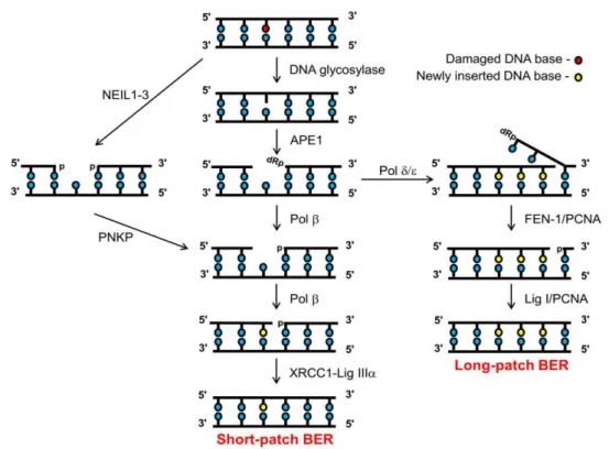
DNA repair pathways are employed depending on the type of DNA damage that needs to be repaired. Base excision repair (BER) is a pathway that repairs simple lesions such as alkylation or oxidation products caused by ROS. There are two major BER sub-pathways in mammalian cells, the long patch BER (LP-BER) and the short patch BER (SP-BER) (figure 4). In the SP-BER a DNA glycosylase initiates the process by removing the damaged base. AP endonuclease-1 (APE1) cuts the phosphodiester backbone and a dRp (DNA single strand break flanked by 3’-hydroxyl and a 5’-deoxyribosephosphate) is formed. Pol β cleaves the 5’-dRP moiety and at the same time adds a single correct nucleotide into the one-nucleotide gap. Lastly, the DNA single strand break (DNA SSB) ends are sealed by the XRCC1-Lig IIIα complex. LP-BER occurs when the 5’-dRP moiety is resistant to cleavage by Pol β. A polymerase switch occurs and Pol δ/ε is recruited adding into the repair gap 2 to 8 correct nucleotides. Pol δ/ε activity creates a 5’-flap structure that is excised by FEN-1 and afterwards the remaining DNA SSB ends are then sealed by Lig I (28,29).
6
Figure 4 - Base excision repair pathways. Adapted from (29).
1.2.3. Heteroplasmy and threshold effect
Within a single cell or tissue all mtDNA copies could be identical in sequence (homoplasmy) or wild-type and mutant mtDNA could exist in different ratios (heteroplasmy). The ratio of wild-type to mutant mtDNA determines the onset of clinical symptoms and a minimum proportion of mutated mtDNA is necessary before biochemical defects and tissue dysfunction become apparent. This is called threshold effect and varies for each mutation and differs amongst tissues, being lower in tissues highly dependent on OXPHOS metabolism such as brain, heart and skeletal muscle. Usually, clinical manifestations occur when the threshold value is between 60% to 90% of mutant to wild-type mtDNA (1,6,30,31) The study of threshold effect in diseases was possible due to the creation of cybrids, cells lacking mtDNA where were introduced mitochondria from cells obtained from patients. Cybrids containing different proportions of mutated mtDNA from 0 to 100% permitted to study the effects of a certain mutant load on the activity of respiratory chain complexes, mitochondrial respiration and cell growth (32).
A powerful tool to treat mitochondrial diseases would be the ability to manipulate mtDNA heteroplasmy and various studies were already made in this field (figure 5). Dai and co-workers studied the effect of rapamycin, an anti-cancer drug, in enhancing mitophagy to select mostly dysfunctional mitochondria with higher levels of mutations, which resulted in decreasing mutation levels over time in a human cybrib expressing a heteroplasmic mtDNA G11778A mutation (33). Also Tanaka and co-workers studied the removal of pathogenic mtDNA by
7
targeting restriction endonucleases to mitochondria of heteroplasmic cells, eliminating with success a mutant mtDNA with the Mt8993T>G mutation from cultured cybrids (34). Minczuk and co-workers demonstrated that it is possible to target and alter mtDNA in a sequence-specific manner by using zinc finger technology (35). Moreover, the use of peptide nucleic acid (PNA) oligomers that could bind selectively to complementary mtDNA inhibiting replication and translation was studied by Muratovska and co-workers (36).
Other methodologies like using endurance and resistance exercises shown to be safe and effective in inducing increase of wild-type mtDNA levels in muscles (37).
Figure 5 - Manipulation of mtDNA heteroplasmy. Heteroplasmic mitochondria containing both wild type (brown) and pathogenic (black) mtDNA are presented in the figure. Strategies to manipulate mtDNA heteroplasmy: A) Endurance or resistance training; B) Targeting restriction endonucleases to mitochondria; C) peptide nucleic acid (PNA) oligomers; D) Zinc-finger binding proteins (ZFB) that could methylate a specific mtDNA mutation. Adapted from (38).
1.2.4. Inheritance of mtDNA
Although nuclear genes display Mendelian segregation, mtDNA is inherited in a non-Mendelian manner. MtDNA is inherited from the mitochondria in the oocyte and this inheritance is generally referred to as “maternal inheritance of mtDNA” (39). MtDNA from sperm is removed by ubiquitination during mammalian zygote formation. Up till now only a single case of paternal transmission in humans has been recorded (21).
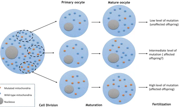
MtDNA mutation is believed to occur either in the female germ line or early in embryonic development but, unexpectedly, only a few mutations are transmitted to each successive generation (40). The mitochondrial genetic bottleneck theory states that, before
8
oogenesis, a size decrease occurs in the population of maternal mtDNA molecules, causing a genetic bottleneck where randomly different mtDNA are segregated. The random segregation of wild type and mutant mtDNAs creates different levels of heteroplasmy (figure 6) (41).
Figure 6 - Mitochondrial genetic bottleneck theory. Mitochondria are randomly segregated through cell division to the primary oocytes. Then, depending on the amount of mutated mithocondria acquired, mature oocytes could have different levels of mutation. Adapted from (69).
1.3. Role of mitochondria in the living cell
1.3.1. Electron transfer system and ATP synthesis
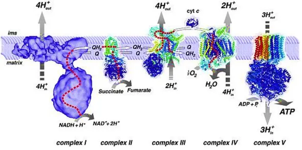
By a process of oxidative phosphorylation (OXPHOS) mitochondria converts biochemical energy stored in food, through the oxidation of nutrients to produce adenosine-5′-triphosphate (ATP) (5). OXPHOS is a result of the mitochondrial respiratory chain that has four multisubunit polypeptide complexes located in the IMM (Figure 7). This polypeptide complexes are NADH-Q oxidoreductase, succinate-Q reductase, Q-cytochrome c oxidoreductase, and cytochrome c oxidase also designated by Complex I, II, III, and IV, respectively.
Electrons are transferred from NADH to O2 in chain by the three large protein complexes
(I, III and IV). The electron flow within these transmembrane subunits leads to the transport of protons across the IMM. Ubiquinone (Q) carries electrons from NADH-Q oxidoreductase to Q-cytochrome c oxidoreductase and electrons from FADH2 (generated in the citric acid cycle) to
Q-cytochrome c oxidoreductase (generated through succinate-Q reductase). Cytochrome c shuttles electrons from Q-cytochrome c oxidoreductase to cytochrome c oxidase.
9
The electrochemical gradient generated across the IMM is then used by ATP synthase to catalyze the conversion of adenosine diphosphate (ADP) and inorganic phosphate (Pi) to ATP(14)(42).
Figure 7 - Mitochondrial respiratory chain. Adapted from (14).
1.3.2. Production of Reactive oxygen species (ROS)
Redox-dependent processes influence many cellular functions, as differentiation, proliferation, and apoptosis. Mitochondria has a great role in this processes, because generates ROS and respond to ROS-mediated changes in the cellular redox state (43).
ROS generation occurs within complex I and III of mitochondrial respiratory chain. When oxygen is reduced by one electron (e-), superoxide anion (O2•−) is formed. Superoxide is a
short-lived free radical that reacts rapidly with a wide range of chemical substrates, including itself. Superoxide engages spontaneously in dismutation, catalyzed by superoxide dismutase. Dismutation of O2•− produces hydrogen peroxide (H
2O2), posteriorly reduced to water or to
hydroxyl radical (OH•). OH• formation is catalysed by reduced transition metals, which in turn
may be re-reduced by O2•− propagating this process. Also, O2•− may react with other radicals
like nitric oxide (NO•) which product is peroxynitrite (ONOO−), a very powerful oxidant. The
oxidants derived from NO• are called reactive nitrogen species (RNS) (44,45).
Excess of ROS production causes damage in key components of cells like lipids, proteins and nucleic acids, the former including production of thymine dimers (figure 8). Due to the mutagenic nature of many of the ROS-induced lesions, mitochondrial free radicals are thought to be an important source of mtDNA mutations and DNA instability (3,28,45).
10
Figure 8 - Effects of reactive oxygen species.
1.3.3. Ca
2+homeostasis
Mitochondria’s capacity to accumulate Ca2+ was described in the 1960’s (46,47).
Calcium is an important regulator of mitochondrial function (figure 9) and stimulates ATP synthesis. Dysregulation of mitochondrial Ca2+ homeostasis is recognized to play a key role in
several pathologies (46,48,49). Matrix Ca2+ is a positive allosteric regulator of dehydrogenases
in the tricarboxylic acid (TCA) cycle and influences the activity of the electron transport chain complexes. When the mitochondrial Ca2+ concentration rises, ATP production increases
however, an overload of Ca2+ can induce the opening of the mitochondrial permeability
transition pore (mPTP) in the inner mitochondrial membrane (IMM). This phenomenon changes the mitochondrial membrane potential and caspase cofactors are released into the cytoplasm triggering an apoptotic cascade (46,50).
Figure 9 - Mitochondrial Ca2+ channels/transporters. The role of Ca2+ in mitochondrial function is also
11
1.3.4. Programmed Cell Death (PCD)
Apoptosis is essential in tissue homeostasis. It has two main pathways, the extrinsic and the intrinsic apoptosis (figure 10) (52).
The extrinsic pathway involves signaling through cell surface receptors of the tumor necrosis factor (TNF) receptor family. These receptors have an intracellular protein domain called death receptor (DR). The ligand-induced activation of DR causes the formation of the death-inducing signaling complex (DISC) by recruiting procaspase-8. Procaspase-8 is cleaved and the mature protease (caspase 8) activates caspase-3 leading to cell death (53-56).
The Intrinsic pathway is the mitochondrial pathway of apoptosis. It is activated as a response to stress conditions, like DNA damage or oxidative stress, resulting in mitochondrial outer membrane permeabilization (MOMP) (54). Pro-apoptotic Bcl-2 family regulate MOMP by forming external membrane pores (57) leading to the release of mitochondrial intermembrane proteins as cytochrome c and second mitochondria-derived activator of caspases (Smac) into the cytosol (58). Cytochrome c forms a protein complex composed of cytochrome c, Apaf-1 and caspase-9, activating caspase-9 that subsequently activates caspase-3 leading to apoptosis (56).
Figure 10 - Molecular pathways that lead to apoptosis. Extrinsic pathway: A lethal ligand triggers DR leading consequently to caspase 8 activation by DISC complex. Caspase 8 stimulates caspase 3 leading to apoptosis and Bid leading to MOMP. Intrinsic pathway: Intracellular stress induces MOMP, regulated by Bcl-2 family. MOMP causes the externalization of Cytochrome c forming a protein complex (Apoptosome) with Apaf-1 and procaspase 9. Therefore caspase 9 is activated leading to caspase 3 activation and apoptosis. AIF (apoptosis-inducing factor), Smac and Omi are also released from mitochondria. Adapted from (54).
12
1.3.5. Mitochondrial dynamics: Fission and Fusion
Mitochondria’s shape is very dynamic. Under respiratory conditions mitochondria undergo frequent cycles of fusion and fission to allow spreading of metabolites and macromolecules throughout the entire compartment (59).
In mitochondrial fusion the four lipid bilayers are merged together. It requires two isoforms of mitofusin1 (Mfn1) and mitofusin2 (Mfn2), which are both anchored to the OMM. They have two transmembrane domains, a cytosolic N-terminal GTPase domain and two cytosolic hydrophobic coiled-coil domains. The coiled-coil domains of Mfn1 and Mfn2 help in binding adjacent mitochondria in both homo-oligomeric and hetero-oligomeric manner. Fusion-deficient cells have greatly diminished respiratory capacity and reduced cell growth.
Mitochondrial fission occurs by interaction of two proteins: dynamin-1-like protein (Dnm1l) and fission protein 1 (Fis1). With the help of Fis1, Dnm1l (localized in the cytosol) is recruited to the constriction sites of the membrane. Once inside, Dnm1l oligomerizes into a ring around the mitochondria. The self-assembly of DLP1 stimulates the final step of fission (60,61).
Cycles of fusion and fission (Figure 11) occur for mitochondria to adapt to the energetic requirements of the cell. The fusion is preferred when optimal mitochondrial function is needed, and the fission is required for elimination of damaged and inactive organelles by autophagy (59).
13
2. Mitochondrial diseases
Mitochondria are associated with many human inherited disorders and diseases, such as neurodegenerative disorders, cardiomyopathies, metabolic syndrome, cancer, and obesity. Mitochondrial diseases can affect any organ system, manifest at any age, and can be inherited from an autosome, an X chromosome, or maternally. Reports indicate that 1 in 8000 individuals carry an mtDNA genetic disorder or are affected by a pathogenic mtDNA mutation and currently there is no cure for mitochondrial disorders and the existing treatments are directed to the relief of the symptoms (3,4).
The principal function of mitochondria is ATP production through OXPHOS and apart from energy conversion, mitochondria take part in a number of other processes, as previously mentioned. Mitochondrial diseases occur mostly when one or more of the OXPHOS complexes are dysfunctional, where this dysfunction together with other factors promotes a vicious cycle (figure 12) (62).
Figure 12 - Mitochondrial etiology of complex disease. Adapted from (63).
2.1. Cytopathies and mutations
Mitochondrial pathologies (figure 13) present a heterogeneous group of multisystem disorders characterized by abnormalities of the mitochondrial ultrastructure as well as of
14
oxidative phosphorylation functioning. Defects in OXPHOS affect any tissue leading to the concept of mitochondrial cytopathies, which preferentially affect the muscle and nervous systems. They are caused either by mutations in the maternally inherited mitochondrial genome or by nuclear DNA mutations (4,64,65)
Mitochondrial genome is particularly vulnerable to mutations because of its proximity to ROS generation sites and it is not protected by histones or membranes (66). Currently, more than 250 pathogenic mtDNA mutations have been described (67) and the phenotype of the disease depends on the mtDNA heteroplasmy and on its threshold percentage on the cells (60 – 90% mutated mtDNAs) (1,6).
Figure 13 - MtDNA mutations and corresponding diseases. Adapted from (31).
2.2. Aging
The involvement of mtDNA in the aging process is mostly because of its proximity to the mitochondrial respiratory chain, source of ROS generation. ROS create an environment in which spontaneous mtDNA mutations are more possible to occur (28,68).This results in an exponential rise in oxidative damage, where cellular and tissue functions are loss with a combination of energy insufficiency, signaling defects, apoptosis, and replicative senescence. An increased number of COX‐deficient cells and higher levels of mutated mtDNA is observed within the process (69).
15
2.3. Neurodegenerative diseases: Parkinson (PD)
Neurodegenerative diseases are characterized by the progressive loss of neurons and result in memory loss, movement problems, cognitive deficits, emotional alterations and behavioral problems. Environmental factors, genetic mutations and old age, are the major risk factors. Disease-linked mutations in genes provide key clues to the molecular mechanisms involved in the pathology and have been studied intensively over the last decades (61,70).
Parkinson (PD) is a neurodegenerative disease associated with progressive resting tremor, rigidity, bradykinesia, gait disturbance, postural instability and dementia where degeneration of dopamine neurons in the substantia nigra occurs (61,71). It is one of the most-common neurological disorders, affecting about 1-2% of the population over age of 60 years (72). PD movement symptoms are caused by the loss of inhibitory dopamine neurons which causes an increase in the activity of the subthalamic nucleus and the globus pallidus. The formation of Lewy bodies is a second neuropathological trait of PD where these cellular inclusions contain a dense core of filamentous material surrounded by a halo of fibrils comprising mainly α-synuclein (61). The accumulation of α-synuclein contributes to mitochondrial fragmentation and also damages the function of Complex I (CI) (73). In PD patients, deficits in CI activity were detected in the striatum, cortical brain tissue, fibroblasts, blood platelets with a slight variability in skeletal muscle and lymphocytes (74). A few genes associated with Mendelian forms of PD such as autosomal dominant (SNCA, LRRK2) or recessive (PARK2/Parkin, PINK1, DJ-1, ATP13A2) inheritance have been discovered throughout the past fifteen years (75).
The idea that mitochondrial dysfunction could influence PD disease emerged with the discover of the mechanism of action of MPTP in the early 1980s where a Parkinson’s-like syndrome was caused by MPP+ blockage of electron flow in CI (44,76). The reduced CI activity
was also observed in cybrids with mtDNA from PD patients, demonstrating that mtDNA mutations may have influence in a few patients with PD. The cybrids gained features from the PD patients which showed reduced mitochondrial membrane potential, mitochondrial respiration, impaired mitochondrial biogenesis and abnormal Ca2+ handling (70).
Genetic evidences suggest that, besides the mitochondrial CI defect, mutations in mtDNA and mitochondrial dysfunction, including defects in bioenergetics, mitochondrial fusion/fission, mitochondrial movement, and transcription play a role in the pathogenesis of PD (70). Furthermore, diverse CI mutations have been associated with PD (77). Two different mutations in mtND1 gene were identified (table 2) making the development of an mtND1 construct a very interesting approach for gene therapy purposes in PD treatment.
16
Table 2 - Confirmed mtDNA variations associated with PD disease. Adapted from (78).
Variation mtDNA gene
A3397G ND1 T4216C ND1 T4336C tRNA(Gln) A4336G tRNA(Gln) A4917G ND2 G5460A ND2 G9055A ATP6 G10398A ND3 A11084G ND4 G13708A ND5 G15928A tRNA(Thr) G15950A tRNA(Thr) T15965C tRNA(Pro)
2.4. Diagnosis of mtDNA disease
The diagnosis is very complex when it comes to mitochondrial diseases. It is difficult due to the mtDNA heteroplasmy and lack of a clear genotype, phenotype correlations and the complex interactions between the nuclear and mitochondrial genome. Laboratory diagnosis of mtDNA disease often involves to gather many types of information from clinical to histochemical, biochemical and genetic allowing the development of rational diagnostic algorithms (figure 14) (31).
17 Figure 14 - Algorithm for investigation of mitochondrial disease. Adapted from (69).
No specific pharmaceutical drugs show to treat mitochondrial disease effectively in large-scale clinical trials (69). In this sense, both new therapies and approaches need to be developed in order to evolve in the treatment of patients suffering from mitochondrial disorders. Gene therapy for treatment of these diseases offers an interesting alternative to the therapeutic strategies that are currently applied (4).
3. Gene Therapy: Mitochondrial gene therapy (MGT)
The growing interest in gene therapy has become a strong incentive to develop gene vectors with high transfection efficiency and minimized toxicity. Although viral vectors demonstrated more effectiveness, disavantages like toxicity and production difficulties limited their applications. Consequently non-viral vectors are prefered because of their lower immune response and higher safety although transfection efficiency is reduced (80).
Genetic manipulation of mtDNA and its gene products is an important subject for investigation regarding the mtDNA disorders treatment. One important aim should be the correction of OXPHOS’s defects as it is a common feature among mtDNA disorders. Research in gene therapy for mtDNA disorders treatment is an emerging field known as mitochondrial gene therapy (MGT). Currently there are no recombinant human mtDNA constructs available for MGT (4) however successful cloning attempts of the human mtDNA where made in S.cerevisiae (1).
Importation of wild-type copies or relevant sections of DNA or RNA into mitochondria, manipulation of mitochondrial genetic content or rescue of a defect by expressing an
18
engineered product from the nucleus are different approaches that could be taken in MGT (81). For this to occur, biological barriers should be surpassed by using different strategies (figure 15).
To be successful MGT has to follow three key conditions. First, the delivery of nucleic acids has to be made in the correct compartment of mitochondria. Second, when applied to living cells, a beneficial effect on mitochondrial function should be obtained. Third, the modulation of mitochondrial function through MGT should occur in vivo and have a significant beneficial effect of the progress of the disease or disability (7).
Figure 15 - Delivery of therapeutic agents into Mitochondria. The delivery only is achieved when biological barriers are surpassed and the therapeutic agent reaches mitochondria. Several strategies can help to overcome biological barriers avoiding RES (reticuloendothetial system) clearance and enabling target site accumulation. When reaching the cell, mitochondrial localization can be achieved by exploiting the negative membrane potential of mitochondria and targeting mitochondrial receptors with peptides and substrates, or using pro-drugs and characterized mitochondriotropics. Adapted from (82).
3.1. Construction/amplification of a recombinant human
mtDNA construct for MGT
MGT is an emerging field for mitochondrial diseases treatment. When one or more specific genes are identified as the major responsible for a disease they can be used in gene therapy for the treatment of that disease. In this work mtDNA was extracted from human blood and an OXPHOS complex I gene, mtND1, was amplified by polymerase chain reaction (PCR) with specific primers. The plasmid selected was pCAG-GFP that produces the green fluorescent protein when present in the cytoplasm of cells, thus allowing to conclude about the construct presence by confocal microscopy.
19
The enzymatic digestion with the same enzymes in both mtND1 and in pCAG-GFP allowed the desired construct to be obtained by a binding reaction. The amplification of the construct was made by transforming competent E.coli cells. When purified, the construct can be used for the formulation of nanoparticles with specific mitochondriotropic moieties that direct the nanoparticle to mitochondria.
3.2. Nanotechnology
Nanobiotechnology applies principles and techniques at a nanoscale level to comprehend and change biosystems by using biological principles and materials to create new devices and systems (83). In the last decade the increase of therapeutic approaches targeting mitochondria, such as MGT, has resulted in the necessity to create novel delivery systems that have a selective delivery of nucleic acids into mitochondria. Liposomes, DQAsomes, polymeric micelles and lipid based nanoparticles (figure 16) associated with diverse mitochondriotropic moieties as rhodamine and triphenylphosphine (TPP), are examples of new non-viral delivery systems that arouse to fulfill this necessity (8).
Liposomes (LP) are generally biocompatible and non-toxic thus having a great potential for gene delivery. Its surface can be easily modified with mitochondriotropic ligands to target mitochondria and deliver the therapeutic agent. Examples of liposomal systems for delivery to mitochondria include octaarginine (R8) - modified liposomes, cell-targeting liposomes equipped with pH-dependent fusogenic peptides such as GALA and incorporation of (TPP) to the liposomal bilayer (8,28)
DQAsomes are liposome-like cationic vesicles derived from the amphiphile dequalinium. These mitochondriotropic vesicles are capable of condensing pDNA and form DQAsome-pDNA complexes protecting the pDNA from nucleases and localizing mitochondria in living cells (12,84,85).
MITO-Porters are liposome-based nanocarriers that deliver the desired nucleic acids to mitochondria via a membrane-fusion mechanism. This device includes ligands for specific receptors, pH-sensitive fusogenic peptides (DOPE) for endosomal escape and mitochondriotropic residues for enhanced mitochondrial delivery (8,86).
20
Figure 16 - Nanocarriers with potential for mitochondrial targeting. Adapted from (8).
The development of these new non-viral nanocarriers with affinity towards mitochondria has a great importance for mitochondrial disease treatment and approaches by MGT. Thus the main goal of this work was the construction of a novel non-viral nanocarrier with an mtDNA construct, obtained in our laboratory, with mitochondria affinity whose details are described in the next topic.
3.2.1. CaCO
3Nanoparticles
Calcium carbonate (CaCO3) is produced by many organisms forming unique structures
with a variety of functions in their tissues. CaCO3/DNAnanoparticles are produced by
co-precipitation of Ca2+ with DNA in the presence of CO32−. This is an attractive method since it is
a very a simple technique, promotes gene delivery with protection of the encapsulated DNA from degradation and the nanoparticles can be internalized into different histological cell types. However, CaCO3/DNA precipitates are uncontrollable, resulting in large size
nanoparticles which causes negative effects on cellular internalization and gene transfection (11,12,80).
CaCO3 based nanoparticles have many advantages such as biocompatibility,
biodegradability, loading ability of different therapeutic agents, easy and cheap production and pH dependent dissolution (9-11). To improve the stability and the transfection efficiency of CaCO3 nanoparticles, natural compounds such as alginate and cellulose were used in the
formulation of the nanoparticles. By changing the formulation, disadvantages like the uncontrollable growth of the nanoparticles could be slowed thus enhancing the stability of the
21
nanoparticles (11). To create CaCO3/DNA nanoparticles with affinity towards mitochondria
Rhodamine 123, a fluorescent amphiphile with mitochondria affinity was used (12,13). In this work CaCO3/pCAG-GFP-mtND1nanoparticles were developed. The
pCAG-GFP-mtND1 construct was successfully developed and produced in our laboratory for the development of the nanoparticles. The purpose of creating nanoparticles with this construct is the possible development of a therapeutic approach for PD disease, with efficient delivery of the construct to mitochondria. Also, a new polymer and its influence in the nanoparticles features was studied. This new polymer was gelatin, a natural polymer derived from collagen, with great advantages, such as, biocompatibility and biodegradability being suitable to be used in MGT (87).
22
Aims of the Project
Mitochondrial genome is considered very susceptible to mutations due to the lack of protection and repair mechanisms. Mutations in this genome are a frequent cause of mitochondrial cytopathies and conventional treatments, using a combination of different drugs, are in most cases, inefficient. The development of gene therapy to treat mtDNA disorders offers a promising approach; it is a practical and potentially effective tool to treat the vast array of clinical phenotypes as it focuses in the actual cause of the disorder. For mitochondrial gene therapy to be feasible in a clinical setting, the formulation of a carrier with mitochondrial targeting ability is imperative. In line with this, the aim of this project is the design and development of a mitochondrial gene based plasmid DNA vehicle suitable for efficient and targeted mitochondrial delivery. To accomplish this goal, the project was divided in three parts. The first involves the construction of a vector using an OXPHOS complex I gene, mtND1 and the pCAG-GFP plasmid. The second aim of this project was focused in the study of different hosts transformed with the pCAG-GFP-mtND1 vector to produce the plasmid. The third and last part, consisted in the formulation of a suitable nanocarrier for efficient and sustained pDNA delivery into mitochondria.
The success of this work is based in the development of a mtND1 gene based plasmid DNA loaded nanomaterial for mitochondrial gene therapy focused in complex I deficiency cytophaties.
23
Materials and Methods
1. Materials
1.1. Reagents
LB broth was obtained from Liofilchem (Roseto degli Abruzzi, Italy) and LB-Agar was obtained from Pronadisa/Conda (Madrid, Sapin). Yeast Extract was obtained from Himedia (Mumbai, India) and Tryptone was obtained from Biokar (France). Glycerol and CaCl2 were
obtained from VWR International (Radnor, PA, USA). KH2PO4 was obtained from Chem-Lab
(Zedelgem, Belgium) and Na2CO3 and K2HPO4 were obtained from Panreac (Barcelona, Spain).
Agarose and Green Safe were obtained from NZYTech (Lisboa, Portugal). Rhodamine 123 and Gelatin was from Sigma-Aldrich (St. Louis, MO, USA). Cellulose powder was obtained from Aldrich Chemical Company (Milwaukee, WI, USA). The plasmid pCAG-GFP was obtained from Addgene (Cambridge, MA, USA).
1.2. Hosts
In this work, different hosts were considered. E. coli XL1B, E. coli DH5α, E. coli JM109,
P. Pastoris X33 and S.cerevisiae NRRL Y-12632 . As the pCGA-GFP gives ampicillin resistance, P.Pastoris X33 and S.cerevisiae were excluded from the study because of its resistance to
ampicillin.
2. Methods
2.1. Vector construction
2.1.1. ND1 gene amplification
In its simplest form, polymerase chain reaction (PCR) based cloning is used to amplify a specific DNA sequence but at the same time it can be used to add restriction sites to the ends of that sequence, so that it can be easily cloned into a plasmid of interest.
Human mitochondrial DNA was extracted from blood samples and the mtND1 gene was amplified by PCR using the extracted mtDNA as template and the following primers: FW: GCAGAGCCCGGTAATCGCATA and RV: GGATTCTCAGGGATGGGTTC. The reagents used for the reaction were 1 U DreamTaq™ Green DNA polymerase (Fermentas, MA, USA), 2 mM MgCl2, 0.2
24
Table 3 - Thermal cycling program for mtND1 PCR amplification.
Step Temperature Time
1 95ºC 5 min
2 95ºC 30sec
3 60ºC 30sec
4 72ºC 40sec
Repeat 35 times steps 2,3 e 4
5 72ºC 8 min
6 12ºC 6min
The primers selected for mtND1 gene PCR created the desired enzyme cutting sites for enzyme digestion with SmaI and XbaI thus preventing the enzymes to cut any part of the gene.
2.1.2. Binding Reaction
For cloning mtND1 gene in pCAG-GFP (both previously digested with SmaI and XbaI) several ratios insert/vector were prepared. The key in these cloning reactions is to vary the insert concentration and maintain the vector concentration. To perform the binding reaction the following formula was used,
(ng vector x kb insert) / kb vector = ng insert (1)
The ratios prepared were 1:1, 3:1, 5:1 and 8:1 using a DNA Ligation Kit (Takara Bio Inc., Otsu, Japan). The mixture consists in 36.7 ng of pDNA with 5 µL of Ligation Mix, ddH20 and a
variable amount of insert (depending on the ratio used) to a total volume of 10 µL. The ligation reaction mixture was incubated at room temperature for about 3h. Afterwards, the preparation of the desired construct was confirmed by sequencing,
2.2. Enzymatic digestion
The purified pCAG-GFP-mtND1 from several colonies was submitted to enzymatic digestion to verify if these colonies had the insert mtND1.
Different concentrations of pDNA (obtained from different selected colonies) were mixed with 2 μL of CutSmart® Buffer, 0.5 μL of SmaI (New Englands Biolabs, Ipswich, MA, USA), 0.5 μL of XbaI (New Englands Biolabs, Ipswich, MA, USA) and ddH2O to a final volume of 10 μL.
25
The conditions for enzyme digestion used are displayed in table 4and were conducted in a T100™ Thermal Cycler (Bio-Rad, CA, USA):
Table 4 - Conditions for enzyme digestion.
Enzyme Temperature Time Cut Type Cutting sites
SmaI 30ºC 1h Blunt ends 5' CCC GGG 3' 3' GGG CCC 5'
XbaI 37 ºC 1h "sticky" ends 5' T CTAGA 3' 3' AGATC T 5'
An agarose gel was done to verify if the digestion was correctly performed.
2.3. Sequencing
The pCAG-GFP-ND1 plasmid was obtained from fermentation of a selected colony and was sequenced by adapting the Beckman Coulter’s GenomeLab™ Dye Terminator Cycle Sequencing protocol.
For preparation of the DNA sequencing reaction, about 50 fmol of purified pCAG-GFP were mixed with 0.3 μL of 0.25 μM primer (RV: EGFP-N CGTCGCCGTCCAGCTCGACCAG), 4 μL of DTCS Quick Start Master Mix and ddH2O to a final volume of 10 μL. The thermal cycling program used is displayed in table 5 and conducted in a T100™ Thermal Cycler (Bio-Rad, CA, USA):
Table 5 - Thermal cycling program for DNA sequencing reaction.
Step Temperature Time
1 98ºC 5 min
2 96ºC 20 sec
3 50ºC 20 sec
4 60ºC 4min
Repeat 30 times steps 2,3 e 4
5 60ºC 10 min
6 4ºC 6 min
After the thermal cycling, ethanol precipitation of the DNA sequencing reaction was promoted. To each reaction tube, 3 μL of Stop Solution/Glycogen mixture (1.2 μL of 3 M Sodium Acetate, pH 5.3, 1.2 μL of 100 mM Na2-EDTA, pH 8.0, and 0.6 μL of 20 mg/mL of glycogen) and 60 μL of chilled 95% (v/v) ethanol were added. This mixture was centrifugated at 3,000 rpm for 30 min. The obtained pellet was washed two times with chilled 70% (v/v) ethanol, with a centrifugation at 3,000 rpm for 5 min between each step. When dried, the pellet was
26
resuspended in 10 μL of Sample Loading Solution. The samples were transferred to the appropriate wells of the sample plate. Each of the samples were covered with a drop of light mineral oil. The samples for plasmid sequencing were loaded into the GenomeLab™ GeXP Genetic Analysis System (Beckman Coulter, California, USA). The primers used were RV: EGFP-N CGTCGCCGTCCAGCTCGACCAG for pCAG-GFP-mtEGFP-ND1 and FW: GCAGAGCCCGGTAATCGCATA and RV: GGATTCTCAGGGATGGGTTC for mtND1 gene. The primers used for the ligation product allowed to verify the existence of an insert in pCAG-GFP. MtND1 was sequenced to verify the successful cloning of the gene of interest in the plasmid.
2.4. Cell transformation
Transformation occurs when a cell captures foreign DNA from its surroundings. It can occur in nature in certain types of bacteria but when competent bacterial cells are used, they can be artificially transformed by a procedure called by heat shock method.
The ligation reaction mixture was used for transformation of E.coli DH5α competent cells by adding 10 µL of the ligation mixture to 100 µL of cells. After a short incubation in ice, the mixture was placed at 42°C for 45 seconds and then placed back in ice. LB medium was added and the transformed cells were incubated at 37 ºC for 2h in an orbital shaker at a shaking frequency of 250 rpm. Afterwards, the transformed cells were plated in LB agar plates (15g/L Agar, 10g/L Tryptone,5 g/L Yeast Extract, 5 g/L NaCl) complemented with 100 µg/mL ampicillin and 50 µg/mL neomycin and grew overnight at 37 ºC. After choosing the best insert/vector ratio (as indicated in section 2.1.2) E.coli JM109 and XL1B strains were also transformed with the vector pCAG-GFP-mtND1.
2.5. Cell Banks
Transformed strains were selected and grew overnight at 37 ºC in 25 mL LB medium (10g/L Tryptone,10 g/L NaCl, 5 g/L Yeast Extract; Sigma-Aldrich) complemented with 100 µg/mL ampicillin and 50 µg/mL neomycin in an orbital shaker at a shaking frequency of 250 rpm. When 0.7 of optical density (OD) at 600 nm was achieved, bank cells were made by adding 30% of glycerol, previously autoclaved, and stored at -80⁰C for 3 to 4 days to stabilize.
2.6. Plasmid DNA production studies
These studies were carried out to verify which of the strains produced the pCAG-GFP-mtND1 with a better yield. The pCAG-pCAG-GFP-mtND1 plasmid was produced in E. coli JM109, XL1B and DH5α strains in 125 mL of Terrific Broth (TB) medium (20 g/L tryptone, 24 g/L yeast extract, 4 mL/L glycerol, 0.017 M KH2PO4, 0.072 M K2HPO4) in 500 mL erlenmeyer complemented with 100
27
of 250 rpm. The fermentations were carried out for 9.5 hours and samples of the culture media with an OD of 0.4 were taken every one and half hour. The samples were centrifuged at 13,000 rpm for 10 minutes in a Mikro 20 centrifuge (Hettich Centrifuges, UK), the supernatant was removed and cells were stored at -20 ºC forfollowing purification. Three independent studies were made for each one of strains for triplicates.
2.7. Growth of pCAG-mtND1 for nanoparticle synthesis
The pCAG-mtND1 plasmid was produced in E. coli JM109 in 250 mL of Terrific Broth (TB) Medium (20 g/L tryptone, 24 g/L yeast extract, 4 mL/L glycerol, 0.017 M KH2PO4, 0.072 M K2HPO4) in 1000 mL erlenmeyer complemented with 100 µg/mL ampicillin and 50 µg/mL neomycin at 37 ºC in an orbital shaker at a shaking frequency of 250 rpm. After approximately 7 hours of fermentation, the cells were harvested by centrifugation for 10 min at 4 ºC and 4500 rpm in an Allegra 25R centrifuge (Beckman Coulter, CA, USA).
2.8. Plasmid Purification
2.8.1. Purification of the samples for yield studies
The purification was carried out using the GeneJet Plasmid Miniprep Kit (Thermo Scientific, Waltham, MA USA). This kit is designed for rapid and cost-effective small-scale preparation of high quality plasmid DNA from recombinant E.coli cultures.
Samples obtained from the culture media (described in section 2.6) were ressuspended and the cells were subjected to a SDS/alkaline lysis. The lysate was neutralised to create the conditions for posterior binding of pDNA on the silica membrane of the spin column. A centrifugation was made to obtain the cell debris and SDS in the pellet and the pDNA on the supernatant. The supernatant was loaded in the spin column membrane and, after elution, the membrane was washed a few times to remove contaminants. Finally the adsorbed pDNA was eluted with the elution buffer. The plasmid yield was determined by Uv-Vis analysis at 260 nm in a NanophotometerTM (Implen, Munich, Germany) and its integrity was confirmed by agarose gel electrophoresis. The purified pDNA was stored at -20ºC.
2.8.2. Purification of pCAG-GFP-mtND1 for nanoparticles
production
NZYtech maxiprep (Nzytech, Lisboa, Portugal) kit is designed for the rapid, large-scale preparation of highly pure plasmid DNA from recombinant E.coli strains.
28
Previously harvested cells (described in 2.7 section) were ressuspended and a SDS/alkaline lysis was promoted. A neutralization solution was added and the sample was centrifuged for 30 min at 20.000 x g at 4ºC. The supernatant was transfered to a new tube and a 15 min centrifugation was made. The lysate was applied to the NZYTech Maxi Colunm and the elution occurred by gravity flow. The column was washed to remove contaminants and finally the pDNA was eluted. Precipitation of the eluted pDNA was promoted by adding 0.7 volumes of room-temperature isopropanol. A 20 min centrifugation at 15.000 x g was made and after, the pDNA was ressuspended in a pH 7.5 TE buffer. The plasmid yield was determined by spectrophotometry at 260 nm and its integrity was confirmed by agarose gel electrophoresis. 500 µL aliquots of pDNA at 100 µg/mL were prepared and stored at -80ºC.
2.9. Agarose gel electrophoresis
Agarose gel electrophoresis was performed to evaluate the size and conformation of the purified pCAG-GFP plasmid, to evaluate the size of mtND1 gene, the ligation product and to verify the enzymatic digestion of the ligation product.
The electrophoresis was carried out using a gel with 1% agarose and 1 μg/mL Green Safe and it was run at 150 V for 30 min in TAE buffer (40 mM Tris base, 20 mM acetic acid and 1 mM EDTA pH 8.0). The gel visualization was made in a UVItec Gel documentation system under UV light (UVItec Limited, Cambridge, United Kingdom).
2.10. Nanoparticles studies
2.10.1. CaCO
3Nanoparticles Synthesis
For the nanoparticles synthesis, two different solutions were prepared: a solution A with 5 μg or 10 μg of pDNA, 120 μL of CaCl2 (0.03 mg/mL) and 7.5 or 15 μL of Rhodamine123
diluted with mili-Q water to a total volume of 290 μL and a solution B with 255 μL NaCO3 (0.0425
mg/ml) and 5 μL of mili-Q water or cellulose (1mg/mL) or gelatin (1mg/mL) to a total volume of 260 μl.
Solution A was added dropwise with a micropipette to solution B to form the nanoparticles by a co-precipitation method. The final solution, called solution C, was centrifuged at 10,000 rpm for 15 min. The pellet contained the nanoparticles.
2.10.2. Particles Morphology
20 µL of tungsten 1% were added to the pellet obtained in the preparation of the nanoparticles by the method described in 2.10.1 and 1:20 and 1:50 dilutions were made. The