Contents lists available atScienceDirect
Environment International
journal homepage:www.elsevier.com/locate/envintIoxynil and diethylstilbestrol disrupt vascular and heart development in
zebra
fish
Yi-Feng Li
a,b,c,d, Adelino V.M. Canário
a,b,c,d, Deborah M. Power
a,b,c,d, Marco A. Campinho
d,⁎aInternational Research Centre for Marine Biosciences, Ministry of Science and Technology, Shanghai Ocean University, Shanghai, China bKey Laboratory of Exploration and Utilization of Aquatic Genetic Resources, Ministry of Education, Shanghai Ocean University, Shanghai, China cNational Demonstration Center for Experimental Fisheries Science Education, Shanghai Ocean University, Shanghai, China
dCentre of Marine Sciences, University of Algarve, Faro, Portugal
A R T I C L E I N F O Handling Editor: Hefa Cheng
A B S T R A C T
Background: Endocrine disruption is one of the consequences of industrialization and chemicals released into the environment have a profound impact on organisms. Waterborne micromolar concentrations of ioxynil (IOX) and diethylstilbestrol (DES) infish affect the development of the heart, vasculature and thyroid gland.
Objectives: The present study aimed to determine how IOX and DES disrupt the crosstalk between the developing thyroid gland and cardio-vascular system in zebrafish.
Methods: Twelve hours post fertilization (hpf) wild type, Tg(fli1:GFP) or Tg(cmalc2:GFPCaaX) zebrafish embryos were exposed to 0.1μM IOX or DES for 36 h (up until 48 hpf) or 60 h (up until 72 hpf). Embryos were used for vascular endothelial cell sorting, whole-mount immunohistochemistry, tissue selective transcriptomics, selected gene expression analysis by quantitative real-time polymerase chain reaction analysis and determination of heart rate by live imaging.
Results: Exposure of zebrafish embryos to IOX and DES (0.1 μM) increased heart beat frequency and reduced ventricle volume and aorta diameter. The transcriptome of endothelial cells from blood vessels of hypertrophic, dilated and arrhythmogenic right ventricular cardiomyopathy was significantly changed and compound-specific toxic effects were found in IOX and DES exposed embryos. Both DES and IOX directly affected vascular and heart development and this indirectly impaired thyroid gland development in zebrafish. Even though the toxicity end-point of the two chemicals was similar, their action seemed to be via different gene regulatory pathways and physiological mechanisms.
Conclusion: IOX and DES directly disrupt cardiovascular development and there is an associated disruption of thyroid tissue that most likely has long term consequences for this endocrine axis.
1. Introduction
Endocrine disrupting chemicals (EDCs) are commonly defined as chemicals that can interfere with or disrupt endocrine systems and have a broad negative spectrum of effects on animals, including humans, and ultimately ecosystems (Schug et al., 2016;White et al., 2017;Patisaul et al., 2018). Diethylstilbestrol (DES) was the first synthetic estrogen prescribed for preventing miscarriages and other complications during pregnancy (Hunt et al., 2016). Like other synthetic estrogens DES in-teracts with the estrogen endocrine pathway (reviewed by (Kiyama and Wada-Kiyama, 2015)) and evidence that DES is an EDC has been ac-cumulating. The evidence indicates that DES is hazardous for maternal health (Tournaire et al., 2016) and is associated with reproductive, cardiac and urogenital anomalies in offspring (Yamamoto et al., 2003;
Titus-Ernstoff et al., 2010). A recent study revealed the incidence of heart defects is increased in children of DES exposed women, including ventricular septal disease, tetralogy of fallout, and atrial septal defects (Titus-Ernstoff et al., 2010). In rats, prenatal exposure to DES increases thyroid function by stimulating the pituitary-thyroid axis (Yamamoto et al., 2003). DES has been banned for human use in the USA since 1971 (Reed and Fenton, 2013), but remains a threat to human health due to its widespread use in livestock production. Concentrations as high as 24.9–102 ng L−1 and 7.2–16.9 μg L−1 are detected in some Chinese
rivers andfisheries waters, respectively (Chen et al., 2009;Qu et al., 2012;Zhang et al., 2012) and in sediments of Mediterranium aquatic environments (Pojana et al., 2007).
Ioxynil (IOX) is an herbicide extensively used in agriculture (Otsuka et al., 2014) and in 2015 the use of IOX in Japan was 107.1 tons (METI,
https://doi.org/10.1016/j.envint.2019.01.009
Received 18 October 2018; Received in revised form 17 December 2018; Accepted 4 January 2019
⁎Corresponding author at: CCMAR, Campus de Gambelas, Universidade do Algarve Ed8 Lab3.32, 8005-139 Faro, Portugal.
E-mail address:[email protected](M.A. Campinho).
Available online 25 January 2019
0160-4120/ © 2019 Elsevier Ltd. This is an open access article under the CC BY-NC-ND license (http://creativecommons.org/licenses/BY-NC-ND/4.0/).
T
2015). IOX may interfere with the human thyroid system by binding to transthyretin (TTR), a thyroid hormone (TH) binding protein (THBP) that transports TH in the blood (Ogilvie and Ramsden, 1988) and one of its toxicological effects is to provoke thyroid tumours in rats (European Commission, 2004). In teleostfish, the effect of IOX and DES on the thyroid system has received more attention (Morgado et al., 2007, 2009;Campinho and Power, 2013). In vitro studies reveal that IOX and DES competitively bind to sea bream (Sparus aurata) transthyretin (TTR) (Morgado et al., 2007). Exposure to 1 mg kg−1of IOX or DES for 21-days did not change TH levels or thyroid follicular morpho-histology in adult sea bream but caused down-regulation of some hypothalamus-pituitary-thyroid (HPT) axis genes, suggesting homeostasis was affected (Morgado et al., 2009). In zebrafish exposed to IOX and DES, the mRNA expression of the essential thyroid gland developmental gene nk2.1a decreased as did that of the thyroglobulin (Tg) gene indicating that thyroid gland development was impaired (Campinho and Power, 2013). Moreover, exposure of zebrafish embryos to IOX and DES significantly changed heart morphology (Campinho and Power, 2013). Strikingly the effect of IOX and DES on thyroid gland development in zebrafish em-bryos resembles that of the cardiac troponin I (tnni1) gene morpholino knock-down (MO). The similar cardiac phenotype led us to hypothesise that the action of IOX and DES on thyroid gland development might be indirect and a consequence of their effect on the development of en-dothelial cells or cardiac function or both.
The heart is the first functional organ to form during vertebrate embryogenesis (Bakkers, 2011) and in zebrafish heart contraction starts approximately 24 hours post-fertilization (hpf) when a regular heart-beat is observed (Chen et al., 1996;Vogel and Weinstein, 2000). The ventral aorta (VA) of the heart is adjacent to the thyroid gland and it plays a role in guiding thyroid gland morphogenesis along the phar-yngeal region (Alt et al., 2006). Crosstalk between the two tissue sys-tems was revealed by the zebrafish cloche mutant (Thompson et al., 1998), which lacks all blood vessels and in which the thyroid gland fails to develop properly (Opitz et al., 2012). The preceding studies suggest that the heart-angiogenic-thyrocyte interaction may be essential for correct development and function of the thyroid gland, HPT-axis and thyroid homeostasis.
The present study aims to uncover the mode of action of IOX and DES on the heart, angiogenic system and thyrocytes during zebrafish embryogenesis. We analysed the function and morphology of the heart and ventral aorta and demonstrate that the abnormal development of these tissues is directly related to impaired thyrocyte development. The genetic basis of the modified vascular system caused by embryonic exposure to IOX and DES was revealed by the transcriptome of 48 hpf zebrafish embryonic endothelial cells. To the best of our knowledge, this study provides thefirst multi-level analysis of cardiac, vascular and thyroid system development in IOX and DES exposed animals and ex-tends our previous observations (Campinho and Power, 2013) by re-vealing these chemicals indirectly disrupt the thyroid in zebrafish. 2. Methods
2.1. Zebrafish maintenance
Adult zebrafish (AB strain) were fed twice a day with dry pellets (Tetramin, Germany) and once a day with live two-day old Red Pepper enriched Artemia. Animals were raised at 28 °C in a recirculating system (Technoplast, Italy) and under a 14 h: 10 h (light: dark) cycle. The embryos were collected and reared at 28.5 °C in an incubator (Sanyo, Japan). All experiments carried out in this work were approved by CCMAR's ethical committee and are in accordance with the regula-tion of Directive 2010/63/EU and Portuguese Naregula-tional legislaregula-tion on experimental animal use.
2.2. Zebrafish Tg(cmlc2:GFPCaaX) transgenic line generation
The proximal promoter (−800–1+) of zebrafish cardiac myosin light chain 2 (cmlc2) was isolated by PCR (zfClmc2PrFw: CGATCCGTGACC AAAGCTTAAATCAGT; zfCmlc2PrRv: CTAGCTAGCGTTCACTGTCTGCT TTGCTGT) using zebrafish genomic DNA as the template and Fusion high-fidelity polymerase (Thermo Scientific). The amplified cmlc2 promoter was isolated from agarose gel using a kit (EZNA) and then cloned into a pCS2+ vector linearized with StuI and the construct confirmed by sequencing. A GFPCaaX sequence isolated from a Tol2kit vector (Kwan et al., 2007) by digestion with the restriction enzymes, EcoRV and EcoRI (Thermo Scientific), was subcloned downstream of the cmlc2 promoter in pCS2+. After sequence confirmation of the appro-priateness of the pCS2 + Zfcmlc2::GFPCaaX construct, it was excised from the pCS2+ vector using the restriction enzymes HindIII and NotI (Thermo Scientific) and subcloned into the pBR322Tol2 vector (Kwan et al., 2007). After confirming the correct integration of the cmlc2::GFPCaaX construct into the pBR322Tol2 vector, plasmid DNA was extracted with phenol/chloroform and diluted in water (Kawakami, 2007). For Tol2 mediated transgenesis, 50 pg of the pur-ified pBR322Tol2-Zfcmlc2::GFPCaaX DNA was co-injected (1 nL) with 25 pg of Tol2 mRNA, prepared with an Ambion mESSENGER RNA kit, into the cytoplasm of one cell-stage zebrafish embryos of the AB strain. Embryos with a mosaic cardiac expression of GFP were selected and grown for crossing with WT zebrafish in order to generate heterozygous transgenic Tg(cmlc2::GFPCaaX) zebrafish lines.
2.3. Chemical exposure
IOX and DES exposure was carried out as described inCampinho and Power (2013). Briefly, 12 hpf zebrafish embryos were incubated in 25 mL of E3 medium (Control) or to 25 mL of E3 medium containing 0.1μM IOX or DES for 36 h (up until 48 hpf) or 60 h (up until 72 hpf) at 28.5 °C in an incubator (Sanyo, Germany). For 36 h treatment assays, the E3 medium was not exchanged. In the 60 h exposure experiments 20% of the bathing E3 medium was substituted in each group with a fresh solution of the appropriate composition at 48 hpf. The chemical exposure experiments were carried out using WT, Tg(fli1:GFP) (Lawson and Weinstein, 2002) or Tg(cmlc2:GFPCaaX) zebrafish embryos.
Zebrafish embryos at 48 hpf (36 h-exposure to IOX or DES) or larvae at 72 hpf (60 h-exposure) werefixed overnight at 4 °C in 4% PFA/1× PBS, pH 7 and used for whole-mount immunohistochemistry (IHC) analysis. For relative gene expression analysis by quantitative reverse transcriptase polymerase chain reaction (qPCR), WT zebrafish embryos or larvae (n = 5 pools each containing 20–25 embryos) were trans-ferred to a 1.5 mL tube after removing excess water and frozen im-mediately in liquid nitrogen and stored at−80 °C until analysis. 2.4. Heart rate determination
Forty-eight hpf (36 h-exposed to IOX or DES) Zebrafish Tg (cmlc2::GFPCaaX) embryos (n = 12–15) were placed under a stereo-scope (Olympus AZX7) coupled to an Hammamatsu ORCAv2 digital camera and video images of the heart collected for 2 min at 28.5 °C. Heart rate was determined by counting heart beat recorded in the video images.
2.5. Fluorescent immunohistochemistry
Embryos from WT and transgenic lines used for control or IOX or DES treatments were hydrated through a methanol/1× PBS series from 100% to 0% methanol, followed by 3 × 5 min in PBTr (1 × PBS + 0.1% Triton X-100). Hydrated embryos were preincubated with 1 × PBTr/10% sheep serum for 2 h at room temperature to block non-specific binding of antisera. The primary antibodies incubated with the zebrafish embryos were, rabbit anti-GFP (1:1000 dilution; Abcam),
Mouse anti-GFP (1:100 dilution; Developmental Studies Hybridoma Bank), Zn8 (1:20 dilution; Developmental Studies Hybridoma Bank) and rabbit anti-thyroglobulin (Tg) (1:1000 dilution; Dako). Fluorescently tagged secondary antibodies were used to detect the primary antisera: goat rabbit 488 (1/400; Jackson Lab), goat anti-mouse 594 (1/400; Anaspec) and goat anti-anti-mouse 488 (1/400; Anaspec). After whole mount staining, zebrafish embryos were stored in 1× PBS at 4 °C andfluorescent microscope images were obtained using a Zeiss Z2fluorescent microscope coupled to a Zeiss HRm digital camera.
2.6. Image analysis
Heart ventricle image volumes were determined using 48 hpf Tg (cmlc2::GFPCaaX) transgenic embryos. The images were deconvoluted in SVI Huygens software (v2.13, Holland) and heart reconstruction and 3D models were generated. Morphometric analysis to determine aorta diameter was carried out in FIJI (Schindelin et al., 2012) using linear measuring tools and the heart ventricle volume was established using the Volumest plug-in (Merzin, 2008).
2.7. Vascular endothelia cells sorting byflow cytometry
Pools of GFP-positive Tg (fli1:EGFP) embryos (200−300) were used for cell sorting in each treatment group. The embryo yolk was dissolved in 55 mM NaCl, 1.8 mM KCl and 1.25 mM NaHCO3 by pipetting the
eggs up and down in the solution. Subsequently, the embryo and yolk solution was centrifuged at 310g for 1 min at 4 °C. The pellet was wa-shed with 1 mL of 0.5 × Danieau's solution (29 mM NaCl, 0.35 mM KCl, 0.2 mM MgSO4·7H2O, 0.43 mM Ca(NO3)2, 2.5 mM HEPES, pH 7.6) and
then the animals were dissociated using a trypsin-EDTA solution (0.5 mg ml−1 trypsin, 0.22 mg mL−1 EDTA). Trypsinization was stopped after adding heat-inactivated fetal bovine serum (HI-FBS) to a final concentration of 5%. Embryos were re-suspended in 1 mL FACSmax solution (AMS Biotechnology, Abingdon, UK) and transferred into a 40μm nylon cell strainer (BD Biosciences, San Jose, CA, USA). A 1-mL syringe plunger was used to force the trypsinized embryos through the cell strainer into a petri dish. Cell suspensions were col-lected and sorted in a FACSAriaII cell sorter (FACSDiva software v6.1.3, BA Biosystems, Germany) using the GFP fluorescent signal to select endothelial cells. Around 80,000 to 100,000 GFP-positive cells and 260,000 to 370,000 GFP-negative cells in each treatment group were collected from each independent experiment. The experiments were performed in duplicate.
2.8. Transcriptome analysis of endothelial cells
RNA was isolated from the sorted cells using a total RNA Extraction Kit (Omega Bio-Tek, Norcross, GA). Extracted RNA was treated with DNAse (DNA-free Kit, Ambion, UK) using a Turbo DNA-free kit (Ambion, UK) and the RNA was precipitated with ethanol and quanti-fied using a Nanodrop (1000 Spectrophotometer, Thermo Fisher Scientific, USA). The total RNA extracted from sorted cells harvested from control, IOX and DES treated embryos was prepared from pools of RNA extracted from 2 independent experiments using the same broodstock (n = 1 transcriptome per treatment).
The sequencing libraries were prepared using a Nextra XT DNA li-brary kit (Illumina) according to the manufacturer's instructions (RNA input was 1 ng). Sequencing was performed using an Illumina MiSeq platform at Majorbio Bio-Pharm Technology Co., Ltd. (Shanghai, China) with 150 bp paired-end reads and a total of 189 million sequences were generated.
2.9. Bioinformatics analysis
The raw sequence data was filtered to remove the adapter
sequences, low quality reads, sequences with a high content of N or reads < 20 bp long by using SeqPrep (https://github.com/jstjohn/ SeqPrep) and Sickle (https://github.com/najoshi/sickle). Thefiltered data was aligned against the zebrafish reference genome (ftp://ftp. ensembl.org/pub/release-84/fasta/danio_rerio/dna/) using TopHat (V2.0.13) with default parameters (Trapnell et al., 2009, 2012). The aligned read files were entered into the Cufflinks (v2.2.1) software (Trapnell et al., 2010) (http://cole-trapnell-lab.github.io/cufflinks/). The gene expression levels were calculated by the method of fragments per kilobase of exon per million mapped reads (FRKM) (Trapnell et al., 2010). Differential expression (DE) of transcripts was established using Cuffdiff (http://cole-trapnell-lab.github.io/cufflinks/cuffdiff/index. html) (Trapnell et al., 2013). The R package Venndiagram (https:// cran.r-project.org/web/packages/VennDiagram/) was used to generate the Venn diagram to obtain an overview of the DE gene transcripts present in one or more of the experimental groups (Chen and Boutros, 2011). A heatmap of DE gene transcripts was generated using the function heatmap.2 available in the R package gplots ( https://cran.r-project.org/web/packages/gplots/) and the distance between samples and genes were determined by the R function cor with Pearson and dist with the Euclidean distance, respectively. Gene ontology enrichment was analysed by GOatools (https://github.com/tanghaibao/GOatools) and was based on a Fisher's exact test and multiple corrections were adjusted by Bonferroni, Holm, Sidak and false discovery rate (Lu et al., 2012). KEGG pathway enrichment of the DE genes was performed using KOBAS (http://kobas.cbi.pku.edu.cn/home.do) (Benjamini and Hochberg, 1995;Xie et al., 2011).
2.10. Real-time quantitative polymerase chain reaction validation Total RNA from zebrafish WT embryos (n = 5 pools of 20–25 em-bryos each) was isolated with an Omega Total RNA kit following the manufacturer's instructions and treated with DNase (Ambion Turbo DNase kit). The cDNA was synthesised using 500 ng of DNA free total RNA, random hexamer primers (50 ng/μL) and a RevertAid kit (Thermo Scientific) following the manufacturer's instructions.
The genes selected for real-time quantitative polymerase chain re-action (qPCR) were selected among those with the most significant differential expression levels (log2-fold change; P < 0.05, FDR > 95%) in the RNA-seq data obtained for endothelial cells iso-lated from zebrafish embryos exposed to IOX or DES (Table S1). The qPCR primer pairs were designed using the assembled RNA-seq se-quence data.
The qPCR reaction was carried out using 1 × FastStart Essential DNA Green Master (Roche Diagnostics), 300μM of the specific forward and reverse primers (Table S1) and ~200 ng of template cDNA. All samples were analysed in duplicate in a CFX Connect Real-Time in-strument (Bio-Rad). The qPCR amplification protocol was as follows: initial denaturation for 3 min at 95 °C followed by 45 cycles of 10 s at 95 °C and 15 s at 60 °C. A melting curve analysis with a temperature gradient of 0.5 °C/s from 65 °C to 95 °C was conducted for each set of primer pairs to confirm that a single amplified product was obtained. Two technical replicates per sample were performed in each qPCR assay. For normalization, 18S was used as the reference gene. The re-lative mRNA expression was determined using the absolute quanti fi-cation method using gel isolated amplicons as standards.
2.11. Statistical analysis
Data were analysed for statistical significance using Prism software and statistical significance was considered when P < 0.05. One-way ANOVA followed by a Bonferroni corrected post-hoc test (significance considered if P < 0.05) was used to determine the effect of EDCs on heart function (heart rate), the volume of the ventricle, the ventral aorta diameter, thyroid follicle number, thyroidfield and the thyroid follicle area. The relative mRNA expression was analysed using
One-Way ANOVA analysis followed by a Turkey post-hoc test (significance considered if P < 0.05).
3. Results
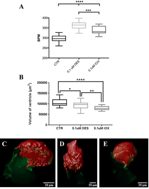
3.1. IOX and DES affect heart function and ventricle morphology Live image analysis of 48 hpf (36 h-exposed) Tg(cmlc2::GFPCaaX) embryos revealed the heart rate increased significantly in both 0.1 μM IOX and DES exposed embryos relative to control siblings (Fig. 1A; P≤ 0.001) and that the heart rate of DES exposed embryos was sig-nificantly higher than the IOX exposed embryos (Fig. 1A; P≤ 0.001). The change in heart rate was associated with a decreased ventricle volume in the DES and IOX treated zebrafish embryos (Fig. 1B, Sup-plementary movies S1–S3). IOX and DES treatments significantly af-fected the ventricle morphology relative to the control groups after 36-h exposure to the chemicals (Fig. 1B; P < 0.05). IOX had the most pro-found effect and strongly reduced the volume of the ventricle relative to the control zebrafish embryos (P < 0.0001) (Fig. 1B, Supplementary movies S1–S3). Exposure of zebrafish embryos to DES also decreased the volume of the ventricle when compared to control embryos (Fig. 1B; P < 0.05) but less than in IOX-exposed sibling (Fig. 1B; P < 0.01).
3.2. IOX and DES affect aorta and thyrocyte development
In zebrafish, the ventrally localized aorta (VA) is the major artery adjacent to the thyroid and the development of the VA is crucial for thyroid morphogenesis and localization in the pharyngeal region (Alt et al., 2006). To characterize the effect of IOX and DES on VA mor-phology and thyrocyte development, zebrafish embryos at 72 hpf were analysed by whole-mount immunofluorescence staining (Fig. 2). Con-sidering that the thyroidfield is distributed along the VA, we measured the diameter of the VA in an anterior, middle and posterior position relative to the thyroid tissue (Fig. 2A). The VA diameter in all three positions measured was significantly decreased in the IOX and DES treated groups compared to the control group (Fig. 2B; P < 0.01). IOX and DES exerted a similar effect on VA diameter (Fig. 2B, P > 0.05).
The effects of IOX and DES on thyroid morphology was analysed by determining the thyroid follicle number, thyroidfield and thyroid fol-licle area (Fig. 2C). The number of thyroid follicles significantly de-creased in IOX treated zebrafish embryos compared to the control groups (Fig. 2C; P < 0.05) but DES did not change thyroid follicle number (P > 0.05). The thyroid field length in DES exposure was significantly lower than the control group (Fig. 2C; P < 0.05) but was not significantly different from the IOX treated siblings (P > 0.05). The total thyroid follicle area was significantly decreased in both IOX and Fig. 1. Box and whisker plot (minimum and max-imum) of (A) heart beats per minute (BPM) and (B) ventricle total volume in 48 hpf zebrafish Tg (fli1:EGFP) control (CTR) and 36 h-exposure embryos to DES and IOX. Different letters indicate statistically significant difference (P < 0.05). C–E Image of the heart with the ventricle (Red) at the top and atrium (Green) at the bottom, (C) control, (D) DES treated and (E) IOX treated. One-way ANOVA was used to test for statistical significance followed by Bonferroni's multiple comparisons test *P < 0.05; **P < 0.01; ***P < 0.001; ****P < 0.0001, the same as below. (For interpretation of the references to colour in thisfigure legend, the reader is referred to the web version of this article.)
DES groups relative to the control embryos (Fig. 2C; P < 0.05). DES had a more pronounced effect on total follicle area (P < 0.01) than IOX (P < 0.05) even though the decrease was not significantly dif-ferent between treatments (Fig. 2C; P > 0.05). The effect of the IOX and DES on the VA and thyroid development was highly specific and the growth of zebrafish embryos was unaffected at 72 hpf (Fig. S1; P > 0.05).
3.3. Transcriptomics of zebrafish endothelial cells exposed to IOX and DES Analysis of the global gene regulation profiles of endothelial cells in
zebrafish embryos treated with IOX and DES revealed 1688 DE gene transcripts relative to the control embryos. KEGG pathway analysis showed that 28 and 19 pathways respectively were significantly changed in the IOX and DES exposure groups relative to the control embryos (Tables S2 and S3; P < 0.05). Several of the pathways sig-nificantly changed in DES and IOX treated embryos were related to blood vessel endothelial cell function and three cardiomyopathy path-ways were identified including hypertrophic cardiomyopathy, dilated cardiomyopathy and arrhythmogenic right ventricular cardiomyopathy (Table 1, P < 0.05). IOX and DES also had an effect on the pathways of calcium signalling, cell adhesion molecules, GABAergic synapse Fig. 2. Exposure to DES and IOX affects thyroid gland and ventral aorta development. (A) Fluorescent images of the pharyngeal region of Tg(fli1:EGFP) zebrafish embryos at 72 hpf. Red and green represent, respectively, thyroglobulin and GFP immunostaining. (B) Determination of aorta diameter in three different regions as depicted in A. From left to right VA-1, VA-2 and VA-3 are represented. The lower panel below the graphs represents examples of Z-slices at each region of the aorta measured. The scale bars in the lower panel below the graphs correspond to 5μm. (C) Measurements of thyroid follicle number, thyroid field length and the thyroid follicle area. (For interpretation of the references to colour in thisfigure legend, the reader is referred to the web version of this article.)
(Table 1, P < 0.05). The type I diabetes mellitus pathway was sig-nificantly affected by DES exposure (Table 1, P < 0.05), while IOX had a significant impact on the complement and coagulation cascades, vascular smooth muscle contraction, amyotrophic lateral sclerosis, platelet activation, retrograde endocannabinoid signalling and ABC transporters (Table 1, P < 0.05).
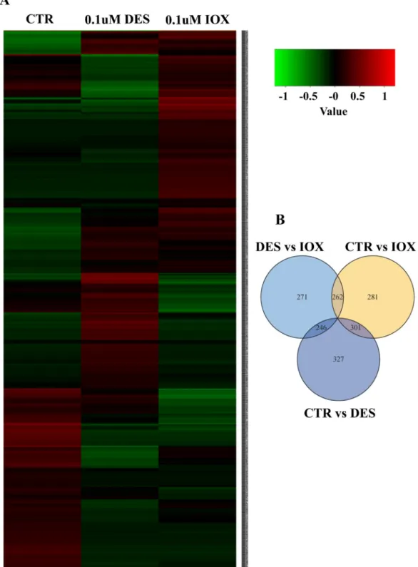
The DE gene transcripts identified by RNAseq were used to generate a heatmap and the pattern of gene expression in the embryos exposed to IOX and DES was compared to the control group (Fig. 3A). The ex-pression signature of DE gene transcripts in IOX and DES were different from the control but also from each other (Fig. 3A). DE analysis re-vealed that some transcripts were affected by both treatments whereas other transcripts were affected by IOX but not DES and vice-versa (in comparison to the control embryos;Fig. 3B).
Seventeen genes related to the function of endothelial cells, in-cluding vascular function, cardiac function, calcium homeostasis and other genes were selected to validate the transcriptome data by qPCR (Table S1; Fig. 4). Most of genes such as hypoxia inducible factor (HIF1aa),β1-adrenergic receptor (Adrb1), von Willebrand factor (Vwf), prostacyclin (Ptgir) and voltage-dependent T-type calcium channel subunit alpha-1H (Cacna1ha) were significantly increased in the IOX treated embryos compared with the control embryos (Fig. 4, P < 0.05). However, no significant differences were found for the gene transcripts analysed between the DES exposed embryos and the control embryos (Fig. 4, P > 0.05).
4. Discussion
This study provides insight into the mechanisms by which DES and IOX affect zebrafish heart and vascular development and how these changes impinge on thyroid development. The data obtained in the present study, together with our previous results (Campinho and Power, 2013), reveal a direct disruptive effect of IOX and DES on the heart and vascular development and an indirect effect on the thyroid. Recently, we found that the action of DES and IOX on zebrafish thyroid devel-opment is directly linked to their effect on normal heart develdevel-opment (Campinho and Power, 2013) and that this eventually leads to impaired thyroid development in zebrafish embryos. Our data also show that exposure to IOX and DES seems to have a similar morphological and functional endpoint in zebrafish development but that the underlying genetic, cellular and molecular mechanisms differ.
The effects of DES and IOX on the heart are related to increased heartbeat and impaired morphology. The ventricular size and shape are
crucial for determining cardiac function (Hu et al., 2001). The observed changes in heartbeat in embryos exposed to IOX and DES were further supported by a decrease in ventricle volume when compared to control siblings and this strongly supports the notion that the heart is a target tissue for these two chemicals. It is unclear if the effect of IOX or DES treatments impact more on the definition of heart morphology or heart function. However, it is likely that this is a reciprocal effect given the relationship between form and function in the developing zebrafish heart (Yelon, 2001;Torrent-Guasp et al., 2005;Miura and Yelon, 2011). It was recently reported that in third generation DES-exposed women there is an increase in the incidence of cardiac defects asso-ciated with the atrial and ventricular septal (Titus-Ernstoff et al., 2010; Tournaire et al., 2016). This data from human studies is in concordance with the results from our previous (Campinho and Power, 2013) and the present study. However, so far no data is available about the thyroid function and physiology in third generation DES-exposed women. In-deed, little is known about the effects of DES on heart development and function in vertebrates in general. This is relevant because our studies in zebrafish (Campinho and Power, 2013) showed that alongside the changes in heart function and ventricle volume there were significant changes in thyroid gland development. Here we provide thefirst evi-dences on the pernicious effects of low exposure doses of DES in zeb-rafish, a vertebrate and important biomedical model and thus hint that these observations are conserved in other vertebrates, including hu-mans (Titus-Ernstoff et al., 2010).
It was not only heart development that was affected in the IOX and DES treated zebrafish embryos as the ventral aorta diameter was also reduced in both experimental groups. A direct effect of IOX and DES on the heart is evident from the morphological and functional data (Fig. 1). The transcriptome data also indicates that developing endothelial cells are differentially affected by these chemicals. The response of en-dothelial cells to IOX and DES exposure was specific given the restricted subset of genes that responded in both groups when compared to the control embryos. Moreover, the response of endothelial cells to IOX or DES was quite different with different subsets of genes and gene net-works being affected in each group.
Three main cardiomyopathy pathways were represented in the DE genes of DES and IOX exposed embryos, including hypertrophic car-diomyopathy, dilated cardiomyopathy and arrhythmogenic right ven-tricular cardiomyopathy together with other pathways such as the calcium signalling pathway (Table 1). qPCR validation of the tran-scriptome data showed that a cluster of genes such as Acvr1bb, Agtr1a, HIF1aa, Notch1b, Ptgir, Adrb1 and Vwf, involved in cardiovascular Table 1
Selected enriched KEGG pathways calculated for modulated genes in DES and IOX treated zebrafish embryos at 48 hpf. “—” indicates ‘not significantly different’. Comparison with control
DES IOX
P value No. of genes P value No. of genes
Up Down Up Down
Calcium signalling pathway 2.63E−04 11 12 3.09E−04 14 11
Neuroactive ligand-receptor interaction 0.0166 18 14 1.03E−04 27 18
GABAergic synapse 0.0064 8 3 0.0354 6 4
Cell adhesion molecules 0.0276 9 4 0.0348 10 4
Hypertrophic cardiomyopathy 0.0116 4 6 0.0258 4 6
Dilated cardiomyopathy 0.0233 5 5 0.0100 6 6
Arrhythmogenic right ventricular cardiomyopathy 0.0349 4 4 0.0118 3 7
Retrograde endocannabinoid signalling 0.0013 7 7 0.0220 8 4
Type I diabetes mellitus 0.0122 2 4 — — —
Vascular smooth muscle contraction — — — 0.0323 7 6
Complement and coagulation cascades — — — 0.0029 9 2
Amyotrophic lateral sclerosis — — — 0.0410 3 7
Platelet activation — — — 0.0476 10 5
function and form were up-regulated in IOX treated groups (Fig. 4). More specifically changes in Adrb1 may explain modified heart rate and atrial cardiac muscle contractility (Parvez et al., 2012) and changes in Agtr1a together with Ptgir (a vasodilator effect mediator) may explain the vasoconstriction observed (Whittle et al., 2012;Dinh et al., 2017). Calcium homeostasis genes (Cacna1da, Calcr and Itpr1b), cell adhesion molecule (Cadm1a) and cholinergic receptor gene (Chrnb3a) were
up-regulated by IOX exposure. Although morphological effects on the heart caused by DES exposure were clear, no differences in the genes involved in cardiovascular function and analysed by qPCR were identified. These findings suggest that IOX and DES induce some different genetic pathways but some genetic responses are similar (Supplementary tables S4, S5 and S6). This data might also indicate that IOX has a stronger endocrine disruption potential.
Fig. 3. Transcriptome analysis of differentially expressed genes in 48 hpf control, DES and IOX treated zebrafish embryos. (A) Heatmap showing the gene expression pattern in control, DES and IOX treated zebrafish; The colour represents the gene expression level (log10FPKM); Red represents high expression and low expression is
indicated in green (B) Venn diagram presenting the differential expressed genes; the number in circles represents the differential expressed transcripts between two groups; the overlapping section of two circles means the common number of differential expressed transcripts. (For interpretation of the references to colour in this figure legend, the reader is referred to the web version of this article.)
Fig. 4. qPCR validation of selected RNA-seq differential expressed transcripts between control, DES and IOX-treated zebrafish embryos 48 hpf (36 h-exposure to chemicals). Analysed genes were related to cardiovascular function (A), calcium homeostasis (B) and others (C). Acvr1bb: activin receptor type-1B-like; Agtr1a: angiotensin II type I receptor; Angptl1b: angiopoietin-related protein 2; HIF1aa: hypoxia inducible factor (HIF-1); Ptgir: prostacyclin receptor-like; Vwf: von Willebrand factor; Adrb1:β1-adrenergic receptor; Mhc1lia: hereditary hemochromatosis; Mybpc2a: myosin binding protein C, fast type a; Cacna1da: de-pendent L-type calcium channel subunit alpha-1D; Cacna1ha: dede-pendent T-type calcium channel subunit alpha-1H-like; Cacng2b: calcium channel, voltage-dependent, gamma subunit 2b; Calcr: calcitonin receptor; Itpr1b: inositol 1,4,5-trisphosphate receptor, type 1b; Cadm1a: cell adhesion molecule 1a; Chrnb3a: cho-linergic receptor, nicotinic, beta polypeptide 3a. Different letters indicate statistically significant difference (P < 0.05).
The disruption of the heart and VA growth in DES and IOX treated groups may affect the morphology of the vessels or may themselves be affected by the impaired vasculature development. The effect on blood vessel development and homeostasis of the sheer stress of blood cells as they circulate and the intensity of cardiac function has been extensively demonstrated (Shiojima et al., 2005;Lu and Kassab, 2011;Boselli et al., 2015). Together with increased heartbeat and lower chamber volume (Fig. 1) it is expected that bloodflow will be affected in both IOX and DES-treated embryos when compared to controls. Although not vali-dated by qPCR, genes involved in angiogenic mechanosensing (Boselli et al., 2015) were DE in the transcriptome (> 2-fold, P < 0.05; FDR 0.05) of IOX and DES embryos relative to the control (Excel Table S1, S2 and S3). In both IOX and DES, expression of Early Growth Response protein 1 (egr1) was up-regulated whereas piezo-type mechanosensitive ion channel component 1 (piezo1) and endothelin 1 (edn1) were down-regu-lated. In contrast, other genes (e.g. klf2, s1p1, pkd2 and cxcr4) involved in mechanosensing and response in endothelial cells were either up-regulated in IOX (vs control) or downup-regulated in DES or vice-versa (Supplementary tables S4, S5 and S6). Taken together, our data seem to suggest that IOX and DES affect blood flow. What is not clear is if the effect is through a direct effect on endothelial cells, cardiomyocytes (and hence heart-function) or both. Of the two chemicals tested IOX presented clearer effects both in relation to endothelial factors (most notably the angiotensin receptor agtr1a) as well as Ca2+-signalling re-lated genes (Fig. 4A and B). Another important observation was that endothelial cells responded differently to IOX and DES, even though similar morphological endpoints were identified they seem to point out for vessel contraction/stiffness implications.
Blood vessel development is known to affect thyroid gland mor-phology and modulates thyroid morphogenesis in zebrafish (Alt et al., 2006; Opitz et al., 2012). Given that thyroid gland development is highly dependent on the development of the heart and aorta (Alt et al., 2006;Wendl et al., 2007;Opitz et al., 2012), the data from the present study revealing that IOX and DES disrupt normal cardiovascular de-velopment in the zebrafish embryos probably explains the observed modifications in thyroid gland development and homeostasis. We have shown that this is likely mediated by changes in the expression of the thyroid gland master developmental factor, nkx2.1a (Campinho and Power, 2013). Taken together our data indicates that thyroid gland development was affected in IOX and DES exposed embryos probably due to disruption of normal cardiovascular development and that this might have repercussions on TH homeostasis in later life stages.
These observations are not entirely unexpected. 2,3,7,8-tetra-chlorodibenzo-p-dioxin (TCDD) has been shown to cause severe heart morphology anomalies by altering looping and also provoked defects in heart function such as blood regurgitation and ventricular standstill in zebrafish embryos (Antkiewicz et al., 2005). In the other only study in vertebrates where it was studied the effect of a anthropogenic derived chemical exposure on cardiac development and thyroid gland devel-opment, it was reported that clofibrate impacts on heart morphology and contractility in zebrafish leading to altered thyroid gland mor-phogenesis (Raldúa et al., 2008). Similarly, a short ventral aorta was observed in clofibrate-exposed zebrafish larvae with disrupted thyroid tissue morphogenesis (Raldúa et al., 2008). Taken together the dis-ruption of thyroid gland morphogenesis was due to impairment of ventral aorta development in clofibrate-exposed zebrafish larvae (Raldúa et al., 2008).
While more work is needed to further dissect the effects of DES and IOX on zebrafish thyroid gland development and homeostasis, the present study shows that DES and IOX are thyroid disrupting com-pounds. We propose that DES and IOX directly affect vascular and heart development and that through this action they indirectly impair the developing thyroid gland and presumably the thyroid axis and disrupt thyroid homeostasis. Notably, the toxicological effects of IOX and DES on zebrafish and humans seem to be remarkably well conserved and highlights the zebrafish as good model for understanding the effect of
environmental exposure to IOX and DES in human populations. 5. Conclusion
Ourfindings suggest that low micromolar levels of waterborne IOX and DES are capable of altering normal cardiac and vascular physiology of zebrafish embryos and have the potential to give rise to thyroid endocrine disruption indirectly. Taken together, the subtle but sig-nificant morphological, functional and gene expression changes at very low levels of IOX and DES, suggest that what are considered safe limits for these chemicals in the environment are lower than previously thought.
Supplementary data to this article can be found online athttps:// doi.org/10.1016/j.envint.2019.01.009.
Conflict of interest
The authors declare they have no actual or potential competing fi-nancial interests.
Acknowledgments
This study was supported by the National Key Research and Development Program of China (2018YFD0900601) and the Shanghai Sailing Program (18YF1410000) and received Portuguese national funds from FCT - Foundation for Science and Technology through project UID/Multi/04326/2016 (CCMAR) and PPBI-POCI-01-0145-FEDER-22122 (Microscopy Unit). MAC is a recipient of a FCT IF2014 Starting Grant (IF/01274/2014).
References
Alt, B., Elsalini, O.A., Schrumpf, P., Haufs, N., Lawson, N.D., Schwabe, G.C., et al., 2006. Arteries define the position of the thyroid gland during its developmental re-localisation. Development 133 (19), 3797–3804.https://doi.org/10.1242/dev. 02550.
Antkiewicz, D.S., Burns, C.G., Carney, S.A., Peterson, R.E., Heideman, W., 2005. Heart malformation is an early response to TCDD in embryonic zebrafish. Toxicol. Sci. 84 (2), 368–377.https://doi.org/10.1093/toxsci/kfi073.
Bakkers, J., 2011. Zebrafish as a model to study cardiac development and human cardiac disease. Cardiovasc. Res. 91 (2), 279–288.https://doi.org/10.1093/cvr/cvr098. Benjamini, Y., Hochberg, Y., 1995. Controlling the false discovery rate: a practical and
powerful approach to multiple testing. J. R. Stat. Soc. B. Methodol. 57 (1), 289–300.
http://www.jstor.org/stable/2346101.
Boselli, F., Freund, J.B., Vermot, J., 2015. Bloodflow mechanics in cardiovascular de-velopment. Cell. Mol. Life Sci. 72 (13), 2545–2559. https://doi.org/10.1007/s00018-015-1885-3.
Campinho, M.A., Power, D.M., 2013. Waterborne exposure of zebrafish embryos to mi-cromole concentrations of ioxynil and diethylstilbestrol disrupts thyrocyte develop-ment. Aquat. Toxicol. 140–141, 279–287.https://doi.org/10.1016/j.aquatox.2013. 06.014.
Chen, H., Boutros, P.C., 2011. VennDiagram: a package for the generation of highly-customizable Venn and Euler diagrams in R. BMC Bioinf. 12, 35.https://doi.org/10. 1186/1471-2105-12-35.
Chen, J.N., Haffter, P., Odenthal, J., Vogelsang, E., Brand, M., van Eeden, F.J., et al., 1996. Mutations affecting the cardiovascular system and other internal organs in zebrafish. Development 123, 293–302.
Chen, H.C., Kuo, H.W., Ding, W.H., 2009. Determination of estrogenic compounds in wastewater using liquid chromatography–tandem mass spectrometry with electro-spray and atmospheric pressure photoionization following desalting extraction. Chemosphere 74 (4), 508–514.https://doi.org/10.1016/j.chemosphere.2008.09. 089.
Dinh, Q.N., Drummond, G.R., Kem-Harper, B.K., Diep, H., De Silva, T.M., Kim, H.A., et al., 2017. Pressor response to angiotensin II is enhanced in aged mice and associated with inflammation, vasoconstriction and oxidative stress. Aging 9 (6), 1595–1606.https:// doi.org/10.18632/aging.101255.
European-Commission, 2004. Review report for the active substance ioxynil. In: EC Review Reports SANCO/4349/2000.
Hu, N., Yost, H.J., Clark, E.B., 2001. Cardiac morphology and blood pressure in the adult zebrafish. Anat. Rec. 264 (1), 1–12.https://doi.org/10.1002/ar.1111.
Hunt, P.A., Sathyanarayana, S., Fowler, P.A., Trasande, L., 2016. Female reproductive disorders, diseases, and costs of exposure to endocrine disrupting chemicals in the European union. J. Clin. Endocrinol. Metab. 101 (4), 1562–1570.https://doi.org/10. 1210/jc.2015-2873.
Kawakami, K., 2007. Tol2: a versatile gene transfer vector in vertebrates. Genome Biol. 8 (Suppl. 1), S7.https://doi.org/10.1186/gb-2007-8-s1-s7.
Kiyama, R., Wada-Kiyama, Y., 2015. Estrogenic endocrine disruptors: molecular me-chanisms of action. Environ. Int. 83, 11–40.https://doi.org/10.1016/j.envint.2015. 05.012.
Kwan, K.M., Fujimoto, E., Grabher, C., Mangum, B.D., Hardy, M.E., Campbell, D.S., et al., 2007. The Tol2kit: a multisite gateway-based construction kit for Tol2 transposon transgenesis constructs. Dev. Dyn. 236 (11), 3088–3099.https://doi.org/10.1002/ dvdy.21343.
Lawson, N.D., Weinstein, B.M., 2002. In vivo imaging of embryonic vascular development using transgenic zebrafish. Dev. Biol. 248 (2), 307–318.https://doi.org/10.1006/ dbio.2002.0711.
Lu, D., Kassab, G.S., 2011. Role of shear stress and stretch in vascular mechanobiology. J. R. Soc. Interface 8 (63), 1379–1385.https://doi.org/10.1098/rsif.2011.0177. Lu, J., Peatman, E., Tang, H., Lewis, J., Liu, Z., 2012. Profiling of gene duplication
pat-terns of sequenced teleost genomes: evidence for rapid lineage-specific genome ex-pansion mediated by recent tandem duplications. BMC Genomics 13, 246.https:// doi.org/10.1186/1471-2164-13-246.
Merzin, M., 2008. Applying Stereological Method in Radiology. Volume measurement (Bachelor's thesis). University of Tartu, Estonia. http://lepo.it.da.ut.ee/~markkom/ volumest/.
METI, 2015. Report of Pollutant Release and Transfer Register: Agricultural Chemicals. Minister of Economy, Trade and Industry (METI), Japan. http://www.meti.go.jp/ policy/chemical_management/law/prtr/h27kohyo/05todokedegai/syousai/2.pdf(in Japanese).
Miura, G.I., Yelon, D., 2011. A guide to analysis of cardiac phenotypes in the zebrafish embryo. Methods Cell Biol. 101, 161–180. https://doi.org/10.1016/B978-0-12-387036-0.00007-4.
Morgado, I., Hamers, T., Van der Ven, L., Power, D.M., 2007. Disruption of thyroid hormone binding to sea bream recombinant transthyretin by ioxinyl and poly-brominated diphenyl ethers. Chemosphere 69 (1), 155–163.https://doi.org/10. 1016/j.chemosphere.2007.04.010.
Morgado, I., Campinho, M.A., Costa, R., Jacinto, R., Power, D.M., 2009. Disruption of the thyroid system by diethylstilbestrol and ioxynil in the sea bream (Sparus aurata). Aquat. Toxicol. 92 (4), 271–280.https://doi.org/10.1016/j.aquatox.2009.02.015. Ogilvie, L.M., Ramsden, D.B., 1988. Ioxynil and 3,5,3′-triiodothyronine: comparison of
binding to human plasma proteins. Toxicol. Lett. 44 (3), 281–287.https://doi.org/ 10.1016/0378-4274(88)90167-1.
Opitz, R., Maquet, E., Huisken, J., Antonica, F., Trubiroha, A., Pottier, G., et al., 2012. Transgenic zebrafish illuminate the dynamics of thyroid morphogenesis and its re-lationship to cardiovascular development. Dev. Biol. 372 (2), 203–216.https://doi. org/10.1016/j.ydbio.2012.09.011.
Otsuka, S., Ishihara, A., Yamauchi, K., 2014. Ioxynil and tetrabromobisphenol A suppress thyroid-hormone-induced activation of transcriptional elongation mediated by his-tone modifications and RNA polymerase II phosphorylation. Toxicol. Sci. 138 (2), 290–299.https://doi.org/10.1093/toxsci/kfu012.
Parvez, B., Chopra, N., Rowan, S., Vaglio, J.C., Muhammad, R., Roden, D.M., et al., 2012. A commonβ1-adrenergic receptor polymorphism predicts favorable response to rate-control therapy in atrialfibrillation. J. Am. Coll. Cardiol. 59 (1), 49–56.https://doi. org/10.1016/j.jacc.2011.08.061.
Patisaul, H.B., Fenton, S.E., Aylor, D., 2018. Animal models of endocrine disruption. Best Pract. Res. Clin. Endocrinol. Metab. 32 (3), 283–297.https://doi.org/10.1016/j. beem.2018.03.011.
Pojana, G., Gomiero, A., Jonkers, N., Marcomini, A., 2007. Natural and synthetic endo-crine disrupting compounds (EDCs) in water, sediment and biota of a coastal lagoon. Environ. Int. 33, 929–936.https://doi.org/10.1016/j.envint.2007.05.003. Qu, K., Zhang, X., Lv, Z., Li, M., Cui, Z., Zhang, Y., et al., 2012. Simultaneous detection of
diethylstilbestrol and malachite green using conductive carbon black paste electrode. Int. J. Electrochem. Sci. 7 (2), 1827–1839.http://www.electrochemsci.org/papers/ vol7/7031827.pdf.
Raldúa, D., André, M., Babin, P.J., 2008. Clofibrate and gemfibrozil induce an embryonic malabsorption syndrome in zebrafish. Toxicol. Appl. Pharmacol. 228 (3), 301–314.
https://doi.org/10.1016/j.taap.2007.11.016.
Reed, C.E., Fenton, S.E., 2013. Exposure to diethylstilbestrol during sensitive life stages: a legacy of heritable health effects. Birth Defects Res. C Embryo Today 99 (2), 134–146.https://doi.org/10.1002/bdrc.21035.
Schindelin, J., Arganda-Carreras, I., Frise, E., Kaynig, V., Longair, M., Pietzsch, T., et al.,
2012. Fiji: an open-source platform for biological-image analysis. Nat. Methods 9 (7), 676–682.https://doi.org/10.1038/nmeth.2019.
Schug, T.T., Johnson, A.F., Birnbaum, L.S., Colborn, T., Guillette, L.J., Crews, D.P., et al., 2016. Minireview: endocrine disruptors: past lessons and future directions. Mol. Endocrinol. 30 (8), 833–847.https://doi.org/10.1210/me.2016-1096.
Shiojima, I., Sato, K., Izumiya, Y., Schiekofer, S., Ito, M., Liao, R., et al., 2005. Disruption of coordinated cardiac hypertrophy and angiogenesis contributes to the transition to heart failure. J. Clin. Invest. 115 (8), 2108–2118.https://doi.org/10.1172/JCI24682. Thompson, M.A., Ransom, D.G., Pratt, S.J., MacLennan, H., Kieran, M.W., Detrich III,
H.W., et al., 1998. The cloche and spadetail genes differentially affect hematopoiesis and vasculogenesis. Dev. Biol. 197 (2), 248–269.https://doi.org/10.1006/dbio. 1998.8887.
Titus-Ernstoff, L., Troisi, R., Hatch, E.E., Palmer, J.R., Hyer, M., Kaufman, R., et al., 2010. Birth defects in the sons and daughters of women who were exposed in utero to diethylstilbestrol (DES). Int. J. Androl. 33 (2), 377–384.https://doi.org/10.1111/j. 1365-2605.2009.01010.x.
Torrent-Guasp, F., Kocica, M.J., Corno, A.F., Komeda, M., Carreras-Costa, F., Flotats, A., et al., 2005. Towards new understanding of the heart structure and function. Eur. J. Cardiothorac. Surg. 27 (2), 191–201.https://doi.org/10.1016/j.ejcts.2004.11.026. Tournaire, M., Epelboin, S., Devouche, E., Viot, G., Le Bidois, J., Cabau, A., et al., 2016.
Adverse health effects in children of women exposed in utero to diethylstilbestrol (DES). Therapie 71 (4), 395–404.https://doi.org/10.1016/j.therap.2016.01.006. Trapnell, C., Pachter, L., Salzberg, S.L., 2009. Tophat: discovering splice junctions with
RNA-Seq. Bioinformatics 25 (9), 1105–1111.https://doi.org/10.1093/ bioinformatics/btp120.
Trapnell, C., Williams, B.A., Pertea, G., Mortazavi, A., Kwan, G., van Baren, M.J., et al., 2010. Transcript assembly and quantification by RNA-Seq reveals unannotated transcripts and isoform switching during cell differentiation. Nat. Biotechnol. 28 (5), 511–515.https://doi.org/10.1038/nbt.1621.
Trapnell, C., Roberts, A., Goff, L., Pertea, G., Kim, D., Kelley, D.R., et al., 2012. Differential gene and transcript expression analysis of RNA-seq experiments with TopHat and Cufflinks. Nat. Protoc. 7 (3), 562–578.https://doi.org/10.1038/nprot. 2012.016.
Trapnell, C., Hendrickson, D.G., Sauvageau, M., Goff, L., Rinn, J.L., Pachter, L., 2013. Differential analysis of gene regulation at transcript resolution with RNA-seq. Nat. Biotechnol. 31 (1), 46–53.https://doi.org/10.1038/nbt.2450.
Vogel, A.M., Weinstein, B.M., 2000. Studying vascular development in the zebrafish. Trends Cardiovasc. Med. 10 (8), 352–360.https://doi.org/10.1016/s1050-1738(01) 00068-8.
Wendl, T., Adzic, D., Schoenebeck, J.J., Scholpp, S., Brand, M., Yelon, D., et al., 2007. Early developmental specification of the thyroid gland depends on han-expressing surrounding tissue and on FGF signals. Development 134 (15), 2871–2879.https:// doi.org/10.1242/dev.02872.
White, J.W., Cole, B.J., Cherr, G.N., Connon, R.E., Brander, S.M., 2017. Scaling up en-docrine disruption effects from individuals to populations: outcomes depend on how many males a population needs. Environ. Sci. Technol. 51 (3), 1802–1810.https:// doi.org/10.1021/acs.est.6b05276.
Whittle, B.J., Silverstein, A.M., Mottola, D.M., Clapp, L.H., 2012. Binding and activity of the prostacyclin receptor (IP) agonists, treprostinil and iloprost, at human prostanoid receptors: treprostinil is a potent DP1and EP2agonist. Biochem. Pharmacol. 84 (1),
68–75.https://doi.org/10.1016/j.bcp.2012.03.012.
Xie, C., Mao, X., Huang, J., Ding, Y., Wu, J., Dong, S., et al., 2011. KOBAS 2.0: a web server for annotation and identification of enriched pathways and diseases. Nucleic Acids Res. 39 (web server issue), W316–W322.https://doi.org/10.1093/nar/gkr483. Yamamoto, M., Shirai, M., Sugita, K., Nagai, N., Miura, Y., Mogi, R., et al., 2003. Effects of
maternal exposure to diethylstilbestrol on the development of the reproductive system and thyroid function in male and female rat offspring. J. Toxicol. Sci. 28 (5), 385–394.https://doi.org/10.2131/jts.28.385.
Yelon, D., 2001. Cardiac patterning and morphogenesis in zebrafish. Dev. Dyn. 222 (4), 552–563.https://doi.org/10.1002/dvdy.1243.
Zhang, X., Zhang, D., Zhang, H., Luo, Z., Yan, C., 2012. Occurrence, distribution, and seasonal variation of estrogenic compounds and antibiotic residues in Jiulongjiang River, South China. Environ. Sci. Pollut. Res. 19 (5), 1392–1404.https://doi.org/10. 1007/s11356-012-0818-z.