Ciências
Cancer gene therapy: design, development and in
vitro evaluation of a plasmid DNA delivery system
Ana Raquel Bastos Neves
Dissertação para obtenção do Grau de Mestre em
Biotecnologia
(2º ciclo de estudos)
Orientadora: Professora Doutora Diana Rita Barata Costa
Coorientadora: Professora Doutora Ângela Maria Almeida De Sousa
iii
Acknowledgments
The elaboration of this theses concludes this stage of my academic journey. This work would not be possible without the support of some people.
First of all, thank the doctor professor Diana Costa and doctor professor Angela Sousa for the opportunity to join to your investigation team so I can research in a so interesting and actual scientific area. Thank you for all the support, effort and scientific information transmission during the entire year. Your criticisms and work suggestions were fundamental to growth as student and future professional.
I thank the University of Beira Interior, specially the Health Science Research Centre, for the facilities, equipment’s and material available. Thanks to all researchers of this centre, namely of the Biopharmaceuticals and Biomaterials group, that contributed for a better work environment and that help me and shared with me important knowledge. A special acknowledgment to Ana Borges for their support in confocal microscopy experiments.
A big thank you to Margarida Almeida that was a grateful help at the beginning and to Tânia Albuquerque and Rúben Faria. Both of you helped me tremendously during the entire year, teaching me the techniques applied in this word.
To my laboratory and academic colleagues thank you for all the support and friendship. To my friends, Diana Pereira, Diana Gomes, Adriana Pinto, Rita Carapito, Rita Proença, Rosa Sequeira, Mariana Flor and Micaela Riscado, that become my family for last 5 years, a huge thank you because you made this year even more relaxed, funny and special.
Last but not least, thank to my family for the transmitted force during the year, for always believe in me. To my parents, Cristina Bastos and Francisco Neves, and brother, Francisco Neves, I have no words to write, they know how grateful I am for having them in my life. For all the efforts, education, dedication, love and support, thank you!
v
Resumo Alargado
O cancro é uma das principais causas de morbidade e mortalidade em todo o mundo. Envolve mudanças genéticas que afetam uma variedade de genes, como os oncogenes e genes supressores de tumor. Esta alteração leva a uma proliferação celular desregulada, angiogénese, invasão e formação de metástases. As abordagens tradicionais para o tratamento do cancro incluem quimioterapia, cirurgia e radiação. A quimioterapia representa a principal escolha de tratamento na maioria dos casos, no entanto, as terapias tradicionais são ineficazes no combate de metástases, na recorrência do tumor e no tratamento do cancro num estado mais avançado. Em alguns casos estes tratamentos são ineficazes, inespecíficos, podem danificar células saudáveis, causar resistência a fármacos e serem acompanhados por efeitos secundários indesejáveis. Portanto, novas estratégias precisam de ser desenvolvidas de forma a aumentar a eficácia terapêutica.
A terapia genética proporciona uma abordagem promissora e única à medicina devido à sua ampla aplicação no tratamento de várias doenças, incluindo doenças hereditárias e patologias adquiridas (infeção ou cancro). É uma técnica que usa ácidos nucleicos exógenos (DNA ou RNA) para reparar, substituir ou regular genes que expressem uma proteína terapêutica de interesse de forma a prevenir ou tratar uma doença. O gene supressor de tumor da proteína p53 é responsável por manter a integridade do genoma sobre situações de stress e está envolvido em várias vias celulares como a reparação do DNA, regulação do ciclo celular e indução da apoptose. No entanto, este gene encontra-se mutado ou inexistente em mais de 50 % dos cancros humanos, e, portanto, é fundamental criar protocolos de terapia génica que permitam restaurar o nível e função desta proteína. Para a terapia génica ser viável num cenário clínico é necessário desenvolver um sistema de entrega de genes eficiente. A conceção de sistemas de entrega baseados em péptidos capazes de penetrar nas células é vantajosa e pode contribuir para a evolução da terapia do cancro. Neste contexto, foi desenvolvido um novo sistema de entrega de DNA plasmídico (pDNA) codificante para a p53 com base no péptido RALA de modo a obter um sistema de entrega intracelular adequado e capaz de entregar o gene e restabelecer os níveis de p53 nas células cancerígenas.
O péptido RALA é um péptido anfipático composto por 30 aminoácidos, com uma estrutura em hélice-α que possui baixa citotoxicidade. Este péptido quando conjugado com ácidos nucleicos estabelece interações eletrostáticas e forma partículas com tamanho nanométrico adequadas para a captação celular e internalização nuclear. No presente trabalho, estes transportadores foram, numa fase inicial, caracterizados em termos de morfologia, tamanho, carga superficial e eficiência de encapsulação por microscopia eletrónica de varrimento (SEM) e através da Dispersão da Luz Dinâmica (DLS) usando o Zetasizer Nano Zs., respetivamente, e a sua estrutura analisada através de espectroscopia de infravermelho transformada por Fourier (FTIR). Os resultados mostraram que as nanopartículas formadas possuem uma forma esférica/oval, um tamanho adequado, carga global positiva e eficiências de encapsulação do plasmídeo elevadas o que indica que são
vi
adequadas para a captação celular, internalização e libertação de genes. Além disso, estudos de estabilidade demonstraram que o péptido RALA é capaz de proteger o pDNA encapsulado das nucleases séricas e o ensaio de brometo de [3-(4,5-dimetiltiazol-2yl) -2,5-difenil tetrazolium (MTT) mostrou que estes sistemas são biocompatíveis. Experiências de microscopia confocal e de live
cell imaging confirmaram a localização intracelular de nanopartículas e a co-localização do
plasmídeo no núcleo, logo após 2 h de transfecção. Além disso, a transfecção in vitro de células HeLa mediada pelos vetores RALA/pDNA permitiu a deteção de transcritos de mRNA por reação em cadeia da polimerase via transcriptase reversa (RT-PCR) e expressão da proteína p53 por western blot. Um kit de ensaio de imunoabsorsão enzimático (ELISA) permitiu quantificar os níveis de proteína produzidos. A partir destes progressos, a indução de apoptose pelos sistemas RALA/pDNA nestas células cancerígenas do colo do útero foi avaliada. A ativação da caspase-3 foi monitorizada por meio de um ensaio colorimétrico e o ensaio de deteção de células em apoptose por TUNEL permitiu confirmar a fragmentação do DNA nuclear após transfecção com os poliplexos. Por último, um ensaio de western blot para marcação da proteína BAX permitiu perceber que a morte celular observada nas células tumorais transfetadas pelos nanos sistemas RALA/pDNA foi induzida pela via de apoptose intrínseca mediada pela BAX que por sua vez foi ativada pela suplementação do nível do supressor de tumor p53 nestas células. Em suma, o conjunto de resultados apresentados no presente trabalho revelam que o vetor RALA/pDNA parece ser adequado para aplicação na terapia génica do cancro.
Palavras-chave
DNA plasmídico codificante para a p53; nanopartículas; péptido RALA; proteína p53; terapia génica do cancro.
vii
Abstract
Cancer is one of the major causes of morbidity and mortality worldwide. It involves genetic changes that affect a variety of genes, namely tumour suppressor genes. The traditional approaches for cancer treatment include chemotherapy, surgery and radiation. Chemotherapy represents the main choice of treatment in most cases, however, traditional therapies seemed unvalued to fight against metastasis, recurrence of tumour, and the treatment of advanced cancer. Therefore, new strategies need to be developed to increase therapy efficacy.
Gene therapy has brought a promising and unique approach to medicine because of its wide application in the treatment of various diseases, including hereditary diseases to acquired (infection or cancer) pathologies. p53 suppressor gene mutation or degradation is found in more than 50% of human cancers, therefore, gene therapy protocols focussed on the restauration of p53 protein are a priority in this field. For gene therapy viability in a clinical setting, the development
of an efficient gene delivery system is imperative.The conception of delivery systems based on
cell penetrating peptides represents an incredible asset and may deeply contribute for the evolution of cancer therapy. In this context, a new system for p53 encoding plasmid DNA (pDNA) delivery based on RALA peptide was designed and developed in order to produce a suitable intracellular delivery platform capable of gene delivery and restoration of p53 levels within the cancer cells. These carriers were characterized in terms of morphology, size, surface charge, loading and encapsulation efficiency and the fine structure was analyzed by Fourier-transformed infrared (FTIR) spectroscopy. The results showed that formed nanoparticles are suitable for cell uptake, internalization and gene release. Furthermore, stability studies demonstrated that RALA is capable of protect encapsulated DNA from serum nucleases and MTT assay showed that these systems are biocompatible. Confocal microscopy and live cell imaging experiments confirmed intracellular localization of nanoparticles, resulting in enhanced sustained pDNA uptake. Moreover, in vitro transfection of HeLa cells mediated by RALA/pDNA vectors allowed the detection of mRNA transcripts by RT-PCR and p53 protein expression by Western Blot. An ELISA kit assay quantified the produced protein levels. From these progresses, apoptosis in cancer cells was investigated. Caspase 3 activation was monitored by means of colorimetric assay and TUNEL assay enabled to confirm nuclear DNA fragmentation post transfection with the carriers. Lastly, a western blot assay with BAX antibody permitted to figure out which apoptosis pathway is responsible for cancer cells death. Taken together, the presented results revealed that the RALA/pDNA vector seems to be suitable as an innovative platform for p53 mediated cancer gene therapy.
viii
Keywords
ix
Table of Contents
Chapter I – Introduction ... 1
1.1. Cancer ... 1
1.2. Cell apoptosis ... 1
1.2.1. Extrinsic and intrinsic pathways ... 2
1.3. Cervical cancer ... 3
1.4. Human Papillomavirus (HPV) ... 3
1.5. Prevention and risks ... 5
1.6. Actual therapies ... 5
1.7. Gene Therapy ... 6
1.7.1. Approaches of cancer gene therapy ... 7
1.7.1.1. Viral vectors ... 8
1.7.1.2. Non-viral vectors ... 9
1.7.1.2.1. Plasmid DNA ... 9
1.7.2. Delivery systems ... 12
1.8. Delivery vehicle types ... 12
1.8.1. Cellular barriers ... 15
1.9. Cell penetrating peptides ... 15
1.9.1 GALA and KALA peptides ... 16
1.9.2. RALA peptide ... 17
Chapter II - Aim of the thesis ... 19
Chapter III - Materials and Methods ... 21
3.1. Materials ... 21
3.1.1. Plasmid DNA ... 21
3.2. Methods ... 22
3.2.1. Bacterial growth conditions and plasmid production ... 22
3.2.2. Plasmid extraction and purification ... 22
3.2.3. Preparation of RALA/pDNA complexes ... 23
3.2.4. Determination of encapsulation efficiency (EE) ... 23
3.2.5. Scanning Electron Microscopy (SEM) ... 24
3.2.6. Particle size and zeta potential measurements ... 24
3.2.7. Fourier-Transformed Infrared Spectroscopy (FTIR) analysis ... 24
3.2.8. Circular Dichroism (CD) ... 25
3.2.9. Plasmid DNA protection studies ... 25
3.2.10. Cell culture ... 25
3.2.11. Cytotoxicity analysis ... 25
3.2.12. In vitro transfection studies ... 26
3.2.12.1. Plasmid labelling with FITC ... 26
3.2.12.2. Fluorescence confocal microscopy ... 26
3.2.12.3. Live Cell Imaging ... 27
x
3.2.13.1. RNA extraction ... 27
3.2.13.2. cDNA synthesis ... 28
3.2.13.3. Reverse transcription polymerase chain reaction (RT-PCR) ... 28
3.2.14. Western Blot ... 28
3.2.14.1. Protein extraction ... 28
3.2.14.2. Protein quantification ... 29
3.2.14.3. Polyacrylamide gel electrophoresis ... 29
3.2.14.4. Electroblotting ... 29
3.2.15. Protein quantification ... 30
3.2.16. Caspase-3 activity ... 30
3.2.17. TUNEL assay ... 30
Chapter IV - Results and discussion ... 33
4.1. The properties of RALA/p53 complexes ... 33
4.2. pDNA protection studies ... 38
4.3. Cytotoxicity analysis ... 41
4.4. Cellular uptake and intracellular location of complexes ... 43
4.5. p53 gene and protein expression ... 50
4.6. Vector-mediated apoptosis ... 52
4.6.1. Caspase-3 activity assay ... 52
4.6.2. HeLa cells - MTT assay ... 53
4.6.3. TUNEL assay ... 53
Chapter V - Conclusions and future perspectives ... 57
Chapter VI - Bibliography ... 59
xi
List of figures
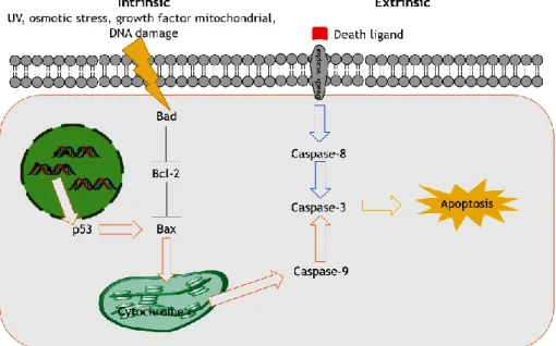
Figure 1: Apoptosis intrinsic and extrinsic pathways. (Adapted from Bio Rad and Cell Signalling). 2
Figure 2: Structure of papilloma virus capsid from cryoelectron microscopy. (Protein Data Bank,
May 2018) ... 3
Figure 3: The lifecycle of a typical hr-HPV. (Chabeda et al. 2018)... 4
Figure 4: HPV E6 and E7 early proteins action. ... 5
Figure 5: Current gene therapy strategies. ... 7
Figure 6: Viral vectors examples used to deliver nucleic acids for gene therapy. (Adapted from (Caffery, Lee, and Alexander-Bryant 2019) ... 9
Figure 7: Schematic representation of a plasmid vector. ... 10
Figure 8: Schematic representation of the biotechnological approach used in the plasmid DNA production. ... 11
Figure 9: Some delivery vehicles types. (Adapted from (Lembo and Cavalli 2010)) ... 13
Figure 10: Barriers to intracellular trafficking of gene delivery systems. (Adapted from (Teo et al. 2016)) ... 15
Figure 11: RALA primary and secondary structure. (Adapted from (Udhayakumar et al. 2017)) .. 17
Figure 12: Plasmid pcDNA3-FLAG-p53. (Adapted from Addgene) ... 22
Figure 13: Calibration curve obtained with protein standards ( 25-2000 µg/mL). ... 29
Figure 14: RALA/pDNA complexation behaviour at various N/P ratios investigated by agarose gel electrophoresis. ... 33
Figure 15: FTIR spectra (Transmittance (%) versus Wavenumber (cm-1)) of pDNA (A), RALA (B), and RALA/pDNA nanoparticles formulated at N/P ratio of 10 (C). ... 34
Figure 16: Representation of circular dichroism spectra of RALA peptide (40 µM) (A) and RALA/pDNA vectors at N/P ratio of 2, 5 and 10 (B). ... 35
Figure 17: Scanning electron micrographs of RALA/p53 nanoparticles formulated at N/P ratios of 1, 2, 5, 10, 15, 20 and 50. ... 36
xii
Figure 18: Electrophoretic analysis of the vector’s protection of pDNA after its incubation with serum supplemented DMEM + 10 % FBS. ... 38
Figure 19: Electrophoretic analysis of the vector’s protection of pDNA after its incubation with DNase I and DNase I + 10 % SDS for 1 h. ... 39
Figure 20: Electrophoretic analysis of the vector’s protection of pDNA after its incubation with trypsin. ... 40
Figure 21: Electrophoretic analysis of the vector’s protection of pDNA after its incubation with 10 % FBS. ... 41
Figure 22: Cellular viability of fibroblast cells after 24 h or 48 h of transfection with RALA/pDNA nanoparticles at various N/P ratios. ... 42
Figure 23: RALA/pDNA vectors transfection ability and intracellular co-localization investigated by fluorescence confocal microscopy after 2 h of transfection. ... 44
Figure 24: RALA/pDNA vectors transfection ability and intracellular co-localization investigated by fluorescence confocal microscopy after 4 h of transfection. ... 44
Figure 25: RALA/pDNA vectors transfection ability and intracellular co-localization investigated by fluorescence confocal microscopy after 6 h of transfection. ... 45
Figure 26: Representative live cell images of the transfection mediated by RALA/pDNA nanoparticles at N/P ratio of 2. ... 47
Figure 27: Representative live cell images of the transfection mediated by RALA/pDNA nanoparticles at N/P ratio of 5. ... 48
Figure 28: Representative live cell images of the transfection mediated by RALA/pDNA nanoparticles at N/P ratio of 10. ... 49
Figure 29: PCR analysis of p53 mRNA in HeLa cells after 24 h of transfection mediated by RALA/pDNA nanoparticles prepared at N/P ratios of 2, 5 and 10. ... 50
Figure 30: Evaluation of p53 and BAX protein expression by western blot analysis after 48 h transfection with RALA/pDNA nanoparticles prepared at N/P ratios of 2, 5 and 10. ... 51
Figure 31: Caspase-3 activity in HeLa cells after 48 h of transfection mediated by RALA/pDNA vectors at N/P ratios of 2, 5 and 10. ... 52
xiii Figure 32: Cell viability of HeLa cells after 24 and 48 h of transfection with RALA/pDNA complexes at different N/P ratios. ... 53
Figure 33: In situ cell death detection in HeLa cells assayed by terminal deoxynucleotidyl transferase-mediated dUTP nick end labelling (TUNEL). ... 54
Figure 34: Abstract submitted to XIV Annual CICS-UBI Symposium. ... 63
xv
List of Tables
Table 1: Example of practical applications of carriers in gene therapy. ... 14
Table 2: Examples of other studies using RALA peptide as carrier. ... 18
Table 3: Mean size, polydispersity index, average zeta potential and pDNA encapsulation efficiency (EE) for RALA/pDNA complexes formulated at several N/P ratios. ... 37
Table 4: Quantification of p53 protein levels in HeLa cells after 24 h of transfection mediated by the formulated RALA/pDNA systems formulated at N/P ratios of 2, 5 and 10. ... 51
xvii
List of Abbreviations
µg: Microgram µm: Micro millimetre µM: Micro molar 3D: Three-dimensional A: AmpereBAK: Bcl-2-antagonist killer Bax: Bcl-2-associated X protein Bcl-2: B-cell lymphoma 2 BSA: Bovine serum albumin CD: Circular Dichroism
cDNA: Complementary Deoxyribonucleic acid CICS: Health Sciences Research Centre cm: Centimetre
CO2: Carbon Dioxide
CpG: Cytosine-phosphate-guanine CPP: Cell Penetrating Peptides DAPI: 4’,6-diamino-2-fenillindol DEPC: Diethylpyrocarbonate DLS: Dynamic Light Scattering
DMEM-HG: Dulbecco´s Modified Eagle Medium, high glucose DNA: Deoxyribonucleic acid
dUTP: Deoxyuridine Triphosphate
E. coli: Escherichia coli
e.g.: for example
ECL: Enhanced Chemiluminescence EDTA: Ethylenediamine tetraacetic acid EE: Encapsulation Efficiency
EGTA: Ethylene Glycol Tetraacetic Acid ELISA: Enzyme-Linked Immunosorbent Assay EMA: European Medicines Agency
etc.: et cetra EU: Endotoxin Unit FBS: Fetal bovine serum
FDA: Food and Drug Administration FITC: Fluorescein Isothiocyanate FMOC: Fluorenylmethyloxycarbonyl
FTIR: Fourier-transform infrared spectroscopy g: G Force
xviii g: Grass
gDNA: Genomic DNA h: Hour
HCl: Hydrochloric acid He-Ne: Helium-Neon
HEPES: 4-(2-hydroxyethyl)-1-piperazineethanesulfonic acid HPV: Human Papillomavirus
hr: high risk
IgG: Immunoglobulin G
K2HPO4: Potassium hydrogen phosphate
kb: kilobase kbp: Kilo base pair kDa: Kilo Dalton
KH2PO4: Potassium dihydrogen phosphate
kV: Kilovolt L: Litter LB: Luria-Bertani M: Molar MgCl2: Magnesium chloride min: Minute mL: Millilitre mm: Millimetre mM: Millimolar MM: Molar Mass
MOPS: 3-(N-morpholino)propanesulfonic acid mPa: Megapascal
mRNA: Messenger RNA
MSNs: Mesoporous silica nanoparticles
MTT: 3-(4,5-Dimethylthiazol-2-yl)-2,5-Diphenyltetrazolium Bromide mV: Millivolts
Mw: Molecular weight N: Amine groups N: Normality
NaCl: Sodium chloride NaOH: Sodium hydroxide ng: Nanogram
NHDF: Normal Human Dermal Fibroblasts NIBS: Non-Invasive Backscatter Optics nm: Nanometer
xix O2: Oxygen
ºC: Degree Celsius oc: open circular OD: Optic Density OH: Hydroxyl
Ori: origin of replication P.I.: Polydispersive index P: Phosphate groups PAGE: Polyacrylamide gel
PALS: Phase Analysis Light Scattering PBS: Phosphate buffer solution pcDNA: Plasmid cloning DNA pDNA: Plasmid DNA
PFA: Paraformaldehyde
PMSF: Phenylmethylsulfonyl fluoride pNA: p-Nitroaniline
pRb: retinoblastoma protein PVDF: Polyvinylidene difluoride RNA: Ribonucleic acid
rpm: Revolutions per minute
RT-PCR: Reverse transcription polymerase chain reaction s: Second
sc: super coiled
SDS: Sodium dodecil sulphate SEM: Scanning electron microscope siRNA: Silencing RNA
spp.: species
ST: Standard deviation TAE: Tris-acetate-EDTA TB: Terrific Broth
TdT: Terminal deoxynucleotidyl transferase
TUNEL: Terminal deoxynucleotidyl transferase dUTP nick end labeling UBI: University of Beira Interior
UV: Ultraviolet
v/v %: Volume per volume percentage V: Voltage
Vis: Visible
VLPs: Virus like particles
xxi
List of Scientific Communications
Oral presentation
Bastos, A.; Sousa, A; Faria, R.; Albuquerque, T.; Queiroz, J; Costa, D. Cancer gene therapy: design, development and in vitro evaluation of a RALA peptide/pDNA vector. XIV Anual CICS-UBI Symposium. 4-5 July 2019, Covilhã, Portugal.
List of Scientific Publications
A. R. Neves, A. Sousa, R. Faria, T. Albuquerque, J. A. Queiroz, D. Costa. 2019. Cancer gene therapy mediated by RALA/plasmid DNA vectors: Nitrogen to Phosphate groups ratio (N/P) as a tool for tunable transfection efficiency and apoptosis. ACS Applied Materials & Interfaces (Submitted).
1
Chapter I – Introduction
1.1. Cancer
Cancer is one of the major causes of morbidity and mortality worldwide.(El-Deeb et al. 2018) The knowhow of cancer biology has grown tremendously, and it is now widely understood to originate from genetic instability as well as microenvironment factors. (Teo et al. 2016) It involves genetic changes that affect oncogenes, tumour suppressor genes and modifier genes, which enable them to sustain proliferative signalling, evade growth suppressors, resist cell death, induce angiogenesis, enable replicative immortality and activate invasion and metastasis.(Costa et al. 2014; Teo et al. 2016)
The tumour suppressor gene p53 keeps the genome integrity under oncogenic stress or other stress signals and it is involved in several cellular pathways such as DNA repair, regulation of the cell cycle and induced apoptosis (programmed cell death). At the molecular level, mutation of the p53 gene is found in greater than 50 % of human tumours. Mutations in p53 abrogate normal tumour suppressor functions, contributing to the survival and/or proliferation of abnormal cells with poorly differentiated phenotypes. Cancer cells containing mutant p53 are associated with more aggressive disease, increased resistance to chemotherapy and radiation therapy, and poor prognosis. In this sense, restoration of p53 functions in tumour cells could result in tumour inhibition, normal cell division and adequately responses to DNA damaging and to impede angiogenesis. (Guidotti, Brambilla, and Rossi 2017; Sharma et al. 2011; Costa et al. 2014)
1.2. Cell apoptosis
In apoptosis, internal and/or external stimuli initiate a series of highly controlled reactions, which ultimately lead to cell death. The two most important groups of proteins involved in apoptosis, caspases and the Bcl-2 (B-cell lymphoma 2) family of proteins, participate in all pathways of apoptotic cell death. (Grilo and Mantalaris 2019)
Caspases are cysteine-dependent endoproteases that catalyse the breaking of the peptide bond.Most caspases can be classified, according to their role, in initiator caspases, executioner caspases and caspases involved in inflammation. Caspase-2 shares structural elements with both initiator and executioner caspases.(Grilo and Mantalaris 2019)
Proteins of the Bcl-2 family are heavily involved in the regulation of apoptosis both with pro- and anti-apoptotic activities.(Grilo and Mantalaris 2019)
2
1.2.1. Extrinsic and intrinsic pathways
The extrinsic pathway is triggered by stimuli external to the cells, designed death ligands. (Figure 1) (Grilo and Mantalaris 2019) These death ligands bind to a death receptor family member with a broad of cell death and survival, differentiation or immune regulation. (Fulda and Debatin 2006; Venere et al. 2018) Once signal transduction have begun externally, cytoplasmic adaptor proteins are recruited to the intracellular domain of the receptors and serve to bind and aggregate procaspase-8 to create a death-inducing signalling complex (DISC) that serve to activate downstream effector caspases, caspase 3 and/or 7, and induce apoptosis. (Venere et al. 2018)
As depicted in Figure 1, the intrinsic pathway is activated by internal stimuli by the mitochondria in recognition of cellular stress. (Venere et al. 2018) These stimuli promote the activation of caspases and pro-apoptotic members of the Bcl-2 family leading to mitochondria outer membrane permeabilization via BAX (Bcl-2-associated X protein) channels. (Grilo and Mantalaris 2019)
The tumour suppressor p53 is an example of protein that can directly trigger permeabilization of the outer mitochondrial membrane via BAX activation. Upon disruption of the outer mitochondrial membrane, a set of proteins normally found in the space between the inner and outer mitochondrial membranes is released, including cytochrome c. The release of cytochrome c directly triggers caspase-3 activation. Then, caspase-3 cleaves key substrates in the cell to produce many of the cellular and biochemical events of apoptosis. (Fulda and Debatin 2006)
3
1.3. Cervical cancer
It is estimated that Human papillomavirus (HPV) related cancers account for 5% of all human cancers. Cervical cancer is an important disease, more so than other cancers (breast, colorectal) as it affects women below the age of 45 and it is the second most common cancer in women. (Ali et al. 2017) This cancer originates in cervix region, the narrow portion of the uterus where it joins with the top of the vagina. Most cervical cancers are squamous cell carcinomas, arising in the squamous epithelial cells that line the cervix. (Medina-Alarcon et al. 2017)
HPV is the most common cause of cervical cancer and the 4th most common cancer in women
worldwide. (Chabeda et al. 2018) In low- and middle-income countries is the second most common cancer and is responsible for approximately 85 % of total new cases worldwide. Most recent data suggest that 90 % of cervical cancer deaths occurred in these countries.As a global health priority, cervical cancer control serves as an example of the substantial differences between countries regarding public health priority, healthcare resources and infrastructure, cultural barriers, technology, and ability to address prevention and treatment strategies. (Vu
et al. 2018)
In 2017, cancer was the second most frequent cause of death in women. 0.4 % of this malignant tumour’s cases were caused by HPV virus. (Direção-Geral Saúde, Instituto Nacional de Estatística, Causas de morte - 2017)
1.4. Human Papillomavirus (HPV)
Human papillomavirus are small non-enveloped double-stranded DNA viruses with a genome size of approximately 8 kb. The capsid is 50–60 nm in diameter and is arranged in a quasi-icosahedral formation (Figure 2). (Chabeda et al. 2018)
4
There are at least 170 HPV genotypes described, which are categorised into two groups: the low-risk types which cause genital, common and flats warts, verruca’s or myrmecia as well as many other skin lesions,and high risk HPV (hr-HPV) types which are responsible for 99.7 % of cervical cancer cases. HPV-16 and -18 are the most prevalent types, causing more than 70 % of cases. (Chabeda et al. 2018)
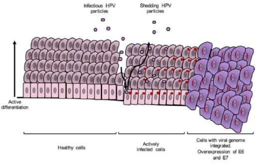
HPV infects basal epithelial cells through anatomically accessible points such as microlesions in the skin, genital organs and oropharyngeal areas. Most infections are cleared by the immune system; however, some benign cervical lesions can progress to cancer. When the viral genome integrates into the host genome and replicate until it reaches a huge number of copies (Figure 3). (Chabeda et al. 2018; Ali et al. 2017)
Figure 3: The lifecycle of a typical hr-HPV. (Chabeda et al. 2018)
HPV infection process starts when capsid proteins L1 and L2 attach to epithelial cell receptors and a long process of entry commences, resulting in cytoplasmic uncoating of the virus and entry of its genome into the nucleus of the infected cell, where it is transcribed and then replicated. The early proteins are expressed first and regulate the host cell life cycle and genome replication (Figure 4). The E6 protein promotes the degradation of the host apoptosis regulator protein p53 via cellular ubiquitination and activates telomerase resulting in disruption of the control of cell cycle progression and extended cell life. The E7 protein targets and disturbs the tumour suppressor retinoblastoma protein (pRb) by repressing regulation of replication-associated genes and leading to the transition of the cell life cycle to the S-phase and subsequent host cell genome replication. E6 and E7 disrupt the cell cycle regulation and promote prolonged host cell life, leading to genomic instability. Maturation of virions occurs after terminal differentiation of epithelial cells, and their release coincides with natural
5 shedding of senescent cells at the end of the epithelial cell life cycle. (Chabeda et al. 2018; Ali
et al. 2017)
Figure 4: HPV E6 and E7 early proteins action.
1.5. Prevention and risks
Testing with a “Pap smear,” more technically referred to as cytology, has been the most pervasive and effective primary screening method used in high-resource settings. (Rizzo and Feldman 2018)
Not all HPV infections confer the same risk of malignancy. Many factors, behind (epi)genetic changes within the cell, can affect the risk of developing cervical cancer, including host and environmental factors which allow a patient to clear the viral infection, such as age, immunocompetence, or tobacco use, as well as the oncogenic potential of different HPV types. (Rizzo and Feldman 2018) The microbiome at individual body sites can have profound effects on cancer development too. Interestingly, changes in the cervicovaginal microbiome occur during progression from HPV-positive lesions to cervical cancer, which include an increased diversity of resident bacteria and a reduction in Lactobacillus spp. (Hoppe-Seyler et al. 2018)
1.6. Actual therapies
Currently, there are three commercially available prophylactic HPV vaccines on the market: Cervarix®, a bivalent HPV-16/18 vaccine; Gardasil®, a tetravalent HPV-6/11/16/18 vaccine; and
6
L1 protein can form virus-like particles (VLPs) when expressed alone in a variety of cell types, that are morphologically and antigenically highly similar to native virions. These three vaccines effectively prevent HPV infections caused by the targeted types by eliciting the production of neutralising antibodies that bind to the viral particles and block their entrance into host cells. However, these vaccines are not effective at eliminating pre-existing infections, since the target antigens, L1 capsid proteins, are not expressed in infected basal epithelial cells. Therefore, individuals already infected with HPV do not benefit from the current vaccines. (Chabeda et al. 2018)
There are strong efforts to develop next-generation vaccines based on the viral L2 minor structural protein, which are expected to protect against a broader range of HPV types. However, there are still important issues to be resolved. Firstly, widespread application of vaccines in less-developed-countries is substantially hindered by financial and logistic hurdles. Indeed, only a small minority (7.5 %) of females worldwide, aged 10-20 years, are estimated to have received at least one shot of an HPV vaccine. In Portugal, in Nacional Programme of Vaccination there are available only the Gardasil®9 vaccine. Complete vaccination implies the
administration of two doses by intramuscular via to girls with 10 years old. (Direção-Geral de Saúde, 2018) Secondly, HPV-induced carcinogenesis results from a persistent infection with oncogenic HPV types in a process lasting several decades. Since the introduced vaccines are prophylactic, they do not eliminate persistent HPV infections or interfere with their progression to malignancy. (Hoppe-Seyler et al. 2018; El-Deeb et al. 2018)
The traditional approaches for cancer treatment include chemotherapy, surgery and radiation. Chemotherapy represents the main choice of treatment in most cases. Thus, HPV-linked carcinogenesis will remain a major health problem for the next decades and new treatment options are urgently required. (Hoppe-Seyler et al. 2018; El-Deeb et al. 2018)
1.7. Gene Therapy
Although traditional therapies can improve the curing effects in the early stage of cancer in some extent, they seem unvalued to fight against metastasis, recurrence of tumour, and the treatment of advanced cancer. (Liu et al. 2015) In some cases these aggressive therapies are unsuccessful, unspecific and can damage normal healthy cells, can cause multidrug resistance and they are always accompanied by inevitable side effects. (Guidotti, Brambilla, and Rossi 2017; Costa et al. 2014)
Gene therapy has brought a promising and unique approach to medicine because of its wide application in the treatment of various diseases, including hereditary diseases to acquired (infection or cancer) pathologies. (Wang, Dou, and Bao 2014; Costa et al. 2019) This technology uses, permanent or transient, exogenous nucleic acids to repair, replace or regulate genes to
7 express a protein of therapeutic interest and prevent or treat several diseases. (Kim et al. 2015; Hardee et al. 2017) It has been demonstrated that therapeutic nucleic acids delivered into target cells can treat various diseases including cancers and genetic diseases. (Zhang et
al. 2017) For example, tumour suppressor genes can be upregulated, or mutated oncogenes
downregulated in affected cells, as well as the delivery of suicide genes can signal cell death. (Kim et al. 2015)
Gene therapy can be used alone or as an adjuvant to the other mentioned therapies since certain genes can sensitize tumour cells to radiation or drugs enhancing the effect of the clinical approach. (Costa et al. 2014)
1.7.1. Approaches of cancer gene therapy
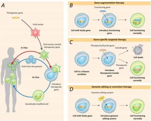
Gene therapy may be carried out by first removing the cells of interest from patients and subsequently reintroducing cells that have been appropriately genetically modified. Alternatively, genetic materials can be directly introduced into the target organs or tissues of the patients (Figure 5A). Different types of gene therapy, including gene augmentation, gene-specific targeting, and most recently, genome editing, have been developed over the prior few decades (Figure 5B-D). (Lee et al. 2019)
Figure 5: Current gene therapy strategies.(A) Health-related or therapeutic genes are directly introduced into the body (red line) or into cells that are removed from the body and then returned to the patient (blue line). Gene therapies utilize several mechanisms, including gene augmentation therapy, gene targeting therapy and genome editing therapy. (B) Gene augmentation therapy replaces a disease-causing
8
gene with a healthy copy of the gene. (C) Gene-specific targeted therapy introduces a new gene or inactivates a disease-causing gene in the body to help treat a disease. (D) Genome editing therapy modifies or corrects a disease-causing gene. (Lee et al. 2019)
Gene augmentation therapy refers to the introduction of a new functional gene into the host genome to compensate for a faulty gene. By adding a functional copy of the faulty gene into the genome, this approach aims to express the functional gene at sufficient levels to replace the missing or dysfunctional protein. (Lee et al. 2019) This gene therapy approach has been used to delivery apoptotic and tumour-associated genes to cancer cells. p53 gene is an example. This gene is delivered to cancer cells in order to restore p53 function and kill tumour cells via apoptosis, cell aging, activating immune system of the host, etc. Even at the advanced stage, restoration of damaged p53 pathway can stop tumour growth. The novel findings have suggested a greater role of p53 in clinical application of gene therapy. (Liu et al. 2011; Chen et al. 2018)
Gene-specific targeted therapy refers to the introduction of genetic materials such as DNA or RNA, designed to specifically alter inappropriate gene activity caused by diseases, or the addition of copies of a gene for protective or regenerative purposes. By manipulating genes involved in pathological changes or their related molecular pathways, gene-specific targeted therapies provide attractive therapeutic strategies for the long-term treatment of nongenetic diseases and autosomal dominant genetic diseases. (Lee et al. 2019)
Genome editing or correction therapy directly repairs and transforms the mutated gene into a normally functioning gene. Thus, this approach has the potential to fundamentally correct mutant genes. It is particularly attractive for the treatment of inherited diseases (autosomal dominant, autosomal recessive and X-linked diseases) caused by genes with very specific spatial and stoichiometric expression. (Lee et al. 2019) However, form all these approaches of cancer gene therapy it is fundamental use viral or non-viral vectors to deliver the target genetic information.
1.7.1.1. Viral vectors
Viral vectors (Figure 6) were the first delivery systems used for gene therapy. (Caffery, Lee, and Alexander-Bryant 2019) Despite the high efficacy of viral vectors such as retroviruses, adeno-associated viruses, lentiviruses and adenoviruses at integrating host genome and delivering genes, they can cause immunogenicity, nonspecific insertion and oncogene activation. Besides, their use currently involves high manufacturing costs, restrictions in the DNA size and perhaps most importantly patient perception. They could lead to the generation of neutralising antibodies which restrict repeated therapy. (Teo et al. 2016; Chabeda et al. 2018; Shao et al. 2017; Bennett et al. 2015)
9 Figure 6: Viral vectors examples used to deliver nucleic acids for gene therapy. (Adapted from (Caffery, Lee, and Alexander-Bryant 2019)
1.7.1.2. Non-viral vectors
A variety of non-viral delivery systems, such as liposomes and polymers, have been tested for gene delivery and will be discussed further. Other non-viral gene delivery vectors as naked RNA or DNA molecules are biocompatible, easily manufactured and relatively inexpensive,
reproductible and synthetically modular.However, their lower gene transfection efficiencies
compared to viral vectors act as a hurdle to their clinical usage.So, they are usually complexed with delivery vehicles (e.g., cationic lipids, cationic polymers, etc.) or subjected to forced entry (electroporation, hydrodynamic injection, etc.). For instance, plasmid DNA has been extensively explored in this field because of its modular nature allowing for straightforward molecular cloning, making it easy to manipulate and design for therapeutic use. (Hardee et al. 2017)
1.7.1.2.1. Plasmid DNA
The enormous potential of plasmids as non-viral vectors in gene therapy has been recognized since at least 1990. (Hardee et al. 2017)
Plasmid vectors represent an important platform for gene delivery as they are safe, stable in storage, easy to manipulate and relatively inexpensive to produce. Plasmids are small circular double-stranded DNA molecules capable of self-replicating in bacterial host cells. Their size ranges from 0.8 to 200 kbp. (Pahle and Walther 2016)
Conventional plasmids contain numerous genetic elements necessary for the plasmid DNA (pDNA) production (origin of replication (Ori), antibiotic resistance genes and eukaryotic selection marker) and suitable for gene therapy (promoters, the gene of interest and terminator). (Figure 7) (Pahle and Walther 2016) The ori allows propagation and self-replication in a bacterial host strain and antibiotic resistance-encoding genes allows selection of plasmid-harbouring. Eukaryotic selection marker promotes plasmid stability and improved transcriptional efficacy and selectivity. Promoters plays a key role in transcription regulation
10
of associated genes. The presence of a strong transcriptional terminator downstream of the encoding sequence prevents transcription of vector sequences, enhances plasmid stability and provides protection against degradation. (Hardee et al. 2017; Singha et al. 2017)
Figure 7: Schematic representation of a plasmid vector.
The pDNA has five conformations, the open-circular (oc), relaxed circular, linear, supercoiled (sc) and the supercoiled denatured conformation. Oc and linear conformations arise through single-stranded and double-stranded nicks, respectively. These plasmid forms have been deemed undesirable for clinical purposes, due to a perceived increased risk of recombination events and integration into genomic DNA (gDNA). These forms may also be subject to more rapid intracellular degradation. Sc conformation is the native pDNA isoform, presents a compact conformation and consists of complete double strand with the normal twist and shape. It is the one of choice in gene therapy due to its outstanding stability, eminent antigenicity and the more efficient for gene transfection of all isoforms. (Abdulrahman and Ghanem 2018; Valente et al. 2014; Ghanem, Healey, and Adly 2013)
Plasmid manufacturing can be divided into various steps (Figure 8):
1. Cell culture: after selecting a specific strain and the target plasmid, the host is cloned. This is followed by the amplification of pDNA copies, in an adequate host, by fermentation at specific and optimized growth conditions;
2. Downstream processing: the main aim of this step is to extract and isolate the pDNA in a reasonable purity that meets the pharmaceutical specifications and get rid of the other cell impurities like RNA, proteins and gDNA. The whole process consists of three stages: preliminary purification, intermediate purification and final purification. In the preliminary purification step, the high-density broth of cells harvested from fermentation step was collected by centrifugation or microfiltration. Subsequently, the cells are lysed to liberate the pDNA by alkaline lysis methods. In the second step, clarification and pre-purification procedures are taken to decrease the levels of impurities in the lysates and concentrate the pDNA lysate sample for the final purification stage;
3. Purification: this is the most important and expensive step of all biotechnology pDNA obtention process. In addition to being dependent on the efficiency of the previous production process is essential to ensure the purity recommended by regulatory agencies
11 in order to continue with its application and profitability of the process. Various purification procedures can be taken like precipitation, ultrafiltration and aqueous two-phase systems; nevertheless, liquid chromatography is certainly the most extensively implemented technique. The purification technique must guarantee good solution appearance (clear, colourless), at least 97 % purity of the final obtained sc pDNA, the RNA and protein content should not be detectable, the amount of gDNA needs to stay above 2 ng/µg of pDNA and the level of endotoxins should not be more than 0.1 EU/µg of pDNA. (Abdulrahman and Ghanem 2018; Valente et al. 2014)
Figure 8: Schematic representation of the biotechnological approach used in the plasmid DNA production. However, the use of plasmid vectors for gene therapy are beset with some notable inherent limitations:
− Most plasmid DNA preparations contain also the non-functional open circular and linear isoforms;
− They are non-replicating episomes, transgene expression is transient and diluted by cell division;
− Bacterial sequences in plasmids can contribute to their gene silencing;
− Unmethylated cytosine-phosphate-guanine (CpG) dinucleotides have the potential to be recognized by the mammalian immune system, potentially precipitating not only transgene silencing but also immune response;
− The bacterial origin of replication (ori) potentially allows plasmids meant to deliver therapeutic sequences to also inadvertently transfer into and replicate in other bacteria; − Plasmids encode antibiotic resistance-encoding genes for selection of plasmid-harbouring
bacteria. The use of antibiotics and their resistance genes in the preparation of plasmid vectors is discouraged by regulatory bodies such as the Food and Drug Administration (FDA) and the European Medicines Agency (EMA) because of the risk of transfer and replication of resistance genes to bacteria in the human microbiome and possibly into the environment. Residual antibiotics that remain from vector production may also trigger an immune reaction in patients.(Hardee et al. 2017)
12
− Plasmids in its native form present some instability in the presence of serum nucleases, as well as low transfection efficiency due to the repulsion by the eukaryotic membrane induced by negative charge of phosphate groups, between other, being fundamental to combine the pDNA with an adequate delivery system.
1.7.2. Delivery systems
The application of nucleic acids in gene therapy is a challenge because of its inability in the extracellular matrix and its inability to cross the eukaryotic cell membrane barrier and reach the nucleus in its native solution form. In order to minimize these limitations and maximize the pDNA transfection efficiency, various delivery system types have been investigated. The use of a suitable delivery vector can compact the nucleic acids providing stability, protection from enzymatic degradation and innate immune system, extravasation from the vasculature and efficient translocation inside the cell through attachment to the target cells, cell membrane passage, endolysosomal escape to reachthe cytosol and transport to the nucleus to induce the therapeutic effect. (Jain et al. 2015; McCarthy et al. 2014; Costa et al. 2015) The carriers should have a suitable size (nanometer range), incorporate large amounts of DNA, be capable of release the payload in a sustained manner and must be biocompatible, elicit minimal side-effects and have good biodistribution. (Costa et al. 2015)
While physical methods of delivery enhancement, such as using ultrasound, heat, light, and applied magnetics or electric fields, have been utilized to deliver gene carriers to target cancer sites, these methods rely on precise knowledge of the location of tumours. To reach both known and unknown locations of cancer cells, other research efforts have focused on the development of viral and non-viral vectors that can be combined to obtain the ideal delivery vehicle to ensure site-specific accumulation and cancer-specific transfection through both passive and active targeting. (Kim et al. 2015)
1.8. Delivery vehicle types
Delivery vehicles are engineered so that they are attracted specifically to diseased cells, which allows the target delivery of drugs, proteins or nucleic acids for the direct treatment of those cells, improving efficacy, decreasing side effects and overall improving human health. (Jahangirian et al. 2017)
Cationic lipid-based vectors (liposomes) (Figure 9) are safe, cost-effective, easier to fabricate and have a great potential for solubilizing, encapsulating and administering drugs with a potential to improve drug absorption thereby contributing with their bioavailability and minimizing side effects. For synthesis of these nanocarriers the most materials are biocompatible lipids. One of the biggest problems of their use includes the high toxicity at low
13 specificity and sensitivity to serum components. (Medina-Alarcon et al. 2017; Martinez-Negro
et al. 2018; Teo et al. 2016; Pahle and Walther 2016)
Figure 9: Some delivery vehicles types. (Adapted from (Lembo and Cavalli 2010))
Polymeric nanoparticles are high tailorable, durable, easy to prepare, purify and chemically modify, are stable and protect against enzymatic degradation, allows controlled release, prolonged circulation half-life and high penetration capacity. However, there are some disadvantages as the degradation and high cost to manufacture. (Medina-Alarcon et al. 2017; Teo et al. 2016)
Metallic or inorganic nanoparticles (Figure 9) are used for distribution of targeted drugs, gene delivery and in the diagnosis. Gold nanoparticles due to their favourable physical, chemical and photometric properties are frequently applied in cancer therapy as drug transporters or with other drugs for penetrating tumour cells, thus decreasing the dose of the drug and improving the response to tumour cells. Another very encouraging class of nanoparticles is the multi-walled carbon nanotubes (Figure 9) and magnetic nanoparticles as they carry plasmids which can target tumours. The major drawback by using inorganic particles is their slow biological clearance and unwanted persistence. (Medina-Alarcon et al. 2017; Pahle and Walther 2016)
Dendrimers (Figure 9) are highly branched 3D polymers. They are capable of penetrate cellular and endosomal membranes. (Caffery, Lee, and Alexander-Bryant 2019) Can incorporate small molecules by electrostatic or hydrophobic interactions, and the drugs can be bonded on their
14
surface through polar interchanges. Are used in medical and biotechnological applications due to their biocompatibility. (Medina-Alarcon et al. 2017)
Micelles (Figure 9) are amphiphilic copolymers that have a hydrophobic core/hydrophilic shell structure. (Caffery, Lee, and Alexander-Bryant 2019) Micelles allow a great depth of tissue penetration for targeted drug delivery and they usually disintegrate rapidly in the body. Despite the capability to load drug and gene, micellar systems need to display high thermodynamic and kinetic stability to prevent premature drug release when administered in the blood stream. (Medina-Alarcon et al. 2017; Teo et al. 2016)
Mesoporous silica nanoparticles (MSNs) have been used to deliver small molecules, genes, and proteins. Their large surface area provides space and binding sites to accommodate nucleic acids via non-covalent encapsulation or direct covalent conjugation. Their pores allow the accommodation of small molecules and their surface can be engineered with cationic polymers to encapsulate nucleic acids via electrostatic binding. (Jiang et al. 2016)
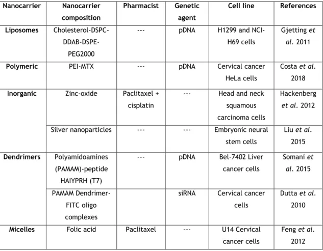
Several nanocarriers have been developed and applied in vitro, some of which are described herein (Table 1).
Table 1: Example of practical applications of carriers in gene therapy.
Nanocarrier Nanocarrier
composition
Pharmacist Genetic
agent
Cell line References
Liposomes
Cholesterol-DSPC- DDAB-DSPE-PEG2000
--- pDNA H1299 and NCI-H69 cells
Gjetting et
al. 2011
Polymeric PEI-MTX --- pDNA Cervical cancer
HeLa cells
Costa et al. 2018
Inorganic Zinc-oxide Paclitaxel +
cisplatin
--- Head and neck squamous carcinoma cells
Hackenberg
et al. 2012
Silver nanoparticles --- --- Embryonic neural stem cells Liu et al. 2015 Dendrimers Polyamidoamines (PAMAM)-peptide HAIYPRH (T7)
--- pDNA Bel-7402 Liver cancer cells Somani et al. 2015 PAMAM Dendrimer-FITC oligo complexes
siRNA Cervical cancer cells
Dutta et al. 2010
Micelles Folic acid Paclitaxel --- U14 Cervical
cancer cells
Feng et al. 2012
15
1.8.1. Cellular barriers
At the cellular level there are various biological barriers that gene carriers need to overcome (Figure 10). These include cellular uptake and endosomal escape, intracellular trafficking, therapeutic molecule unpacking and biocompatibility. Complexes may be internalized via endocytosis after electrostatic binding to the anionic proteoglycans on the target cell membrane. The different endocytic pathways define the intracellular routes. Currently, clathrin-mediated endocytosis is the most characterized pathway, during which clathrin-coated pits pinch of the cell membrane and fuse with endosomes. Vesicle acidification continues when endosomes mature to lysosomes, with reduction of pH to around 5. Thus, the ability for the complexes to escape from enzymatic degradation in the endosomal low pH environment is paramount to successful gene delivery. (Teo et al. 2016)
For the polyplexes that escape out of the endosome into the cytosol, where is abundant nucleolytic enzymes, protection of nucleic acids is mandatory. For the specific case of pDNA, although the exact mechanism by which pDNA travels to the nucleus and is expressed is still under investigation, researchers have suggested that pDNA travels on the microtube network driven by microtubule-associated motor proteins to reach the nucleus. (Teo et al. 2016)
Figure 10: Barriers to intracellular trafficking of gene delivery systems. (Adapted from (Teo et al. 2016))
1.9. Cell penetrating peptides
Many peptides have been developed over recent years because they present the ability to penetrate the plasma membrane without compromising their integrity. These peptides are normally short (less than 30 amino acids), frequently rich in basic amino acids residues like
16
arginine residues, resulting in positively net charged. The cationic profile of cell penetrating peptides (CPP) is the key factor for their interaction with nucleic acids, giving rise to the formation of micro or nanometer complexes. CPPs are advantageous over other translocation methods because they possess high cellular permeability rates, ability to translocate into a wide spectrum of cell types, large cargo capacity and low cell toxicity associated with no immunological response. (Costa et al. 2019; Silva, Almeida, and Vale 2019)
The most publicized peptides have been the anionic membrane destabilizing peptide, GALA, which requires conjugation of a positively charged ligand to facilitate delivery of nucleic acid. On the other hand, CADY, KALA and RAWA are CPP which allow independent delivery of nucleic acids. (Bennett et al. 2015)
1.9.1 GALA and KALA peptides
Perhaps one of the most revolutionary peptides is GALA. It is fusogenic in nature and the seven EALA repeats confers α-helicity linked to pH responsiveness, realized through excellent endosomal disruption. Due to the spacing of the charged residues this peptide is capable of taking on the conformation of an amphipathic α-helix at a lower pH. However, due to its anionic nature, it cannot condense and protect nucleic acids. Coating of cationic complexes with GALA peptide, however, has proven successful at facilitating the endosomal escape of multi-functional delivery systems which are capable of binding and delivering nucleic acids intracellularly. (McCarthy et al. 2014)
To that end, KALA was the first artificially designed peptide of its kind with the glutamate residues of GALA replaced with lysine residues, enabling nucleic acid condensation and protection. The presence of multiple lysine residues in KALA means that in contrast to GALA, it has the ability to permeabilize membranes in a composition and pH dependent manner. The net charge of KALA increases with decreasing pH in the presence of neutral liposomes disrupting the α-helical structure and ablating any membrane permeabilizing activity. In the presence of negatively charged membranes however, the increased positive charge on KALA allows improved binding to the membrane and enhances its membrane permeabilizing activity. (McCarthy et al. 2014) However, circular dichroism (CD) studies showed that KALA undergoes a conformational change from an α-helical conformation to a mixture of α-helix and random coil as the pH is lowered. A decrease in the pH below 7 results in protonation of the histidine, charge neutralization of the glutamic acid side chains, and hence an increase in the net positive charge in the molecule which appears to disrupt the α-helix. In vitro studies also showed that KALA cause membrane destabilization and leakage in low-pH environments similar to that of the endosome. (Wyman et al. 1997)
17
1.9.2. RALA peptide
Arginine is found in naturally occurring DNA binding/condensing motifs with a higher frequency than lysine and has been shown to be a superior transfection agent. As arginine-rich peptide sequences exhibit improved internalization they have been widely exploited to aid the delivery of various gene delivery vehicles including peptide dendrimers, liposomes and polymer systems. McCarthy modified the RALA peptide, a 30-amino acid peptide where the KALA lysine residues were replaced with arginine, with decrease cytotoxicity, pH sensitivity and improved binding to outer leaflets of membranes and nucleic acids. (McCarthy et al. 2014)
RALA is an amphipathic peptide and exhibits an alpha helical structure (Figure 11) in a hydrophilic environment with hydrophobic amino acids on one face (leucine’s) and hydrophilic on the other (arginine’s), making it a suitable candidate for DNA condensation and efficient membrane perturbation. (Jain et al. 2015)
NH
2-WEARLARALARALARHLARALARALRACEA-COOH
Figure 11: RALA primary and secondary structure. (Adapted from (Udhayakumar et al. 2017))
Arginine is positively charged in neutral, acidic and most basic environments, making RALA more responsive to the low pH found in the endosome, causing α-helicity selectively in the endosome and theoretically minimizing toxicity and leading to endosomal disruption and release of the cargo into the cytosol. (Massey et al. 2016; Bennett et al. 2015)
This peptide when exposed to anionic nucleic acids and due to electrostatic interactions self-assembles into nanoscale particles suitable for cell membrane penetration facilitating the intracellular delivery of DNA across the cell membrane and promoting nuclear localisation of the DNA for transcription. (McCrudden et al. 2017; McCaffrey et al. 2016)
Characterization studies indicated that RALA does not affect viability in vitro and successful reporter gene delivery following systemic administration in vivo.(Bennett et al. 2015)
18
RALA has been applied at delivered plasmids encoding reporter genes, mRNA, siRNA, DNA vaccines, mRNA vaccines and small molecules demonstrating broad utility (Table 2). (McCrudden et al. 2018)
Table 2: Examples of other studies using RALA peptide as carrier.
Nanocarrier Genetic agent Cell line References
RALA-PLA-PEG pDNA ZR-75-1 Breast cancer cells Jain et al. 2015
RALA pEGFP-N1 ZR-75-1 Human breast
cancer cells and PC-3 human prostate cancer cells
McCarthy et al. 2014 RALA-poly(vinylpyrrolidone) (PVP) pCMV-Luc and pEGFP-N1
NCTC-929 Fibroblast cells McCaffrey et al. 2016
RALA iNOS Breast cancer cells McCrudden et al.
2016 RALA pFKBPL and siFKBPL ZR-75-1 Breast cancer cells Bennett et al. 2016 RALA pFKBPL ZR-75-1 Breast cancer cells Bennett et al. 2015
RALA iNOS MDA-MB-231 Breast cancer
cells
McCrudden et al. 2017 RALA E6/E7 DNA NCTC-929 Fibroblast cells Ali et al. 2017 RALA pDNA NCTC-929 Fibroblast cells Cole et al. 2018
RALA mRNA Dendritic cells Udhayakumar et al.
2017 RALA Bisphosphonates PC3 Prostate cancer and
MDA-MB-231 breast cancer cells
19
Chapter II - Aim of the thesis
The objective of this thesis consisted in the design and development of a peptide/plasmid DNA delivery system, its biological activity evaluation in vitro and the investigation of its potential cancer therapeutic effect as alternative to conventional therapies applied in clinical setting nowadays. The main idea was to restore the p53 normal function in the cell and regulate their cycle leading to apoptosis.
In order to achieve this objective, it was crucial the formulation of a system composed by plasmid encoding p53 tumour suppressor gene encapsulated by RALA peptide at different N/P ratios and characterize it in terms of morphology, size, surface charge, encapsulation efficiency and pDNA protection and stability. Posteriorly, the capacity of the developed systems for cell uptake and internalization must be evaluated. Genetic expression, p53 protein expression and efficacy in inducing cancer cell apoptosis were also analysed in order to verify the therapeutic potential of this vector.
21
Chapter III - Materials and Methods
3.1. Materials
The pcDNA3-FLAG-p53, plasmid #10838, was purchased from Addgene (Cambridge, MA, USA). For the bacterial cultures, tryptone and yeast extract were obtained from Bioakar Diagnostics and Luria-Bertani (LB) medium in PanReac. For alkaline lysis and pDNA purification a protocol optimized by our research group and described in the literature was used. (Diogo et al. 2000; Sambrook, Fritsch, and Maniatis 1989) In electrophoresis assays, GreenSafe reagent (GreenSafe Premium) was obtained from NZYTech Lda. For systems formulation, RALA peptide
(N-WEARLARALARALARHLARALARALRACEA-C) with average Mw =3328.30 MM and a purity of 95.02
% was obtained from Biomatik. 3-(4,5-dimethylthiazol-2-yl)-2,5-diphenyltetrazolium bromide (MTT) and fluorescein isothiocyanate (FITC) were obtained from Sigma-Aldrich (St Louis, M.O., EUA). 4′,6-diamidino-2-phenylindole (DAPI) was purchased from Invitrogen (Carlsbad, CA). Normal human dermal fibroblasts (NHDF), Ref. C-12302 (cryopreserved cells), and cancer HeLa cells were purchased from PromoCell and Invitrogen, respectively. For PCR methodology, Taq polymerase and MgCl2 were obtained from NZYTech, Lda. For western blot assay anti-p53
antibody (Santacruz Biotechnology, DO-1: sc-126), anti-mouse IgG–peroxidase antibody (Sigma-Aldrich, A3682), BAX antibody (Cell Signalling, #2772), anti-rabbit IgG–peroxidase antibody (Sigma-Aldrich, A0545), monoclonal anti-β-actin−peroxidase antibody (Sigma-Aldrich, A3854), PierceTM ECL Western Blotting substrate (Thermo Scientific) and NZYcolour Protein Marker II
(NZYTech, Lda. - Genes and Enzymes, Lisbon, Portugal) were acquired. All solutions were freshly prepared by using ultra-pure grade water, purified with a Milli-Q system from Millipore (Billerica, MA, USA).
3.1.1. Plasmid DNA
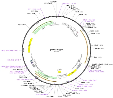
The 6.59 kbp plasmid pcDNA3-FLAG-p53 used in this set of assays encodes for the human p53 protein, for the ampicillin resistance gene and the neomycin selectable marker (Figure 12).
22
Figure 12: Plasmid pcDNA3-FLAG-p53. (Adapted from Addgene)
3.2. Methods
3.2.1. Bacterial growth conditions and plasmid production
The plasmid was amplified intracellularly in Escherichia coli (E. coli) DH5α. Initially, the strain was inoculated in LB-agar plates containing ampicillin (30 µg/mL) and it was left to grow overnight at 37 ºC. After the growth in solid medium, some colonies were transferred to an Erlenmeyer with capacity of 250 mL, containing 62.5 mL of Terrific Broth (TB) liquid pre-fermentation medium, composed by 20 g/L tryptone, 24 g/L yeast extract, 4 mL/L glycerol, 0.017 M KH2PO4 and 0.072 M K2HPO4 and supplemented with 30 μg/mL ampicillin. The cells were
left to grow in an orbital agitator (Agitorb 200 da Aralab, Albarraque, Portugal) at 250 rpm and 37 ºC. When an optic density (OD) of approximately 2.6 was reached, a certain volume of pre-fermentation medium was transferred to 125 mL of TB pre-fermentation medium included in two erlenmeyers of 500 mL. After 16/18 h (OD ~ 9, at the late log phase), bacterial growth was suspended, and the total volume of fermentation divided for 50 mL Falcon tubes for posterior cell collection by centrifugation at 1000 g for 4 min and pellets storage at -20 ºC until use.
3.2.2. Plasmid extraction and purification
Posteriorly to production, plasmid was extracted/recovered by the modified alkaline lysis method and purified according to the optimized protocol by our research group. (Diogo et al. 2000; Sambrook, Fritsch, and Maniatis 1989) The modifications were made to NZYTech Plasmid Maxi kit manufacturer protocol in order to optimize the plasmid recovery rate. Initially, the
23 bacterial pellet was resuspended in 10 mL of P1 buffer (50 mM of Tris-HCl at pH = 8.0, 10 mM of EDTA hydrated and 100 μg/mL RNase A) and then in 10 mL of P2 buffer (200 mM of NaOH and 1 % of SDS (w/v)). To promote cell lysis the tubes were carefully inverted and incubated at room temperature for 5 min. The lysis step was stopped with the addition of 10 mL of P3 buffer (3.0 M potassium acetate at pH = 5.0 and 1 % of SDS (w/v)) and incubation on ice for 20 min. Then, tubes were centrifugated for 30 min (20 000 g and 4 ºC), supernatant was transferred to new tubes and centrifugated 20 min (20 000 g and 4 ºC). Thus, some precipitated contaminants are eliminated, such as cell debris, gDNA and proteins. Afterwards, the columns (anion-exchange resin with L-methionine-agaroses matrix) were regenerated with a regeneration solution (0.15 % Triton X-100 and 3 M NaCl) and equilibrated with QBT buffer (0.75 M NaCl, 50 mM MOPS, 15 % isopropanol (v/v), 0.15 % Triton X-100 (v/v) at pH = 7.0). The supernatants were added to columns and the proteins and low molecular weight molecules were removed by the addition of a buffer with low salt content (0.5 M NaCl). Elution of plasmid DNA was promoted directly to empty centrifuge tubes by the addition of a buffer with higher salt concentration (1.25 M NaCl). The pDNA was precipitated by the addition of 0.7 volumes of isopropanol, followed by incubation on ice for 20 min. The obtained samples were subjected to centrifugation (16 000 g, 4 ºC and 10 min) and the pellet was resuspended in 1 mL of Tris base (10 mM, pH 8.0) and stored at -80 ºC. The 260/280 nm absorbance ratio was found to be above 1.8. The samples were also analysed by electrophoresis for 30 min under 120 V in 1 % agarose gel containing ethidium bromide with Tris-acetate-EDTA (TAE) (40 mM Tris base, 20 mM acetic acid and 1 mM EDTA, pH 8.0) running buffer and GreenSafe as colorant to verify the main conformation of the plasmid and possible contaminations.
3.2.3. Preparation of RALA/pDNA complexes
RALA was synthetized by solid-state synthesis (fluorenylmethyloxycarbonyl, FMOC) and supplied as a desalted lyophilized powder. Reconstitution was in ultrapure grade water to a stock concentration of 0.5 mg/mL. Aliquots were stored at -20 °C until use.
The systems were prepared at various nitrogen/phosphate (N/P) ratios, ranging from 1 to 50, considering the mass per charge ratio of DNA (330 g/mol, relative to one phosphate group) and
RALA (44 g/mol, correspondent to one amine group). Nanoparticles were formedby adding an
appropriate and calculated volume of peptide solution, dropwise at vortex for 60 s, to a fixed amount of pDNA (1 µg) (at ratio N/P 1 were added 2.9 µg of RALA peptide to 1 µg of pDNA) and left for 30 min at room temperature to allow complexes formation.
3.2.4. Determination of encapsulation efficiency (EE)
After complexes formation they were centrifuged at 10 000 rpm for 20 min at 4 ºC and the pellet contained the pDNA based vectors was recovered. The amount of non-bound pDNA in