http://dx.doi.org/10.1590/s2175-97902017000400212
Article
*Correspondence: T. K. Chatterjee. Department of Pharmaceutical Science and Technology, JIS University, Kolkata-700109, West Bengal, India. E-mail: tkchatterjee_81@rediffmail.com / crctkc@gmail.com
Pharmacological and toxicological investigations of etodolac loaded
gum katira microspheres prepared by W
1/O/W
2emulsion solvent
evaporation technique in rats
Biswajit Ruhidas
1, Rajat Ray
2, Debjyoti Naskar
1, Biplab Kumar Chakra
1,
Tapan Kumar Chatterjee
3*
1Department of Pharmaceutical Technology, Jadavpur University, Kolkata, India,2Department of Pharmaceutical
Technology, Adamas University, Kolkata, India, 3Department of Pharmaceutical Science and Technology, JIS University,
Kolkata-700109, India
Etodolac is a non-steroidal anti-inflammatory drug (NSAID) and approved by USFDA as a COX2 inhibitor. Although etodolac therapy provides clinical benefits, it is associated with upper gastrointestinal (GI) tract complications also. Etodolac loaded gum Katira microsphere (ELGKM) was prepared by
W1/O/W2 emulsion solvent evaporation technique. The gastric irritation properties of orally administered
pure etodolac, ELGKM and blank microspheres (without etodolac) were evaluated in experimental rats treated for 6 days. The stomach examination and biochemical investigation of stomach tissue of treated rats indicated that ELGKM formulation remarkably reduced ulcerogenecity as compared to pure etodolac. The anti-inflammatory activities of pure etodolac and ELGKMs were ascertained by the implantation of cotton pellets in rats for 6 days. Based on the results, ELGKMs showed significant anti-inflammatory activities (P<0.01) as compared to control group. The cotton pellets test suggested that ELGKM formulation retained more anti-inflammatory properties among the groups. The hematological changes, biochemical analysis and histopathological studies of subacute toxicity in rats revealed that ELGKM were the effective sustained release formulation in the treatment of chronic pain and inflammation. In conclusion, the physicochemical characterization, pharmacological and toxicological studies suggest that ELGKMs may represent as a potential candidate for sustained drug delivery (10-12 hours) in chronic joint pain related diseases with remarkably diminished gastrointestinal side effects.
Keywords: Etodolac/inflammatory conditions. Etodolac/physicochemical characterization. Gum Katira/ microspheres. Pain therapy.
INTRODUCTION
Non-steroidal anti-inflammatory drugs (NSAIDs) have been used for the treatment of several rheumatic and inflammatory diseases for more than 30 years. Etodolac
is used as an analgesic for the treatment of signs and symptoms of rheumatoid arthritis, osteoarthritis and
the relief of post-operative pain (Craig, Stitzer, 2004).
The pharmacological actions of etodolac are related
to inhibition of prostaglandin bio-synthesis at the site
of inflammation. Etodolac decreases the synthesis of
peripheral prostaglandins involved inflammation by
inhibition of the cyclooxygenase (COX-2) enzyme preferentially (Glaser et al., 1995).
The oral route is one of the most popular routes
of drug delivery because of easy administration, patient compliance, least sterility and flexible design of dosages forms. The most conventional dosage forms (tablets and capsules) for oral drug delivery are formulated to
get the active compound in the systemic circulation
immediately after administration. Now a day, various modified drug release system have been developed to
deliver therapeutic drug at a controlled rate in sustained
the drug for prolong period of time (10-12 hours) in a sustained fashion. There were no significant chemical interaction among the excipients used in the microspheres preparation (Ruhidas et al., 2016).
Etodolac is well absorbed from the gastrointestinal tract following oral administration. The bioavailability of etodolac is about of 80% and plasma peak concentration usually attained within about 1.4 hours (tablet or capsule) or 7 hours (extended-release tablets) (Indian Pharmacopoeia Commission, 2014). In case of rheumatoid arthritis and
osteoarthritis, patients generally forget to take medicine
in the morning and evening or in between. Once or twice daily dosing thus improves therapy by maintaining steady state plasma drug concentration of the drug in the blood, so avoids the peaks of high plasma drug concentration as well as troughs of low plasma drug concentration.
Gastrointestinal side effects are the most frequently occurring adverse effects associated with orally
administered anti-inflammatory drugs. The risks of
gastrointestinal ulceration, bleeding and even perforation with NSAIDs therapy was well known (Rainsford, 1989).
These effects may result from local action of drugs
which caused injuries to the sub-mucosal capillaries with subsequent necrosis and bleeding or from the inhibition
of formation of protective prostaglandins.
ELGKM avoids the side effects associated with high concentration as well as no side effect during troughs, giving better overall therapy and improves patient compliance. Therefore, appreciable amounts of etodolac are prescribed as a special formulation (ELGKMs), which has the potential to minimize its toxicity and improve its pharmacological efficacy as well as bioavailability. ELGKM has been formulated to reduce gastrointestinal
tract irritation and to release the drug over a prolonged
period (10-12 hours). The present pharmacological and toxicological study has shown reduction in ulcerogenic property and better anti-inflammatory activity of ELGKMs
formulation as compared to pure etodolac.
MATERIAL AND METHODS
Chemicals
Etodolac (molecular weight, 287.35g/mol, 95.42%) was gifted by M/S Fleming Laboratory Limited, Dist. Medak, Andhra Pradesh, India. Crude Gum Katira was obtained from Seoni District of Madhya Pradesh. Eudragit®RS75 and Eudragit®RL75 polymer granules were obtained as gift sample from Evonik Rohm, Pharma Polymers, Kirsechenallee, Darmastand, Germany. Span 80 (Loba Chemie Pvt. Ltd, Mumbai, India),
Tri-Sodium Orthophosphate (Loba Chemie Pvt. Ltd. India), Hydrochloric Acid 35% (Merck Life Science Pvt. Ltd. India), Tween 80 (Merck Specialties Pvt. Ltd, India), Dichloromethane (Merck Specialities Pvt. Ltd, India), Potassium dihydrogen phosphate (Merck Specialties Pvt. Ltd, India), and all others analytical grade chemicals were
purchased and used as received.
Animal
Male albino rats (150-200 g) and rabbits (1.5-2 kg) were taken as experimental animal. The animals were acclimatized to laboratory conditions for 3days before the commencement of the experiment and kept under standard condition of temperature (25 ºC), relative humidity (70±10%) and a 12 hours light/12 hours dark
cycle environment. During the study period, guideline of Committee for the Purpose of Control and Supervision of
Experiment on Animal (CPCSEA), Institutional Animal Ethics Committee (IAEC) were properly followed for the maintenance of animal and the experiment protocol was approved by Animal Ethics Committee of Jadavpur University, Ref. No. AEC/PHARM/1601/02/2016 dated 22/04/2016.
Preparation of etodolac loaded gum Katira Microspheres
Etodolac loaded gum Katira microspheres were prepared by double-emulsion solvent evaporation technique. Gum Katira (50 mg) was mixed with 4ml of phosphate buffer (pH-6.8) to form a homogeneous mixture
in a magnetic stirrer for 1hr at a constant temperature of
40-450C. Etodolac was added to the homogeneous mixture
and stirring was continued for another 1hr. The prepared etodolac and gum Katira mixture was then dispersed in a solution of Eudragit®RS100 and Eudragit®RL100 (7:1), Dichloromethane, Acryflow (Lubricating agent)
and Span 80 (30 µL) through a 20-gauge syringe. The
above mixture was homogenized well for 5-10 minutes using magnetic stirrer (1000rpm) to form W1/O emulsion. A separate acidic aqueous solution (100 mL) containing
Tween 80 (50 µL) and a slight amount of polyvinyl alcohol
was subjected to mechanical stirring (900 rpm) and to
it the previously prepared W1/O emulsion was added
Ulcerogenic investigation
Experimental procedure
Ulcerogenic properties of etodolac and ELGKMs formulation were assessed using the method described by Shanbhag et al. (Meier et al., 1950). Rats were randomly distributed into four groups (n = 6 in each group). The control group received no drug while the standard groups
were given pure etodolac (50 and 75 mg/kg b.w., po),
test groups received ELGKMs formulation (50 and 75
mg/kg b.w) respectively, po) and blank microspheres
formulation (equal to 50 mg) for 6 days. The animals were fasted 8 hours pre and 4 hours post-treatment (Vyas et al., 2009). Food and water were available for the rest
of the time. On 7th day, the animals were sacrificed. The
stomachs were removed and placed on the saline soaked filter paper until the examination. A longitudinal incision along the greater curvature was made with fine scissors. The stomachs were or absences of gastric irritation were
determined. Based on the severity of gastric mucosal
damage, each gastric lesion was assigned a score. The scores were averaged and the mean score tabulated as the severity indexes for the drug and the formulated drug
suspension administered. The rating scale of mucosal
damage of stomach tissue has been shown in Table I (Tammara et al., 1993). The overall score was divided by the factor of 10, which was designated as the ‘Ulcer Index’ ( Main, Whittle, 1975).
Preparation of stomach tissue homogenate
After the observation of mucosal damage of stomach of the different groups of animals were kept in So-Low Ultra Low freezer (-80ºC) for 12 hours (So-Low Ultra Low freeze, Model No.- C85-5, USA). The following day, a small portion of stomach tissue was homogenated using a homogenizer (RIMI MOTOR, Type-RQ 127A) with the favorable buffer solution for in different biochemical
testing for assay of ulceration.
Biochemical investigation of ulcerogenic stomach of rats
Estimation of Total Protein (T.P)
To 0.1 mL of tissue homogenate 0.5 mL freshly prepared reagent C (alkaline coper solution: 50 mL of reagent A mixed with 1 mLof reagent B) was added and kept for 10 minutes at room temperature. To 0.5 mL of the solution, 0.5 mL of distilled water and 5.0 mL of reagent C was added. Then 0.5 mL reagent D (Folin and Ciocalteu reagent: diluted 2:1 with distilled water) was added to this solution and kept for 30 minutes at room temperature. Optical density was measured at 660 nm in UV. Total Protein was estimated by the Lowery method (Peterson, 1979) using Bovine serum albumin (BSA) as
standard, at 660 nm.
Assay of thiobarbituric acid reactive substances (TBARS)
The level of Lipid per-oxidation was estimated by measuring the concentration of malondialdehyde in the stomach tissue according to the modified method of
Ohkawa et al. (1979). 0.5 ml of tissue homogenate (0.025
M Tris-HCl buffer, PH-7.8),0.5 mL saline and 1.5 mL of 20% TCA ( Tricyclic acetic acid) were added and mixed well and then centrifuged at 3000 rpm for 20 minutes. To 1.0 mL of the protein raw supernatant, 1.5 mL of 0.8%TBA (Thiobarbituric Acid) reagent was added. The content
mixture was mixed well and boiled for 1 hour at 95 ºC and
pink colour developed. The tubes were then cooled under running tap water. The absorbance of clear supernatant was measured against reference blank at 532 nm (Ohkawa,
Ohishi, Yagi, 1979).
Assay of catalase (CAT)
The stomach tissue homogenate was prepared by using phosphate buffer (0.01 M, PH-7.0). The homogenate was centrifuged at 5000 rpm for 10 minutes. To 0.9 ml phosphate buffer, 0.1ml of tissue homogenate
(supernatant) and 0.4 mL of 2 mM H2O2 were added.
The reaction was arrested after 15, 30, 45, 60 seconds by adding 2.0 mL of dichromate acetic acid mixture (5% potassium dichromate and glacial acetic acid were mixed in 1: 3 ratio). The tubes were kept in a boiling water bath for 10 minutes and cooled. The colour was developed and measured at 620 nm (Sinha, 1972).
Assay of reduced glutathione (GSH)
Reduced glutathione (GSH) in gastric mucosa was assayed by the method of Ellman. At first, stomach was homogenized in phosphate buffer (0.2 M, PH-8.0). Then
TABLE I - Rating scale of mucosal damage of stomach in rats
Observation of mucosal damage Score
No lesion 0.0
Punctiform lesions (less than 1mm) 1.0
Five or more punctiform lesions 2.0
One to five small ulcers (1-2 mrn) 3.0
More than five small ulcers or one large ulcer 4.0
0.5 mL of the tissue homogenized mixture was treated
with 2.0 ml 5% TCA (Trichloroacetic acid).The mixture
was kept on ice for 10-20 minutes and then centrifuged at 3000rpm for 15 minutes. After that 2.0 mL of the supernatant of homogenized mixture was treated with 1.0 ml of Ellman’s reagent and 4.0 mL of 0.3 M disodium hydrogen phosphate. The absorbance of yellow colour developed was measured in a UV spectrophotometer at 412 nm within 2-3 minutes. The amount of glutathione is expressed as Unit/minute/mg-protein (Ellman, 1959).
Investigation of anti-inflammatory activity
Pharmacological investigations of ELGKMs formulation by granuloma tissue formation method through cotton pellet grafting under skin
The animals were deprived of food for 24 hours (water ad libitum) prior to the drug administration and received free access to water during the experiment. They were divided into five groups of six animals in each group. The first group was treated as control group receiving 1% CMC solution. Second and third groups were treated
as standard groups receiving pure etodolac suspension
(50, 75mg/kg b.w. respectively). Test groups were given ELGKMs (50, 75 mg/kg b.w respectively). The animals were anaesthetized with diethyl ether. Then the hair of back skin of lumber region was removed and disinfected with 70% ethanol. A subcutaneous incision was made by scalpel. A sterilized cotton pellet (60-80 mg) impregnated with 1% carrageenan solution was implanted. The wounded skin was then closed with a stainless steel suture clip. The animals were kept in separate cages. Then they were treated with pure etodolac and ELGKMs suspension
per oral for 6days (through oral gavages). On the 7th day,
the animals were sacrificed under anaesthetized and cotton pellets surrounded with granuloma tissue were removed. The weight of wet cotton pellets was taken and recorded. Then they were dried at 60 ºC until the weight remained constant. Finally, weights of dry cotton pellets were recorded. The net weight of cotton pellets was determined by subtracting weight of dry cotton pellets from weight of wet cotton pellets.
Haematological assay
At the end of the experimental, on 7th day the
blood was collected directly from heart and used for the
analysis of hematological parameters such as erythrocyte
count, total and differential leukocyte count, hemoglobin concentration, platelet count and blood clotting time.
Haemoglobin estimation
The heparinized blood was taken in the Sahli Hemoglobinometer and diluted with the 0.1 N HCl until the colour matched with the standard. The data was taken from graduated cylinder and expressed as g/dl of blood.
RBC count
The blood sample was diluted 1:75 with the RBC diluting fluid using Thoma pipette after vigorous mixing, a drop of blood mixture was dropped under cover glass of Neubauer hemocytometer and corpuscles were allowed to settle for 3 minutes. The number of RBC in 80 small squares was determined under light
microscope.
WBC count
The blood sample was diluted 1:20 with the WBC diluting fluid with Thoma pipette after vigorous mixing a drop of blood mixture was dropped under cover glass of Neubauer hemocytometer and corpuscles were allowed to settle for 3 minutes. The number of WBC in 16 small squares was determined under light microscope (Armour,
Blood, Belden, 1965; Wintrobe et al., 1976).
Differential leukocyte count and platelet count
The heparinized blood was analysed by digital automatic hematology analyser/blood cell counter (Care Well Biotech Pvt. Ltd., India) in Nilam-The Complete Care, 26B, Ahiripukur Road (Lower Range- Beck Bagan) Kolkata-700019, India and the results were obtained.
Blood clotting time
The blood sample was taken using a glass capillary from orbital plexus of the eye of each rat and the time was noted down. Small pieces of capillary were broken from one end at every 30s till fibrin threads of blood appeared between the broken ends of capillary (Ghai, 1990).
Biochemical assay
Histopathology
Stomach, Liver, Kidney and Heart of treated rats from control and treated groups were dissected into small sections and preserved in cedar wood oil. Infiltration was done by dipping the tissues in xylene: paraffin wax in 1:1 ratio at 60º C for 1 hour and then tissues were dipped in molten paraffin at 60ºC for 1 hour. The processed tissues were embedded in the molten wax for section cutting. Thin section of the paraffin blocks containing tissue was done by rotary microtome. Then the slides were stained with eosin and hematoxylin and mounted with dextrene polystyrene xylene and examined microscopically for pathological examination.
Statistical analysis
All the results were expressed as mean ± SEM. The results were analyzed by one-way analysis of variance (ANOVA) followed by Dunnett’s test through the computer program Graph Pad Instat 3. P value <0.01and
>0.05 were considered statistically significant whereas p
value <0.05 was considered statistically not significant.
RESULT AND DISCUSSION
Gastric irritation properties of orally taken compounds were evaluated in rats kept in fasting condition (Vogel, 2002). The investigations of ulcerogenic
activity of pure etodolac and etodolac loaded gum katira
microspheres (ELGKM) treated rats indicated that all the ELGKMs formulation treated rats showed a remarkable
decrease in ulcerogenic properties compared to pure etodolac-treated rats. The statistical analysis of ulcer
index of treated rats suggested that there were significant differences among the groups (Table II). Etodolac caused gastrointestinal damages and ulcers in pure form as well as microspheres dosages form (Figure 1). But pure etodolac form (50 mg/kg b.w. and 75 mg/kg b.w. respectively) showed a significant gastric damages and ulcerations whereas the ELGKMs formulation (50 mg/kg b.w. and 75 mg/kg b.w) showed significantly lesser gastric damages
and ulceration. Based on ulcerogenic study on treated
rats, the results indicated that ELGKMs formulation (50 mg/kg and 75 mg/kg) possess markedly minimized
the gastrointestinal damaging tendency. Biochemical analysis of stomach tissue homogenate of treated rats
also supported that ELGKM decreased the gastric
damages properties of etodolac in comparison to pure etodolac-treated rats. The gastric damages, ulcerations
and others toxic conditions of stomach causes marked
reduction in some enzyme concentration (Total protein, Catalase and GSH) and increases the concentration of TBARS (Adeyemi et al., 2015). From the biochemical investigation, it was noted that the both the pure etodolac and ELGKMs formulations increased the level of TBARS
and decreased the level of some enzymes concentrations
in stomach tissues but the increase in level of TBARS was much less with the ELGKMs formulations than the
pure drug. Similarly the decrease in enzyme concentration
was less with the ELGKMs formulations in comparison to the pure drug. From the statistical analysis it was found that most of the biochemical parameters (TP, TBARS, CAT and GSH) of rat stomach tissue homogenates were significantly different at 0.05 and 0.01 level as compared to control group (Table III).
G r a n u l o m a t i s s u e f o r m a t i o n i s t h e k e y
characteristics in chronic inflammatory conditions.
Most of the time, animal models of granuloma tissue formation are being employed for the screening of anti-inflammatory drugs (Paschapur et al., 2009). The cotton pellets test was first described by Meier, Schuler and
FIGURE 1 - Study of Ulcerogenic activity in Rats: A) Control
Group; B) Blank Microspheres (Without etodolac); C) 50 mg of
pure etodolac; D) 50mg equal quantity of Etodolac in ELGKMs;
Desuelles (1950) for the evaluation of non-steroidal anti-inflammatory drugs ( Meier, Schuler, Desaulles, 1950). Granuloma tissue formation was described by Vogel that reflected the chronic proliferative inflammation (Vogel, 1970). According to Finney and Somers, the time interval between implantation of the cotton pellets and removal of granuloma tissue associated cotton pellet should be six days (Finney, Somers, 1958). In case of pure etodolac-treated rats (50, 75 mg/kg b.w), the average weights of cotton pellets were higher in comparison to ELGKMs treated rats of 50 or 75 mg/kg b.w respectively (Table IV). The probable reason behind these results was that ELGKMs delivered the drugs for a longer period of time (10-12 hours) at the inflammatory sites of treated rats. As a result, the granuloma tissue formation was reduced in ELGKMs treated groups. The graphical representation
of cotton pellet test has been displayed in Figure 2. The
results of cotton pellets test suggested that ELGKM, sustained-release formulation was the more efficacious
in the treatment of chronic pain and inflammatory conditions.
WBC cells take part directly in the inflammatory process in the human body mechanisms (Kytridis, Manetas, 2006). The WBC counts such as lymphocytes are significantly increased in response to cytotoxicity (Robin, 1974). The hematological changes such as decrease in RBC count, Hemoglobin concentration, Lymphocytes,
Eosinophil and increase in clotting times and platelet
counts were observed in treated groups.
Paulus and Whitehouse stated that some of the complements took part a vital role in the protective
mechanism of the tissue or organ against exogenous or endogenous injuries (Buer, 2014). The complements
activation results in formation of many more pathogenic
and inflammatory factors which cause smooth muscle contraction, increases capillary permeability, accumulation
of migrated leukocytes and lysis of platelets. The
following experimental results of platelet count in treated
TABLE II - Comparative study of Ulcerogenic activity in treated rats
Treatment Dose
(PO, mg/kg b.w) Total Score Ulcer Index
Control - 0 0
Blank Microspheres Equal quantity on basis of
50mg Etodolac
4.83±0.75 0.48
Pure Etodolac 50mg 17.16±1.16 1.71
ELGKMs 50mg* 10.33±1.21 1.03
Pure Etodolac 75mg 23.16±2.04 2.31
ELGKMs 75mg* 18.50±1.04 1.85
*Dose equal quantity of Etodolac present in ELGKMs. Data are taken as mean ±SD, P<0.01 as compared to Blank group. Table
value (df 5, 25) = 3.86 at 0.01 level. The results were analyzed by one-way ANOVA followed by Dunnett’s Multiple Comparison Test. Each value represents the mean ±SEM of 6 animals.
TABLE III - Biochemical parameters of Stomach of treated rats
Parameters Control Blank
Microspheres
Etodolac (50 mg/kg)
ELGKMs (50 mg/kg)
Etodolac (75 mg/kg)
ELGKMs (75 mg/kg)
TP (μg/ml) 24.35±0.98 22.12±0.90 13.01±1.40** 17.74±1.7** 9.02±1.03** 11.95±0.5**
TBARS
(nMol/μg protein) 0.96±0.66 1.34±0.46* 1.76±0.39 1.22±0.42* 2.36±0.28** 1.86±0.16**
CAT
(U/min/μg protein)
2.66±0.48 2.40±0.22* 1.60±0.34** 2.16±0.24* 0.72±0.38** 1.28±0.39**
GSH
(U/min/μg protein)
2.94±0.19 2.77±0.20* 2.07±0.51** 2.32±0.10** 1.25±0.14** 1.62±0.23**
Data are taken as mean ±SD, P<0.05, P*>0.05 and P**<0.01 as compared to control group. Table value (df 5, 30) =2.53 at 0.05
level and 3.70 at 0.01 level. The results were analyzed by one-way ANOVA followed by Dunnett’s Multiple Comparison Test. Each value represents the mean ±SEM of 6 animals. [P<0.05, Biochemical parameters are not significant compared to respective
rats followed the explanation. The results of hematological analysis among the animal groups indicated that ELGKMs formulation (50 mg/kg and 75 mg/kg) treated groups suggested better results in comparison to the other animal
groups. There were significant differences at 0.05 and 0.01
levels in hematological parameters among the drug treated groups as compared to control group (Table V).
Lysosomal enzymes such as ALT, AST and ALP played an important role in the initiation of inflammation, tissue injury and connective tissue breakdown (Weissmann et al., 1971). Researcher Anderson found that levels of lysosomal enzymes were significantly increased in
inflamed tissue or serum of rats as compare to normal
rats (Anderson, 1970). According to Adam, 1998 and Crook, 2006 (Adam, 1998; Crook, 2006) stated that in toxic stages, the blood level of SGOT and SGPT were known to increase significantly. The higher concentration of SGOT, SGPT and Alkaline Phosphatase may be an
indication of tissue damage in liver and kidney. The
biochemical analysis of pure etodolac and ELGKM treated rats blood indicated that comparatively decrease in amount of total protein, glucose are less with ELGKM than that
TABLE IV - Comparative granuloma tissue weight of implanted cotton pellets in rats
Treatment Dose
(PO, mg/kg b.w) Granuloma Tissue weight in mg ±SD
Control - 131.5± 6.091
Pure Etodolac 50mg 112.3±6.022
ELGKMs 50mg* 102.8±6.242
Pure Etodolac 75mg 78.17±7.985
ELGKMs 75mg* 69.83±5.565
*Dose equal quantity of Etodolac present in ELGKMs. P<0.01 compared with control group. Table F value (df 4,25)= 2.76 at
0.05 level and 4.18 at 0.01 level respectively. The results were analyzed by one-way ANOVA followed by Dunnett’s Multiple Comparison Test. Each value represents the mean ±SEM of 6 animals.
FIGURE 2 - Graphical representation of comparative
Anti-inflammatory efficacy by cotton pellet implantation method
in rat.
TABLE V - Hematological parameters of Microspheres treated rats
Hematological
Parameters Control Group
ELGKM (50 mg/kg b.w)
ELGKM (75 mg/kg b.w.)
Pure Etodolac (75 mg/kg b.w)
Haemoglobin (gm%) 12.94 ± 1.69 10.60 ± 0.61** 9.55 ± 1.83** 11.52 ± 0.53*
R.B.C.
(millions/cu.mm.)
7.77 ± 1.10 7.17 ± 0.27* 6.64 ± 0.29** 7.55 ± 0.09*
W.B.C. (cu.mm.) 14900± 904.99 13176.00 ± 50.29** 12158.33± 640.64** 14816.67± 240.14*
Lymphocyte (%) 71.64 ± 1.31 68.03 ± 0.49** 66.95 ± 0.72** 69.95 ± 0.31**
Monocyte(%) 2.69 ± 0.42 2.05 ± 0.18** 1.82 ± 0.28** 2.49 ± 0.08*
Platelet Count -(lakhs/cu.mm)
1081.17±120.43 1191.68 ±34.30 1246.33 ± 79.35** 1138.00 ± 14.69*
Cloting Time (mins) 2.313 ± 0.38 2.405 ± 0.04* 2.46 ± 0.64* 2.37 ± 0.03*
Data are taken as mean ±SD, P<0.05, P*>0.05 and P**<0.01 as compared to control group. Table value (df 3,20)=3.10 at 0.05
level and 4.94 at 0.01 level. The results were analyzed by one-way ANOVA followed by Dunnett’s Multiple Comparison Test. Each value represents the mean ±SEM of 6 animals. [P<0.05, Hematological parameters are not significant compared to respective
of pure drug. Similarly increase in amount of SGOT SGPT and concentrations of alkaline phosphatase are more pronounce with the pure drug than that of ELGKM
formulations.
Calcium is required for secretion of lysosomal
enzymes from the neutrophils (Rubin, 1970; Woodwin, Wienwke, 1963). Calcium entry into the neutrophil cells provoked the accumulation of intracellular cyclic GMP which enhanced the secretion of lysosomal enzymes in the extracellular environment. Calcium levels in drug-treated rats were lower in compared to non-drug treated rats but negative results were noted on serum iron concentration. The results of biochemical parameters at different drug treated groups were analyzed by one-way ANOVA followed by Dunnett’s Multiple Comparison Test. It was noted that the biochemical variables in formulation treated groups were significantly different in comparison to control group at 0.05 and 0.01 levels (Table VI).
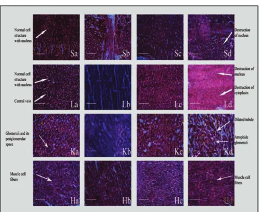
The histological studies confirmed that significant tissue morphology was changed in etodolac treated rats (Figure 3). Pure etodolac caused more toxic environment in the rat than ELGKM formulation treated rats. The present experimental results of both pharmacological and toxicological parameters suggest that ELGKMs will be more beneficiary for the treatment of chronic pain or inflammatory conditions (Mariappan et al., 2011).
CONCLUSION
From the investigation of ulcerogenic activity of etodolac loaded gum Katira microspheres (ELGKMs)
prepared by W1/O/W2 emulsion solvent evaporation
technique showed significant decrease in the stomach ulceration in the rat’s reacted with ELGKMs in comparison with the pure etodolac treated groups. The studies on anti-inflammatory activity prove beyond doubt that ELGKMs has more pronounced anti-inflammatory activity than the pure etodolac. Therefore on the basis
of pharmacological evaluation of anti-inflammatory
activity and ulcerogenecity of ELGKMs compared with pure etodolac, it can be said that ELGKMs showed better
efficacy in comparison to pure etodolac in a chronic
inflammatory condition of rats. The histological analysis of different tissues of drug treated rats indicated that a few number of damaged cells were noticed in ELGKMs treated
rats than that of pure etodolac. The pharmacological
and toxicological investigation of orally administered ELGKMs in rats suggested that etodolac loaded gum Katira microspheres has the potential to be developed into a better and effective sustain release formulation for the
management of rheumatoid arthritis, osteoarthritis and other chronic joint pain related diseases.
ACKNOWLEDGEMENTS
The authors would like to express their gratitude to University Grants Commission [F.No. 10-01/2008(SA-I);
Dated 22nd November’2012], New Delhi, India for the
financial support. The authors greatly acknowledge Evonik India Pvt. Ltd for providing the Polymer Eudragit®RS100 and Eudragit®RL100 and also thankful to M/S Fleming Laboratory Limited for providing etodolac.
TABLE VI - Biochemical parameters of microspheres treated in rats
Biochemical
Parameters Control Group
ELGKMS (50 mg/kg b.w)
ELGKMs (75 mg/kg b.w.)
Pure Etodolac (75 mg/kg b.w.)
Total Bilirubin (mg/dl) 0.482 ± 0.019 0.457 ± 0.021* 0.433 ± 0.047* 0.467 ± 0.029*
SGOT (U/L) 195.50±3.834 292.83± 26.21 304.33± 16.98** 228.33 ± 12.91*
SGPT (U/L) 26.00 ± 3.85 39.33 ± 2.58** 40.33 ± 5.92** 33.50 ± 4.68
Blood Sugar (mg/dl) 108.67±5.125 84.17 ± 5.35** 80.00 ± 7.51** 99.00 ± 6.03
Alkaline Phosphatase (U/L)
287.00± 8.25 337.00 ± 8.94** 372.33 ± 5.05** 303.00 ± 11.10**
Serum Triglyceride (mg/dl)
72.83 ± 4.53 116.67± 11.81** 145.00 ± 6.06** 99.50 ± 14.79**
Serum Calcium (mg/dl) 9.20±2.05 8.20±0.59* 6.70±1.78 8.60±1.26*
Serum Iron (ug/dl) 98.00±3.47 143.00±6.14** 177.00±4.56** 121.00±2.45**
Data are taken as mean ±SD, P<0.05, P*>0.05 and P**<0.01 as compared to control group. Table value (df 3,20)=3.10 at 0.05
level and 4.94 at 0.01 level. The results were analyzed by one-way ANOVA followed by Dunnett’s Multiple Comparison Test. Each value represents the mean ±SEM of 6 animals. [P<0.05, Biochemical parameters are not significant compared to respective
DECLARATION OF INTEREST
The authors state no conflict of interest.
REFERENCES
Adam SEI. Toxic effects of Francoeuriacrispa in rats. Phytother Res. 1998;12(7):476-479.
Adeyemi OT, Osilesi O, Adebawo OO, Onajobi FD, Oyedemi SO, Afolayan AJ. Alkaline phosphatase (ALP), aspartate aminotransferase (AST) and alanine aminotransferase (ALT)
activities in selected tissues of rats fed on processed atlantic
horse mackerel (Trachurustrachurus). Adv Biosci Biotechnol. 2015;6(3):139-152.
Anderson AJ.Lysosomal enzyme activity in rats with adjuvant-induced arthritis. Ann Rheum Dis. 1970;29(3):307.
Armour DFE, Blood FR, Belden DA. In the manual of laboratory work in mammalian physiology. 3rd ed. Chicago: The University
of Chicago, Liilious; 1965. 216 p.
Buer JK. Orginals and impact of the term ‘NASID’. Inflammopharmacol. 2014; 22(5):263-267.
Craig CR,Stitzer RE. Modern pharmacology with clinical
application. 6th ed. USA: Lippincott William and Wilkins; 2004.
832 p.
Crook MA. Clinical chemistry and metabolic medicine. 7th ed.
London: Hodder Arnold; 2006. 426p.
Ellman GL. Tissue sulfhydryl groups. Arch Biochem Biophys. 1959;82(1):70-77.
Finney RS, Somers GF.The anti‐inflammatory activity of
glycyrrhetinic acid and derivatives. J Pharm Pharmacol. 1958;10(1):613-620.
Ghai CL. Textbook of practical physiology. New Delhi: Jaypee Medical Publishers; 1990. 371 p.
Glaser K, Sung ML, O’Neill K, Hartman D, Carlson R, Kreft A, et al. Etodolac selectively inhibits human prostaglandin G/H synthase 2 (PGHS-2) versus human PGHS-1. Eur J Pharmacol. 1995;281(1):107-111.
FIGURE 3 - Study of Histological changes of Stomach(S), Liver (L), Kidney (K) and Heart (H) of Rats: Sa, Sb, Sc, and Sd are
Control, 75 mg of pure etodolac, 50 mg and 75 mg of equal quantity of etodolac in ELGKMs respectively; La, Lb, Lc and Ld are
Control, 75 mg of pure etodolac, 50 mg and 75mg of equal quantity of etodolac in ELGKMs respectively; Ka, Kb, Kc and Kd are
B. Ruhidas, R. Ray, D. Naskar, B. K. Chakra, T. K. Chatterjee
Indian Pharmacopoeia Commission. Indian Pharmacopoeia.7th
ed. Ghaziabad; 2014. v.2, p. 1718.
Kytridis VP, Manetas Y. Mesophyll versus epidermal anthocyanins as potential in vivo antioxidants: evidence linking the putative antioxidant role to the proximity of oxy-radical source. J Exp Bot. 2006;57(10):2203-2210.
Main IH, Whittle BJ. Investigation of the vasodilator and antisecretory role of prostaglandins in the rat gastric mucosa by
use of non‐steroidal anti‐inflammatory drugs. Br J Pharmacol.
1975;53(2):217-24.
Mariappan G, Saha BP, Sutharson L, Singh A, Garg S, et al. Analgesic, anti-inflammatory, antipyretic and toxicological evaluation of some newer 3-methyl pyrazolone derivatives. Saudi Pharm J. 2011;19(2):115-22.
Meier R, Schuler W, Desaulles P. Zurfrage des mechanismus der hemmung des bindegewebswachstumsdurch cortisone. Experientia. 1950;6(12):469-71.
Ohkawa H, Ohishi N, Yagi K. Assay for lipid peroxides in animal tissues by thiobarbituric acid reaction. Anal. Biochem.
1979;95(2):351-8.
Paschapur MS, Patil MB, Kumar R, Patil SR. Evaluation of
anti-inflammatory activity of ethanolic extract of Borassus flabellifer
L. male flowers (inflorescences) in experimental animals. J Med Plant Res. 2009;3(2):49-54.
Peterson GL. Review of the Folin phenol protein quantitation method of Lowry, Rosebrough, Farr and Randall. Anal Biochem. 1979;100(2):201-220.
Rainsford KD. Mechanism of gastrointestinal toxicity of nonsteroidal anti-inflammatory drugs. Scand J Gastroenterol. 1989;24(165):9-16.
Robins SL. Lymph nodes and spleen: pathologic basis of disease. Philadelphia: WB Saunders Co; 1974. 1050 p.
Rubin RP. The role of calcium in the release of neurotransmitter substances and hormones. Pharmacol Rev. 1970;22(3):389-428.
Ruhidas B, Naskar D, Banerjee S, Karan S, Chatterjee TK.
Evaluation of gum katira as a model sustained release adjuvant in the preparation of etodolac loaded microsphere. Indian J
Pharm Educ. 2016;50(1):1-13.
Shojaei AH. Buccal mucosa as a route for systemic drug delivery: a review. J Pharm Pharm Sci. 1998;1(1):15-30.
Sinha AK. Colorimetric assay of catalase. Anal Biochem. 1972;47(2):389-94.
Tammara VK, Narurkar MM, Crider AM, Khan MA.
Synthesis and evaluation of morpholinoalkyl ester prodrugs of
indomethacin and naproxen. Pharm Res. 1993;10(8):1191-9.
Vogel HG, editor. Drug discovery and evaluation: pharmacological assays. 2nd ed. New York: Springer-Verlag;
Berlin: Heidelberg; 2002;1408.
Vogel HG. Das Glasstabgranulom, eine Methode zur
Untersuchung der Wirkung von Corticosteroiden auf
Gewicht, Festigkeit und chemische Zusammensetzung des Granulationsgewebes an Ratten. Arzneim Forsch Drug Res. 1970;20:1911-8.
Vyas S, Trivedi P, Chaturvedi SC. Dextran-etodolac conjugates: synthesis, in vitro and in vivo evaluation. Acta Pol Pharm. 2009;66(2):201-6.
Weissmann G, Zurier RB, Spieler PJ, Goldstein IM. Mechanisms of lysosomal enzyme release from leukocytes exposed to immune complexes and other particles. J Exp Med. 1971;134(3):149-65.
Wintrobe MM, Lee GR, Boggs DR, Bithel TC, Anthens JW, Foerersters J. Clinical Hematology. 7th ed. Philadelphia: Lea &
Febiger; 1976.1896 p.
Woodin AM, Wieneke AA. The accumulation of calcium by the polymorphonuclear leucocyte treated with staphylococcal leucocidin and its significance in the extrusion of protein. Biochem J.1963;87(3):487.
Received for publication on 23th November 2016