R E S E A R C H
Open Access
Effect of phospholipase A
2
inhibitors during
infection caused by
Leishmania
(Leishmania) amazonensis
Maria L. A. C. Bordon
1,3, Márcia D. Laurenti
1, Susan Pereira Ribeiro
2, Marcos H. Toyama
3,
Daniela de O. Toyama
4and Luiz Felipe D. Passero
3*Abstract
Background:Lipid metabolites play an important role in parasite differentiation and virulence. Studies have revealed thatLeishmaniasp. uses prostaglandins to evade innate barriers, thus enabling the parasites to survive inside immune cells. Despite the role of the enzyme Phospholipase A2(PLA2) in prostaglandins production, few studies have investigated the role of parasite PLA2during the interaction betweenL. (L.) amazonensisand the host (in vitro and in vivo) immune cells.
Methods:In the present work, the leishmanicidal effect of PLA2inhibitors, methyl arachidonyl fluorophosphonate (MAFP), bromoenol lactone (BEL) and aristolochic acid (AA) were investigated in vitro (promastigote and
intracellular amastigote forms ofL. (L.) amazonensis) and during in vivo infection using BALB/c mice.
Results:The aforementioned inhibitors were deleterious to promastigote and amastigote forms of theL. (L.) amazonensisand were non-toxic to peritoneal macrophages from BALB/c mice.L. (L.) amazonensis-infected BALB/c mice treated with the inhibitor BEL presented decreased lesion size and skin parasitism; however, BEL treatment induced hepatotoxicity in BALB/c mice.
Conclusions:Results presented herein suggested that PLA2inhibitors alteredL. (L.) amazonensisviability. In spite of liver toxicity, treatment with BEL was the most selective compound in vitro, as well in vivo, resulting in lower skin parasitism in the infected mice. These findings corroborate the role of PLA2in parasite virulence and maintenance in vertebrate hosts, and suggest that molecules structurally related to BEL should be considered when planning
compounds againstLeishmaniasp.
Keywords:Leishmania (Leishmania) amazonensis, Macrophages, BALB/c mice, Phospholipase A2, Phospholipase A2 inhibitors
Background
The Leishmania parasite and its first steps of interac-tions with phagocytic cells have been extensively studied, mainly because the type of interaction and molecules in-volved determine the fate of Leishmania, which may be associated with death by molecules and/or cells from the host; or Leishmania parasites can enter in the main host’s cells, the macrophages, determining disease [1].
The initial interaction between macrophages with the
Leishmaniasp. occurs through the complement receptor (CR), mannose-fucose, fibronectin, and Fcγ macrophage receptors.
Following inoculation ofLeishmaniapromastigotes into the dermis of the mammalian host, a parasite metallopro-teinase of 63 kDa (gp63) is able to cleave the C3b factor of the complement system into an inactive form (iC3b), which is able to bind to leishmanial lipophosphoglycan
(LPG) and even to gp63. These opsonized Leishmania
promastigotes bind to CR1 and CR3 macrophage recep-tors thereby commencing phagocytosis. This main type of
* Correspondence:felipepassero@yahoo.com.br;felipepassero@clp.unesp.br
3São Paulo State University (UNESP), Institute of Biosciences, São Vicente, Praça Infante Dom Henrique, s/n, 11330-900 São Vicente, SP, Brazil Full list of author information is available at the end of the article
phagocytosis seems to impact the course of infection, since the inhibition of respiratory burst and the Th1-driven immune response create favorable conditions forLeishmaniasurvival. Conversely, interactions between
Leishmania and fibronectin receptors will trigger an in-flammatory response associated with parasite death [2,3].
Leishmanial molecules are also critical in the modula-tion of macrophages’intracellular environments. LPG is one of the main glycoconjugate ofLeishmania promasti-gotes, and is involved in the protection of the parasite not only from the acidic parasitophorous vacuoles, but also from inhibition of phagosome maturation and modulation of cytokine production. The gp63 metallo-proteinase has been credited as being a potent inhibitor of the protein kinase C pathways, which, if functioning properly, are responsible for cell proliferation, differenti-ation, apoptosis, and reactive oxygen and nitrogen spe-cies production; this context suggests that gp63 (in addition to LPG) has a deep impact on the modulation of leishmanicidal activity and in the establishment of leish-manial infection in macrophages [4–6]. These types of studies are extremely important for expanding the current knowledge on the physiopathology of leishmaniasis.
Although LPG and gp63 antigens have been identified as vital for parasite survival, other parasitic components are also important during the process of phagocytosis, as well as in the intracellular survival of Leishmania para-sites. In this regard, it was demonstrated that the super-natant of L. (L.) amazonensis presented phospholipase A2 (PLA2) activity, and when an additional source of
PLA2 was added in culture, the pathway of eicosanoid
production was stimulated and prostaglandin E2(PGE2)
was produced at high levels; this was associated with in-creased numbers of intracellular amastigotes [7]. More-over, in vivo studies demonstrated that PLA2-stimulated L. (L.) amazonensis induced tissue injuries when com-pared with the control parasite [7]. This suggests the
in-volvement of PLA2 in the pathway of prostaglandin
production, and that this pathway can be considered
an additional mechanism by which L. (L.)
amazonen-sis parasites infect, modulate inflammation and persist in the host.
Overall PGE2, a major byproduct of the metabolism of
arachidonic acid, has been linked to pathology in leish-maniasis. Farrel and Kirkpatrick [8] were among the first to suggest the participation of this lipid mediator in leish-maniasis, sinceL. major-infected splenocytes from BALB/ c mice produced elevated amounts of PGE2and were
un-able to proliferate under specific stimuli. This is in con-trast to splenocytes isolated from animals treated with indomethacin, a selective inhibitor of the enzyme cycloox-ygenase (COX), which did not produce PGE2 and were
capable of proliferating. Similarly, the human lineage of U937 macrophages presented a time-dependent elevation
in the production of PGE2 following infection with L. donovani [9], and decreased after the addition of COX2
inhibitors. On the other hand, other secreted PLA2
en-zymes were able to eliminate promastigote forms ofL. (L.) infantum,L. (L.) amazonensis[10–12].
In order to evaluate the importance of PGE2 during
experimental leishmaniasis, L. (L.) mexicana-infected BALB/c mice were treated with indomethacin. It was ob-served that treated animals partially controlled the size of lesions and skin parasitism parasite load the skin, a finding that was associated with reduced levels of inter-leukin (IL)-4, IL-10, and PGE2 in the supernatants of
splenocytes [13]. These works clearly demonstrated how prostaglandin production can modulate not only the im-munological response but also the outcome of parasite infection, suggesting that certain inhibitors of this com-plex pathway can serve as useful tools for controlling parasitism – such as the inhibitors of PLA2, which is a
key enzyme responsible for triggering prostaglandin pro-duction. Thus, the main purpose of this work is to inves-tigate the efficacy of inhibitors of cytosolic and secreted PLA2, such as bromoenol lactone (BEL), methyl
arachi-donyl fluorophosphonate (MAFP) and aristolochic acid (AA) during infection (in vitro and in vivo) with L. (L.) amazonensis.
Methods
Parasites
TheL. (L.) amazonensisparasite (MHOM/BR/73/M2269) was kindly provided by Prof. Dr. Fernando T. Silveira from the Leishmaniasis Laboratory Prof. Dr. Ralph Laison Cryobank, Department of Parasitology, Evandro Chagas Institute, Ministry of Health, Belém, Pará, Brazil. Parasite phenotyping was identified by monoclonal antibodies and isoenzyme electrophoretic profiles (at the Leishmaniasis Laboratory of the Evandro Chagas Institute - Belém, Pará, Brazil). Parasites were grown in RPMI 1640 medium (Ros-well Park Memorial Institute - Gibco®; Thermo Fisher Sci-entific, Waltham, MA, USA), supplemented with 10% heat-inactivated fetal bovine serum, 10μg/mL of gentami-cin, and 1000 U/mL of penicillin (R10) at 25 °C. Promasti-gote forms in the stationary phase were used.
PLA2inhibitors
Aristolochic acid (AA) inhibits secretory PLA2, such as
human synovial fluid PLA2and PLA2purified snake and
scorpion venoms. Bromoenol lactone (BEL) is an irre-versible inhibitor of calcium-independent PLA2 able to
inhibit the release of arachidonate from different cell lines. Methyl arachidonyl fluorophosphonate (MAFP) is a selective and irreversible inhibitor of cytosolic PLA2
Determination of leishmanicidal potential of PLA2
inhibitors
Promastigote forms of L. (L.) amazonensis (2 × 10⁶ pro-mastigotes/well) were incubated in 96-well culture plates
in R10 medium with bromoenol lactone (BEL) (0.7–
60μM), methyl arachidonyl fluorophosphonate (MAFP) (4.7–300 μM) or Aristolochic acid (AA) (9.4–600 μM). Miltefosine was used as standard drug (1.9–245μM). A negative control group cultivated in medium and di-methyl sulfoxide (DMSO) was used as a vehicle solution (never exceeding 1% v/v). The parasites were incubated for 24 h at 25 °C. Plates were washed 3 times with 200μL of sodium chloride 0.9% (w/v) and centrifuged at 3000 rpm for 10 min at 4 °C; MTT (3-[4,5-dimethylthia-zol-2-yl]-2,5-diphenyltetrazolium bromide) (5.0 mg/mL) was added for 4 h. Subsequently, 50uL of sodium dodecyl sulfate (SDS -10%) was added to each well. The plates were incubated for 18 h and read in an enzyme-linked im-munosorbent assay (ELISA) reader at 595 nm. In order to access the leishmanicidal potential of PLA2 inhibitors,
50% effective concentrations (EC50) were estimated using
the software Graph Pad Prism 5.0 (GraphPad Software Inc., La Jolla, CA, USA). The EC50is the concentration of
the inhibitors where 50% of leishmanicidal effect is ob-served after the specified exposure time.
Peritoneal macrophage culture and cytotoxicity assay
Approximately 2 × 10⁵ peritoneal macrophages from
BALB/c mice were cultured in R10 medium with BEL (0.7–60μM), MAFP (4.7–300μM), AA (9.4–600μM) or miltefosine (1.9–245 μM). As negative control, macro-phages were cultivated in medium and DMSO (not ex-ceeding 1%v/v). After 24 h, cell viability was analyzed by the MTT method. After evaluation of the surviving curves, non-toxic concentrations were recorded and used in further experiments. In addition, the 50% cyto-toxic concentration (CC50) was estimated using the
soft-ware GraphPad Prism 5.0. The CC50 is defined as the
concentration of the inhibitors where 50% of the host cells were unviable after the specified exposure time.
Effect of PLA2inhibitors during the interaction between
the parasite and host macrophages
Promastigote forms ofL. amazonensis in stationary phase were adjusted to a concentration of 2 × 10⁶promastigotes/ mL and were added to macrophage cultures (1:10 macrophage-to-parasite ratio). The co-cultures were main-tained in a humidified incubator at 5% CO2at 35 °C. The
inhibitors AA (25.0; 50.0; 100.0μM), BEL (1.0; 2.0; 4.0μM) and MAFP (5.0; 10.0; 20.0μM) were added to the infected cells. As standard treatment, miltefosine’s EC50 was used
[14]. After 24 h of incubation, infection indexes were esti-mated [15] and the concentrations able to decrease the
infection index to 50% were estimated via the software GraphPad Prism 5.0.
Efficacy of PLA2inhibitors during experimental cutaneous
leishmaniasis
Thirty male BALB/c mice were subcutaneously infected in the right hind footpad with 106promastigote forms ofL. (L.) amazonensis, while five BALB/c mice received sodium chloride 0.9% (w/v) via the same route (healthy group). Five weeks after infection, L. (L.) amazonensis-infected BALB/c mice were divided into six groups containing 5 animals each: groups 1 and 2 were injected with 10.0 nM (0.012μg/kg) and 30.0 nM (0.036μg/kg) of BEL, respect-ively. The concentration of BEL was chosen based on pre-vious reports of animal treatment [16]; groups 3 and 4 were injected with 0.7 mM (0.015 μg/kg) and 1.4 mM (0.030μg/kg) of MAFP, respectively. All these groups were treated intraperitoneally. Group 5 received Glucantime (50 mg/kg) intralesionally. Group 6 (infected only) was injected with PBS solution. Group 7 (non-infected, non-treated animals) received only the vehicle solution (PBS control by intraperitoneal or subcutaneous routes).
Animals treated intraperitoneally were injected with 50 μL of PLA2 inhibitors or PBS solution; while those
treated intralesionally received 20 μL of Glucantime or PBS solution. Animals were injected with PLA2
inhibi-tors, Glucantime or vehicle solution a total of 15 times, once a day, at 24-h intervals. The animals’physical con-ditions were monitored once a week. Glucantime was injected intralesionally, based on a previous work [17], and this administration route was effective at eliminating tissue amastigotes. The inhibitors BEL and MAFP were injected intraperitoneally to improve their distribution in the animal body. A group of non-infected animals were treated with BEL, MAFP or Glucantime to analyze histo-logical alterations. One week after the last injection, the animals were anaesthetized with thiopental and sacri-ficed by cardiac puncture. There were no deaths prior to the endpoint, and all animals were euthanized to analyze the parasitism in the skin. Animals were not treated with AA because reports in the literature had reported its high toxicity for animals [18].
medium. The skin and lymph node suspensions were subjected to 12 serial dilutions with four replicate wells. The number of viable parasites was determined based on the highest dilution in which the promastigotes could be grown after 10 days of incubation at 25 °C. Biopsies of the heart, lung, spleen, liver and kidney were collected and fixed in buffered 5% formalin for analysis of histo-pathological alterations in treated animals. These organs were collected because they are highly vascularized, which enables drugs in the bloodstream to provoke major changes and impact their physiology.
Statistical analysis
The results were expressed as the mean ± standard deviation of three independent experiments, and the
nonparametric Mann-Whitney U test was used to
compare the results between groups. Differences were considered statistically significant at a 5% significance level (P< 0.05). The software GraphPad Prism 5 was employed to analyze the results.
Results
Leishmanicidal and cytotoxic activities of PLA2inhibitors
BEL was the most active PLA2inhibitor, killing 50% of
promastigote forms of L. amazonensis with 15.1 ±
3.7 μM; MAFP presented intermediate activity (50.5 ±
7.8 μM), followed by AA, eliminating promastigote
forms with EC50 of 450.1 ± 45.6 μM; miltefosine elimi-nated promastigote forms with an EC50of 12.6 ± 2.1μM (Table1). PLA2inhibitors did not induce cytotoxicity to
peritoneal macrophages in the tested range (Table 1). The most selective molecule was MAFP, followed by miltefosine and BEL. The lowest selectivity index was presented by AA (Table1).
It was observed that BEL was the most active against amastigote forms and decreased the infection index by 50% at 2.6 ± 0.8μM, followed by MAFP (17.6 ± 7.9μM), miltefosine (21.6 ± 2.2 μM) and AA (76.9 ± 5.7 μM), as indicated in Table 1. Furthermore, it was observed that BEL was the most selective molecule, followed by MAFP, AA and finally miltefosine (Table1).
Effect of PLA2inhibitors during experimental cutaneous
leishmaniasis
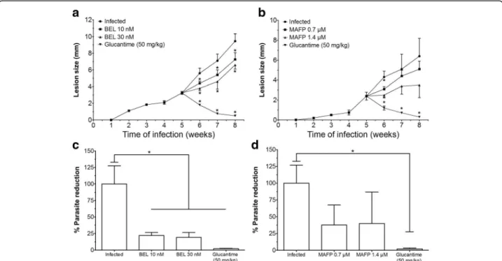
Infected BALB/c mice treated with 10 nM or 30 nM of the inhibitor BEL (Fig. 1a) presented smaller lesion sizes at weeks 6, 7, and 8 post-infection (P< 0.05), when com-pared with the infected control. In contrast, infected ani-mals treated with 0.7 or 1.4μM of MAFP (Fig.1b) did not show significant alterations during the course of infection. Animals treated intralesionally with Glucantime presented small lesion size from 6 weeks post-infection until the end of the experiment, at the 8th week (Fig.1aandb).
In relation to skin parasitism, animals treated with 10 nM or 30 nM of BEL had fewer parasites on the skin when compared with the infected control (Fig.1c). Infected animals treated with MAFP did not change the parasite load in the skin (Figs. 1d). Glucantime-treated animals (50 mg/kg) presented decreased parasite load when com-pared with the infected control group (Figs.1candd). Ani-mals treated with BEL or MAPF presented lymph node parasitism similar to that in the infected control group (data not shown). Parasites were not detected in lymph nodes of Glucantime-treated animals (data not shown).
Histopathological analysis
Histological sections of the heart, liver, kidney, and spleen were analyzed to verify toxic effects of PLA2
in-hibitors on BALB/c mice. Infected control animals did not present significant histolopathological changes in the heart, kidney or lungs; however, in the spleen signals of germinal center activation were verified (data not shown). In the liver, some focal areas of inflammation were observed (black arrow in Fig.2a).
Infected animals treated with MAFP did not show sig-nificant changes in analyzed organs compared with the infected control (data not shown). In comparison with infected controls, the BEL-treated group did not present signs of alterations in the heart, spleen or lungs (data not shown); however, the liver presented extensive areas of hepatocellular necrosis, as illustrated in the insets of one respective image from the group treated with 30 nM of BEL (Fig. 2b). A similar histological pattern was veri-fied in non-infected animals treated with 30 nM of BEL (Fig. 2c). Infected and non-infected animals treated intralesionally with Glucantime did not show changes in histological sections of analyzed organs; for compari-sons, a histological liver section from an infected animal treated with 50 mg/kg of Glucantime is presented in Fig. 2d. Healthy animals did not present alterations in analyzed organs (data not shown); for comparisons a histological liver section is presented in Fig. 2e.
Discussion
In spite of classical mechanisms of innate immunity dur-ing the relation between parasite and host cells, other
Table 1Activity of PLA2inhibitors against promastigote,
amastigote and peritoneal macrophages from BALB/c mice
Compounds EC50(μΜ)a CC50(μM) SIa EC50(μM)b SIb
AA 450.1 ± 45.6 > 600 > 1.3 76.9 ± 5.7 > 7.8
BEL 15.1 ± 3.7 > 60 > 3.9 2.6 ± 0.8 > 23.1
MAFP 50.5 ± 7.8 > 300 > 5.9 17.6 ± 7.9 > 17.0
Miltefosine 12.6 ± 2.1 60.8 ± 3.1 4.8 21.6 ± 2.2 2.8
The efficacies of compounds were analyzed through effective concentration 50 (EC50), and the selective indexes (SI) were estimated
components of the inflammatory process may take place during Leishmania infection. Recently, it was demon-strated that L. (L.) amazonensis-infected macrophages treated with PLA2 presented more intracellular
amasti-gotes when compared with the control group. Further-more, an association between high intracellular parasitism and PGE2production by infected macrophages was
dem-onstrated [7]; PGE2is a lipidic mediator that
downregu-lates the respiratory burst in infected macrophages [20]. Other studies have also demonstrated a pathogenic role of the prostaglandin pathway in leishmaniasis [21]; how-ever, to the best of our knowledge, few studies have fo-cused on the initial enzyme responsible for triggering prostaglandin production, the PLA2enzyme, during
ex-perimental leishmaniasis.
Thus, in the present study, it was demonstrated that the viability ofL. (L.) amazonensispromastigotes was
al-tered when the PLA2 inhibitors BEL and MAFP were
added in culture. In trypanosomatids, PLA2 plays vital
roles in host cell invasion, Ca+ 2 influx, and during lipid turnover [22, 23]. Therefore, these inhibitors should affect the physiology ofL. (L.) amazonensis, thus impact-ing their survival in culture, as evidenced in Table 1. In addition, L. (L.) amazonensis promastigotes were more sensitive to the inhibitor BEL in comparison with other treatments, and this molecule is a specific inhibitor of Ca2+-independent PLA2, since 15.1μM was able to elim-inate 50% of the parasite population; followed by MAFP, an inhibitor of calcium-dependent PLA2, that presented
an EC50 of 50.5 μM. These data suggest that Ca2
Fig. 1BALB/c mice were infected in the right hind footpad with promastigote forms ofL. amazonensisin stationary phase of growth. Five weeks after infection, the treatments were started and the lesion sizes of BEL- and MAFP-treated animals were recorded (aandb, respectively), and the skin parasitism analyzed in the animals treated with BEL (c) and MAFP (d). *P<0.05 indicates significant differences when comparing the treated groups versus infected control group
Fig. 2Histopathology analysis of the liver section. Liver of infected control (a) showed portal inflammation; the inset shows preserved
+
-independent PLA2 may hold vital biological
signifi-cance for the survival of parasites, such that molecules able to target these enzymes can be considered valuable prototype drugs. Moreover, the activities of these inhibi-tors on the parasites seems to be specific, since macro-phages incubated with these same inhibitors, at the same range of concentrations, did not show significant alterations in their viability, suggesting that the inhibi-tors are more selective to the parasite than to the host cells.
In order to verify whether PLA2inhibitors can impact the
survival of intracellular amastigotes,L. (L.) amazonensis- in-fected macrophages were treated with BEL, MAFP and AA. In this case, both BEL and MAFP inhibitors were able to de-crease the intracellular parasitism, suggesting that different families of PLA2enzymes not only may play some role
dur-ing the infection, but also might be important for the intra-cellular survival of amastigote forms. Possibly PLA2s (from
host and parasites) may favor the production of PGE2, which
is suppressive for macrophages [24,25], as demonstrated in different studies [7,9, 26, 27]. In the other hand, AA was shown to be the least active of all inhibitors assayed, despite presenting an intermediate selectivity index. Other PLA2
in-hibitors – such as quinacrine, 4-bromophenacyl bromide and phentermine–were used during the interaction of Try-panosoma cruzi and macrophages. In this case it was ob-served that the inhibitors suppressed parasite surface binding on the host cell and internalization [28], suggesting that PLA2presents a role during intracellular infections, and
thus can be used as a molecular target for designing new leishmanicidal molecules.
Given that the inhibitors BEL and MAFP were able to decrease the intracellular parasitism into host macro-phages, in vivo experiments were carried out to verify whether PLA2could be considered a molecular target to
characterize new prototype drugs. In this aspect, it was verified that BALB/c mice infected with L. (L.) amazo-nensisand treated with 10 nM or 30 nM of BEL exhib-ited decreased lesion size at 6, 7, and 8 weeks post-infection when compared with the infected control, an effect associated with reduced parasitism on the skin, but not in the lymph nodes (data not shown), suggesting that in vivo PLA2 may be responsible, at least in part,
for the induction of pathology in a murine model of cu-taneous leishmaniasis. On the other hand, animals treated with MAFP did not show significant alterations in the course of infection. The inhibitor AA was not assayed in vivo due to its toxicity for animals [14].
In the cellular environment, PLA2 enzymes catalyze the
hydrolysis of phospholipid sn-2 ester bond from cell mem-branes. This reaction is the primary pathway that allows the release of arachidonic acid. Following this biochemical step, the enzyme cyclooxygenase converts arachidonic acid into different classes of prostaglandins, according to the available
enzymes associated with eicosanoid production. In leish-maniasis some studies demonstrated that this pathway is correlated with disease worsening; on the other hand, the use of inhibitors or commercial available drugs able to inter-fere with enzymes pertaining to this pathway can suppress the production of prostaglandins, thus improving the le-sions. In this respect, Pérez-Santos and collaborators [13] showed that the treatment of L. (L.) mexicana-infected BALB/c mice with indomethacin, a selective inhibitor of COX enzymes, was able to restrain skin and lymph node parasitism; moreover, treated animals presented increased amounts of Th1 interleukins [13,29]. Therefore, the block-age of enzymes belonging to this pathway can improve the outcome of infection. Given that PLA2 is the first step in
triggering eicosanoid production, it should be considered an interesting molecular target for designing new therapeutic molecules.
In spite of these positive findings, the inhibitor BEL was toxic to the liver of infected BALB/c mice as well as their non-infected counterparts, as demonstrated in histological sections, where degenerated hepatocytes (inset) and nec-rotic areas were identified. A previous work demonstrated that although BEL was not toxic to peritoneal macrophages, but the constant inhibition of PLA2in the neurons resulted
in a long-term loss of neuronal viability, suggesting that in some organs, (such as the liver), preserving the activity of PLA2s is essential for maintaining host cell viability and
homeostasis [30]. This study also opens future perspectives to use BEL inhibitor as an intralesional treatment, which in turn could reduce liver toxicity.
Despite the toxicity presented by BEL, the relevance of PLA2during the infection was also demonstrated.
Fur-thermore, although BALB/c mice did not clear the L. (L.) amazonensis infection following the BEL treatment, these results suggested that PLA2might represent a
po-tential target for inhibiting Leishmania infection. How-ever, the main challenge is to find or design effective and non-toxic PLA2 inhibitors that are able to block the
entry or survival of the leishmania parasites in host cells.
Conclusions
Taken together, our results demonstrated that promasti-gote and intracellular amastipromasti-gotes (in vitro and in vivo) of L. (L.) amazonensisare more sensitive to BEL inhibi-tor compared with other inhibiinhibi-tors, and that molecules structurally correlated with BEL might serve as an inter-esting alternative for designing new prototypes directed againstLeishmaniaparasites.
Abbreviations
%:Percentage; °C: Celsius; AA: Aristolochic acid; BEL: Bromoenol lactone; COX: Cyclooxygenase; CR: Complement receptor; CR1 and CR3: Complement receptors 1 or 3, respectively; ELISA: Enzyme-linked immunosorbent assay; gp63: Metalloproteinase of 63 kDa; iC3b: Inactive form of C3b fragment; IL: Interleukin; kDa: Kilo Dalton; kg: Kilogram;L.:Leishmania;
MTT: 3-[4,5-dimethylthiazol-2-yl]-2,5-diphenyltetrazolium bromide; nM: Nanomolar; PGE2: Prostaglandin E2; PLA2: Phospholipase A2; R10: RPMI medium; Th1: T helper 1;μg: Microgram;μL: Microliter;μM: Micromolar
Funding
The present study received funds from the São Paulo Research Foundation (FAPESP) (2016/00468–0), HCFMUSP-LIM50 and CNPq-PIBIC (135478/2010–3).
Moreover, this publication was supported by the Coordination for the Im-provement of Higher Education Personnel (CAPES) through Programa Editor-ação CAPES (edital n. 13/2016, auxílio n. 0722/2017, processo n.
88881.142062/2017–01) and by the National Council for Scientific and
Technological Development (CNPq) through Programa Editorial CNPq/CAPES (chamada n. 26/2017, proc. n. 440954/2017–7).
Availability of data and materials
The datasets used during the current study are available from the corresponding author on request.
Authors’contributions
Formal analysis: MLACB, LFDP. Investigation: MLACB, LFDP. Methodology: MLACB, LFDP. MDL, MHT, DOT, SPR, LFDP. Writing: LFDP, MDL, MHT, DOT, SPR, LFDP Funding acquisition: LFD, MDL, MHT. All authors read and approved the final manuscript.
Ethics approval
Animal experiments were carried out using protocols approved by the Ethics Committee for Animal Experimentation of São Paulo University under the number CEP 067/1.
Consent for publication
Not applicable.
Competing interests
The authors declare that they have no competing interests.
Publisher’s Note
Springer Nature remains neutral with regard to jurisdictional claims in published maps and institutional affiliations.
Author details
1
Laboratory of Pathology of Infectious Diseases (LIM-50), Medical School, University of São Paulo (USP), Av. Dr. Arnaldo, 455, São Paulo, SP CEP 01246903, Brazil.2Pathology Department, Case Western Reserve University, 2103 Cornell Rd, room 5503, Cleveland, OH 44106, USA.3São Paulo State University (UNESP), Institute of Biosciences, São Vicente, Praça Infante Dom Henrique, s/n, 11330-900 São Vicente, SP, Brazil.4School of Dentistry, Camilo Castelo Branco University (Unicastelo), Rua Carolina Fonseca, 584, São Paulo, SP CEP 08230-030, Brazil.
Received: 2 March 2018 Accepted: 1 August 2018
References
1. de Morais CGV, Castro Lima AK, Terra R, dos Santos RF, Da-Silva SAG, PML D. The dialogue of the host-parasite relationship:Leishmaniaspp and Trypanosoma cruziinfection. Biomed Res Int. 2015;2015:324915. 2. Arango Duque G, Descoteaux A.Leishmaniasurvival in the macrophage:
where the ends justify the means. Curr Opin Microbiol. 2015;26:32–40.
3. Podinovskaia M, Descoteaux A.Leishmaniaand the macrophage: a multifaceted interaction. Future Microbiol. 2015;10(1):111–29.
4. Olivier M, Gregory DJ, Forget G. Subversion mechanisms by which Leishmaniaparasites can escape the host immune response: a signaling point of view. Clin Microbiol Rev. 2005;18(2):293–305.
5. Forestier CL, Gao Q, Boons GJ.Leishmanialipophosphoglycan: how to establish structure-activity relationships for this highly complex and multifunctional glycoconjugate? Front Cell Infect Microbiol. 2014;4:193. 6. Passero LFD, Assis RR, da Silva TNF, Nogueira PM, Macedo DH, Pessoa NL,
et al. Differential modulation of macrophage response elicited by glycoinositolphospholipids and lipophosphoglycan fromLeishmania (Viannia)shawi. Parasitol Int. 2015;64(4):32–5.
7. Passero LFD, Laurenti MD, Tomokane TY, Corbett CEP, Toyama MH. The effect of phospholipase A2 fromCrotalus durissus collilineatusonLeishmania (Leishmania) amazonensisinfection. Parasitol Res. 2008;102(5):1025–33.
8. Farrell JP, Kirkpatrick CE. Experimental cutaneous leishmaniasis. II. A possible role for prostaglandins in exacerbation of disease inLeishmania major-infected BALB/c mice. J Immunol. 1987;138(3):902–7.
9. Matte C, Maion G, Mourad W, Olivier M.Leishmania donovani-induced macrophages cyclooxygenase-2 and prostaglandin E2 synthesis. Parasite Immunol. 2001;23(4):177–84.
10. Barros GAC, Pereira AV, Barros LC, Lourenço A Jr, Calvi SA, Santos LD, et al. In vitroactivity of phospholipase A2 and of peptides fromCrotalus durissus terrificusvenom against amastigote and promastigote forms ofLeishmania (L.) infantum chagasi. J Venom Anim Toxins incl Trop Dis. 2015;21:48. https://doi.org/10.1186/s40409-015-0049-0.
11. Terra ALC, Moreira-Dill LS, Simões-Silva R, Monteiro JRN, Cavalcante WLG, Gallacci M, et al. Biological characterization of the Amazon coralMicrurus spixiisnake venom: isolation of a new neurotoxic phospholipase A2. Toxicon. 2015;103:1–11.
12. Pereira AV, de Barros G, Pinto EG, Tempone AG, de Oliveira Orsi R, dos Santos LD, et al. Melittin inducesin vitrodeath ofLeishmania(Leishmania) infantumby triggering the cellular innate immune response. J Venom Anim Toxins incl Trop Dis. 2016;22:1.https://doi.org/10.1186/s40409-016-0055-x. 13. Pérez-Santos JL, Talamás-Rohana P.In vitroindomethacin administration
upregulates interleukin-12 production and polarizes the immune response towards a Th1 type in susceptible BALB/c mice infected withLeishmania mexicana. Parasite Immunol. 2001;23(11):599–606.
14. Bezerra-Souza A, Yamamoto ES, Laurenti MD, Ribeiro SP, Passero LFD. The antifungal compound butenafine eliminates promastigote and amastigote forms ofLeishmania(Leishmania)amazonensisand Leishmania(Viannia)braziliensis. Parasitol Int. 2016;65(6 Pt A):702–7.
15. Passero LFD, Sacomori JV, Tomokane TY, Corbett CEP, da Silveira FT, Laurenti MD.Ex vivoandin vivobiological behavior ofLeishmania(Viannia) shawi. Parasitol Res. 2009;105(6):1741–7.
16. Yeo JF, Ong WY, Ling SF, Farooqui AA. Intracerebroventricular injection of phospholipases A2inhibitors modulates allodynia after facial carrageenan injection in mice. Pain. 2004;112(1–2):148–55.
17. Yamamoto ES, Campos BLS, Jesus JA, Laurenti MD, Ribeiro SP, Kallás EG, et al. The effect of ursolic acid onLeishmania(Leishmania)amazonensisis related to programed cell death and presents therapeutic potential in experimental cutaneous leishmaniasis. PLoS One. 2015;10(12):1–19.
18. Shi M, Ma L, Zhou L, Fu P. Renal protective effects of 17β-estradiol on mice with acute aristolochic acid nephropathy. Molecules. 2016;21(10):1391. 19. Campos BLS, Silva TN, Ribeiro SP, Carvalho KIL, Kallás EG, Laurenti MD, et al.
Analysis of iron superoxide dismutase-encoding DNA vaccine on the evolution of theLeishmania amazonensisexperimental infection. Parasite Immunol. 2015;37(8):407–16.
20. Serezani CH, Chung J, Ballinger MN, Moore BB, Aronoff DM, Peters-Golden M. Prostaglandin E2suppresses bacterial killing in alveolar macrophages by inhibiting NADPH oxidase. Am J Respir Cell Mol Biol. 2007;37(5):562–70.
21. Arcanjo AF, LaRocque-de-Freitas IF, Rocha JDB, Zamith D, Costa-da-Silva AC, Nunes MP, et al. The PGE2/IL-10 axis determines susceptibility of B-1 cell-derived phagocytes (B-1CDP) toLeishmania majorinfection. PLoS One. 2015;10(5):e0124888.
22. Opperdoes FR, van Roy J. The phospholipases ofTrypanosoma bruceibloodstream forms and cultured procyclics. Mol Biochem Parasitol. 1982;5(5):309–19.
23. Belaunzarán ML, Lammel EM, de Isola ELD. Phospholipases a in trypanosomatids. Enzyme Res. 2011;2011:392082.
24. Tsuchida K, Ibuki T, Matsumura K. Bromoenol lactone, an inhibitor of calcium-independent phospholipase A2, suppresses carrageenan-induced prostaglandin production and hyperalgesia in rat hind paw. Mediat Inflamm. 2015;2015:605727.
25. Bhattacharjee A, Majumder S, Das S, Ghosh S, Biswas S, Majumdar S. Leishmania donovani-induced prostaglandin E2 generation is critically dependent on host toll-like receptor 2-cytosolic phospholipase A2 signaling. Infect Immun. 2016;84(10):2963–73.
26. França-Costa J, Van Weyenbergh J, Boaventura VS, Luz NF, Malta-Santos H, Oliveira MCS, et al. Arginase I, polyamine, and prostaglandin E2pathways suppress the inflammatory response and contribute to diffuse cutaneous leishmaniasis. J Infect Dis. 2015;211(3):426–35.
28. Connelly MC, Kierszenbaum F. Modulation of macrophage interaction with Trypanosoma cruziby phospholipase A2-sensitive components of the parasite membrane. Biochem Biophys Res Commun. 1984;121(3):931–9.
29. Li J, Padigel UM, Scott P, Farrell JP. Combined treatment with interleukin-12 and indomethacin promotes increased resistance in BALB/c mice with establishedLeishmania majorinfections. Infect Immun. 2002;70(10):5715–20.