BJRS
RADIATION SCIENCES
02 (2014) 01-08EVALUATION OF THE DOSE DUE TO THE
PO-SITION OF THE PROTECTION BLOCKS USED
TO PROTECT THE EYES
FERNANDA L. OLIVEIRA
1,2, FABIANA F. DE LIMA
1and ÊUDICE
C.VILELA
11 Centro Regional de Ciências Nucleares - Comissão Nacional de Energia Nuclear, Av. Prof. Luís Freire, 200, CEP
50740-540 Recife, PE
2 Departamento de Energia Nuclear - Universidade Federal de Pernambuco, Av. Prof. Luís Freire, 1000, CEP
50740-540 Recife, PE
[email protected], [email protected] and [email protected]
ABSTRACT
Several techniques have been proposed to perform the radiation treatment of medulloblastoma. However, each method presents limitations, and problems have occurred during irradiation due to the application of non-coplanar fields, such as the joint protection of fields and nearby organs that may be affected during irradiation. Among these organs is the eye, located 9 mm from the cribriform plate. With the improper positioning of the protection blocks, an underdosage in the cribriform plate could bring about a relapse of the tumor due to the migration of tumor cells, whereas an overdosage in the eye may well cause opacities of the crystalline lens or a total loss of vision. This study aims to evaluate the dose levels in four tech-niques of radiotherapy planning according to the positioning of the protection blocks and the collimator angle. In the first two treatments, the prescribed dose was 1.5 Gy/day, using a lead block with collimator angles of 11.3° and 348.7°. In the final two treatments, the prescribed dose was of 1.8 Gy/day, using the cerrobend block with a single field set-up and collimator angles of 9° and 351°. An ALDERSON-RANDOM anthropomorphic phantom and thermoluminescent dosimeters were used to implement these measures during treatment. Dose levels applied to the eyes were as follows: in the first two treatments, 27% of the prescribed dose; in the third treatment, 41% of the prescribed dose; and in the fourth treat-ment, 55% of the prescribed dose. In the first two treatments the dose are still within acceptable limits to prevent future sequel. In the cribriform plate, the doses were much higher: in the first treatment, doses
were on average 128.4% of the prescribed dose; in the second and third treatments, doses reached 121.7%; and in the fourth treatment, doses reached 108.7%. These values show that the lead blocks pro-tected better than did the cerrobend blocks, and that the collimator angles of 9° and 351° reduced the dose levels in the cribriform plate; however, these remained above the original prescribed dose.
Keywords: Dosimetry, Radiotherapy, Protection blocks
1.
INTRODUCTION
Craniospinal irradiation, performed to treat medulloblastoma, is the most complex radiotherapy technique, as it can affect surrounding organs that are normally spared. One such example is the crystalline lens, which is located beside the cribriform plate, which should receive the maximum prescribed dose due to the high percentage of relapse of this disease in this region (Rene N. J.et al.; 2010).
The gradual slope of the cribriform plate between the eyes, especially in children, causes a sig-nificant and undesired spreading of the dose in the crystalline lens, whereas the other compo-nents of the eye – the lacrimal glands, eyelids, conjunctiva, iris, and retina – are relatively unaf-fected by doses of craniospinal irradiation. These only present adverse effects at doses of greater than 45 Gy. When the crystalline lens is irradiated, the germinal zone of the epithelium on the equator of the crystalline lens is damaged, in turn causing cataract induced by radiation (Cochran D. M.et al., 2008)
With the eye located at less than 9 mm from the cribriform plate, the poor positioning of the pro-tection blocks during planning will cause the maintenance of an error, which will affect both low and high dose levels, which vary according to patient age (Weiss E.et al.; 2001).
Among the most used forms of protection are the lead and cerrobend blocks. Today, the accel-erators come equipped with multileaf collimators which can be duly set up to protect the region chosen during treatment. In Brazil, however, the use of both lead and cerrobend blocks are still quite common. The positioning of these blocks involves both the set-up as well as the angling of the collimator camp and the table.
COCHRAN D.M. et al. (2008) reported that, upon comparing the doses due to their positioning within the camps, lateral and angled opposites, one key factor that contributed to the variation of the dose level within the crystalline lens was the age range of the patients. The anatomical varia-tions of the paranasal sinuses, which change significantly with age, reaching full development at puberty, appear in an angled position for adolescents and present a 60% reduction in the pre-scribed dose in the cribriform plate. By contrast, in older patients, almost no changes could be observed. Nevertheless, these authors recommend that the opposite lateral camps be placed at a beam angle of 15º-20º, given that, with this variation, an average reduction of 75% of the dose level in the crystalline lens occurred, thus, over the long-term, sparing the patient from radio-induced cataract.
Many methods can be applied to minimize the dose level in patients’ eyes, the most common of which are the bolus method and the shading blinding methods or shielding. The bolus method applies the accumulation phenomenon and a lower dose level on the surface associated with high energy beams to protect the surface structure of the eye. The shading methods or shielding blocks are frequently used to reduce the primary beam to a desired level (1%-3%) (Indra J. das; 1990).
Therefore, the present work aims to evaluate the dose levels in the crystalline lens according to the positioning of protection blocks and the collimator angle, using four techniques of radiother-apy planning: I. the half-beam technique; II. the angled camp technique; III. the angled camp technique with a mobile gap; and IV. the angled camp technique without a mobile gap.
2.
MATERIALS E METHODS
By means of a level III dosimetric intercomparison, according to that set forth by the International Atomic Energy Agency (IAEA) in its Safety Report #17 ( IAEA, 2000; Kron et al., 2002), the present study used an ALDERSON-RANDOM anthropomorphic simulator and thermoluminescent dosimeters (TLD-100) which were irradiated using beams generated by the linear accelerators provided by the participating institutions.
Four plannings were formulated using the different techniques mentioned above. Next, the treatments were performed following the parameters established in these plannings. The dosimeters were positioned in the simulator in the regions of interest to carry out the treatments. The first two plannings were executed during five sessions of radiotherapy, while the following two were employed in three subsequent sessions.
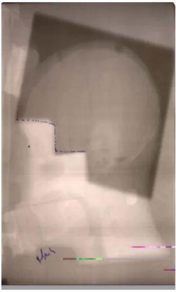
For the first treatment, applying the half-beam technique, and the second, applying the angled camp technique, both executed using conventional planning, the prescribed doses were equal to 1.5 Gy/day, with the angled collimators set at 11.3º and 348.7º, respectively (Figure 1). The lateral camps measured 18 x 15 cm2.
Figure 1: Planning lead block.
In the third treatment, using the angled camp technique with a mobile gap, and the fourth treatment, using the angled camp technique without a mobile gap, the dose was 1.8 Gy/day. The cerrobend blocks, with an individual set-up, and the collimator were measured at 9º and 351º, respectively (Figure 2). The lateral camps measured 19 x 25 cm2.
Figure 2: Planning cerrobend block.
In plannings I and II, the images were taken by X-ray from a 600C Clinac, 6MV, Varian linear accelerator with independent collimators. In the third and fourth plans, the images were taken on a Siemens SOMATOM and were transferred to a CMS XiO Release 4.3.1 computerized
radiation treatment planning system, and the irradiations used a Siemens Mevatron, 6 MV, linear accelerator, with dependent collimators, as shown in Figures 1 and 2.
3.
RESULTS AND DISCUSSION
Table 1 shows the results obtained for the four techniques used in the present study. The values of the obtained doses coincide with the two first plannings. As regards the values obtained resulting from the techniques applied in the third and fourth plannings, it could be observed that the values were mainly higher in the angled gantry technique without a mobile gap (Planning IV).
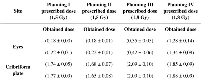
Table 1 - Values of mean doses obtained in the eyes lens and cribriform plate Site Planning I prescribed dose (1,5 Gy) Planning II prescribed dose (1,5 Gy) Planning III prescribed dose (1,8 Gy) Planning IV prescribed dose (1,8 Gy) Obtained dose Obtained dose Obtained dose Obtained dose
Eyes (0,18 ± 0,00) (0,18 ± 0,01) (0,35 ± 0,05) (1,28 ± 0,14) (0,22 ± 0,01) (0,22 ± 0,01) (0,42 ± 0,06) (1,34 ± 0,09) Cribriform plate (1,74 ± 0,05) (1,68 ± 0,07) (2,09 ± 0,10) (1,85 ± 0,09) (1,77 ± 0,09) (1,65 ± 0,08) (2,09 ± 0,10) (1,88 ± 0,09)
In the eye, the doses levels were only 27% of the prescribed dose in the first two treatments, while in the third treatment, the dose levels reached 41% of the prescribed dose and in the fourth treatment, the dose levels reached 55% of the prescribed dose. In the cribriform plate, the doses proved to be quite high: in the first treatment, the doses reached, on average, 128.4% of the prescribed dose, whereas in the second and third treatments, the doses reached 121.7% and in the fourth treatment, 108.7% of the prescribed dose. These values demonstrated that the lead blocks protected better than the cerrobend blocks, and that the angles of 9°and 351° reduced the dose level in the cribriform plate; however, they still remained at a higher dose level than the original prescribed dose.
The use of different types and formats of protector blocks (lead and cerrobend) in the plannings may well have contributed to the increase in the dose level in the crystalline lens. In addition, the
angles of the collimator also varied. In the first and second plan, the angles were 11.3° and 348.7°, while in the third planning, the angles were 171° and 189°, and in the fourth planning, the angles were 9° and 351°.
Cochran D.M. et al. (2008), apud Merriam and Focht, were the first authors to report on the dose-response relationship of cataract-induced radiation in 1958, for a single dose of radiation. In their study, clinically significant opacities after doses of above 4-5 Gy could be observed. Using fractioned irradiation, a 60% frequency of cataract could be observed after 7.5 to 9.5 Gy, given that 100% could be observed after 11.5 Gy. Another study by Cochran D.M. et al. (2008) suggested a dose limit of 20 Gy to avoid damage to the crystalline lens.
The average values of dose levels in the crystalline lens coincide with the first two plans, presenting values of 0.18±0.01 Gy and 0.22±001 Gy, for the right and left eyes, respectively. Concerning the values obtained from the techniques applied in the third and fourth plannings, it could be observed that the values were mainly higher in the angled gantry technique without a mobile gap, with dose levels reaching 1.28±0.14 Gy and 1.34±0.09 Gy in the right and left eyes, respectively.
It is common knowledge that the eye is an organ that presents both acute as well as late effects after radiotherapy. Late effects, such as cataract, appear at an average of 2 to 3 years after radiotherapy, and a dose of 2-10 Gy or fractioned doses of 10-15 Gy produce opacities. By contrast, retinopathy can occur at any time from 6 months to 3 years after radiotherapy, with doses of 30-35 Gy (Pawlicki et al., 2004). Thus, it could be noted that the values of the observed doses in these two plannings are beyond the possible generation of such effects.
It can also be observed, in these cases, that the average dose level in the crystalline lens corresponds to 11.7% and 14.4% of the average dose obtained in the average plane of the brain during the first and second plannings, respectively. Woo et al. (1989), also studying different techniques of cranial irradiation, observed a percentage of the dose on the crystalline lens, as compared to the dose level in the average plane of the brain, which varied from 9.9% to 10.6%. As regards the dose prescribed for the cranial fields, it could be observed that the average dose in the crystalline lens corresponded to 12% and 14.6% in plans I and II.
Greater disagreements, however, could be observed when the third and fourth plans were applied. The third plan presented dose values of 0.35±0.05 Gy and 0.42±0.06 Gy for the left and right eyes, respectively. These values, although nearly 100% greater than the values obtained in the first and second plans, are still within acceptable limits to prevent future sequelae. By contrast, the fourth planning, which uses the angled gantry technique without a mobile gap, presented results of values that were 711% and 610% higher than the results obtained in the two first plannings for the left and right eyes, respectively, and 367% and 319% higher when compared to the third planning (computerized).
This fact may well result from the positioning of the simulator, which can happen in patients undergoing neuro axis treatment, who, due to the symptoms of the disease, can present physical
changes. When considering the use of a fixation mask, small changes can occur when positioning these on the radiotherapy table, which may well have contributed fully or in part to the increase in the dose level in the crystalline lens due to the spread of radiation.
Woo et al. (1989) observed that, due to the divergence of the beams, the most important contributor to the dose level in the crystalline lens is the dose of the exit beam as compared to the lateral beam. The angle of the beam in the posterior field of the eye can minimize the divergence of the camp located geometrically behind the radiation beam. In addition, the use of half-beam blocks can also minimize the divergence of the radiation gantry, in turn reducing the dose level in the crystalline lens.
4.
CONCLUSION
The evaluation of doses by means of a dosimetric intercomparison must be performed more of-ten, including more radiation therapy centers, given that, although the new accelerators already come equipped with multilayered collimators, the protection blocks continue to be used. Moreo-ver, the results from the evaluations in the application of these blocks must be put in practice, as this would lead to improving the services offered to the population as a whole, in addition to ensuring a higher quality of life for patients, protecting them from the sequelae caused by irra-diation.
REFERÊNCIAS
1 - Cochran D. M.; Yock T. I.; Adams J. A. and Tarbell N. J. Radiation dose to the lens during craniospinal irradiation—an improvement in proton radiotherapy technique. Int. J. Radiation Oncology Biol. Phys., vol. 70, no. 5, pp. 1336–1342, 2008.
2 - Geber T.; Gunnarsson M.; Mattsson S., Eye lens dosimetry for interventional procedures e Relation between the absorbed dose to the lens and dose at measurement positions. Radiation Measurements, 46pp. 1248-1251,2011.
3 - Indra J. das, Kase K. R.; Fitzgerald T. J. and Ligon D. A. Study of dose perturbation parameters for eye shielding in megavoltage photon beam therapy. In. J. Radiation Oncology Biol. Phys., Vol. 19, pp. 461-467, 1990.
4 - Kron T.; Hamilton C.; Roff M. AND Denhaam J. Dosimetric intercomparison for two Australian clinical trials using an anthropomorphic phantom. Int. J. Radiat. Oncol. Biol. Phys. Vol.52, n°2, pp. 566-579, 2002.
5- Oiveira F. L.; Lima F. F. e Viela Ê. C.. Avaliação da dose em radioterapia crânio-espinhal para meduloblastoma . Dissertação de mestrado; 2008.
6-Pawlicki T.; Luxton G.; LE Q.-T.; Findley D. and MA C.-M. Lens dose in mlc-based IMRT treatments of the hear and neck. Int. J. Radiat. Oncol. Biol. Phys. Vol. 59, n° 1, pp. 293-299, 2004.
7 - Rene N. J.; Brodeur M.; Parker W.; Roberge D.; Freeman C. A comparison of optic nerve dosimetry in craniospinal radiotherapy planned and treated with conventional and intensity modulated techniques. Radiotherapy and Oncology. Vol.97, pp. 387–389, 2010
8 - Weiss E.; Krebeck M.; k¨ohler B.; Pradier O. and Hess C. F. Does the standardized helmet technique lead to adequate coverage of the cribriform plate? An analysis of current practice with respect to the icru 50 report. Int. J. Radiation Oncology Biol. Phys., vol. 49, no. 5, pp. 1475– 1480, 2001.
9 - Woo S .Y.; Donaldson S. S.; Heck R.J.; Nielson K.L. and Shostak C. Minimizing and measuring lens dose when giving cranial irradiation. Radiotherapy and Oncology. Vol. 16, pp. 183-188, 1989.