Outubro de 2013
Tese de Mestrado
Biologia Molecular, Biotecnologia e Bioempreendedorismo em
Plantas
Trabalho efetuado sob a orientação da
Professora Doutora Susana Pereira
e
Doutora Cláudia Pereira
Universidade do Minho
DECLARAÇÃO
Nome: VANESSA DE SOUSA VIEIRA
Endereço eletrónico: vieira.aad@gmail.com Telefone: 918229557
Nº do Bilhete de Identidade: 13184335
Título da Tese de Mestrado:
Cardosins trafficking and sorting events in Arabidopsis and Tobacco plants Orientadoras:
Professora Doutora Maria Susana Pereira e Doutora Cláudia Pereira Ano de conclusão: 2013
Designação do Mestrado:
Biologia Molecular, Biotecnologia e Bioempreendedorismo em Plantas
É AUTORIZADA A REPRODUÇÃO INTEGRAL DESTA TESE/TRABALHO, APENAS PARA EFEITOS DE INVESTIGAÇÃO, MEDIANTE DECLARAÇÃO ESCRITA DO INTERESSADO, QUE A TAL SE COMPROMETE,
Universidade do Minho, / / Assinatura:
i Acknowledgements
À Doutora Cláudia Pereira por todo o apoio, pelo encorajamento, pela dedicação e sobretudo por acreditares na minha capacidade e competência para realizar este trabalho. Muito obrigada pela orientação e amizade.
À Professora Doutora Susana Pereira um agradecimento especial por toda a motivação, disponibilidade, e sobretudo por me receber no “lab 2.61”. Muito obrigada por toda a ajuda, discussão de ideias e orientação.
Ao Professor Doutor José Pissarra pelo apoio e motivação transmitidos através da sua orientação e exigência. Por estar sempre disponível para ouvir os nossos desabafos e preocupações. Obrigada por me integrar neste grupo que se tornou uma família.
Obrigada a todos os meus colegas por criarem esta família. Ao Bruno que partilhou comigo todos os “negativos e positivos” durante este ano de trabalho. À Marta pela amizade, motivação e por tantas conversas. Ao Alberto (proíbo-te de comentares o teu aparecimento aqui!) obrigado por todo o apoio, preocupação, orientação, ajuda e por seres tu mesmo. Ao lab 2.60 por estarem sempre ali ao lado quando precisamos. Em especial à Ana Marta por todos os conselhos sobre as nossas ‘plantinhas’ e por reabasteceres os meus stocks de mel que sempre tornam a vida mais doce.
Aos “meus estagiários” que vão ser sempre os meus estagiários, desejo-vos um futuro de sucesso e muitas alegrias. Ao Luís, que também fez parte deste projeto, pela dedicação e empenho. Em especial, ao Rui pela amizade que ficou depois de tantas batalhas com o cardo.
Aos meus pais, que fizeram todo o esforço para eu chegar até aqui, por acreditaram em mim e por me amarem desde sempre. Adoro-vos e tenho muito orgulho em vocês.
À Pretinha, a minha pantera, pelos momentos de mimo e brincadeira que ajudam aliviar a má disposição que às vezes decorre deste trabalho.
ii Cardosins trafficking and sorting events in Arabidopsis and Tobacco plants
Summary
Cardosin A and B are aspartic proteinases from Cynara cardunculus L. and a role during several important developmental stages of the plant have been suggested for cardosins namely during plant reproduction and seed germination. In order to participate in these events, cardosins must accumulate and traffic to different compartments inside the cell. However, not much information is available regarding cardosin A biogenesis, sorting or trafficking to the different compartments, mainly because the transformation protocols available are difficult to apply in cardoon plants. To solve this obstacle we obtain transgenic Arabidopsis thaliana lines’ stably expressing cardosins constructs. In order to validate these mCherry-based constructs in Arabidopsis we succeed in their transient expression, adapting a method described by Marion and co-workers (2008). Moreover, this technique also allowed us to conclude that the cardosins trafficking events are conserved between the Nicotiana tabacum leaf and Arabidopsis thaliana cotyledon epidermal cells. Despite the great achievements using mCherry, the possibility of using photoinducible fluorescent proteins is a powerful addition to track cardosins fusions. Therefore, we obtained mEos-fusions both to cardosins and to the Plant-Specific Insert (PSI) even though these fusions will need further optimization. Despite the similarity between cardosins protein sequences the N-glycosylation pattern is different. The presence of a glycosylated PSI is a conserved characteristic of plant aspartic proteases that is absent from cardosin A, contrarily to cardosin-B PSI. The absence of a conserved glycosylation site in the PSI seems to be related with the already described PSI-dependent Golgi bypass. To understand the relevance of N-glycosylation, one of the main goals of this work was to obtain point mutations of the glycosylation sites. The results obtained during this work strongly suggest that glycosylation seems to be a key feature in determining the PSI-driven route and renew the debate about the importance of glycosylation for protein trafficking. Furthermore, the transgenic plants preliminary analysis are the starting point for a detailed study of cardosin A and cardosin B expression, localisation, sorting and trafficking routes, in the whole-plant context and to explore the role of cardosins during plant development.
iii Trânsito e direcionamento intracelular das cardosinas em Arabidopsis e tabaco
Resumo
A cardosina A e a cardosina B são proteinases aspárticas de Cynara cardunculus L. e foi sugerido um papel importante das cardosinas durante vários estados de desenvolvimento da planta, nomeadamente durante a reprodução da planta e a germinação da semente. Para participarem nestes eventos, as cardosinas devem acumular-se e deslocar-se para diferentes compartimentos dentro da célula. Contudo, não há muita informação disponível sobre a biogénese, direcionamento e trânsito para os diferentes compartimentos principalmente, porque os protocolos de transformação existentes são difíceis de aplicar no cardo. Para ultrapassar este obstáculo, obtivemos linhas de Arabidopsis thaliana a expressar estavelmente os constructos das cardosinas. Para validar estas fusões com a proteína fluorescente mCherry em Arabidopsis, obtivemos com sucesso, a sua expressão transiente utilizando um método adaptado de Marion e colaboradores (2008). Para além disso, esta técnica permitiu-nos concluir que os eventos no trânsito das cardosinas são conservados entre as células da epiderme da folha de Nicotiana tabacum e dos cotilédones de Arabidopsis thaliana. Apesar dos excelentes resultados obtidos usando a mCherry, a utilização de proteínas fluorescentes foto-indutiveis é uma ferramenta valiosa para seguir in vivo a síntese e trânsito das fusões das cardosinas. Por conseguinte, obtivemos fusões da mEos às cardosinas e ao Plant-Specific Insert (PSI) de cada uma, contudo, estas fusões precisam de mais optimização. Apesar das semelhanças entre as sequências proteicas das cardosinas o padrão de N-glicosilação é diferente. A existência de um PSI glicosilado é uma característica conservada entre proteases aspárticas mas que está ausente na cardosina A, contrariamente ao PSI da cardosina B. A ausência de um local de glicosilação conservado no PSI parece estar relacionado com a via já descrita que evita o Golgi dependente do direcionamento pelo PSI. Para perceber a relevância da N-glicosilação, um dos principais objetivos deste trabalho era obter mutações pontuais dos locais de glicosilação. Os resultados obtidos durante este trabalho sugerem fortemente que a glicosilação parece ser uma característica-chave na via dependente do Golgi e reacendem o debate sobre a importância da glicosilação para o trânsito das proteínas. Para além disto, a análise preliminar das plantas transgénicas é um ponto de partida para um estudo detalhado da expressão, localização, direcionamento e vias de trânsito das cardosinas em toda a planta e para explorar o papel das cardosinas durante o desenvolvimento da planta.
iv Table of Contents Acknowledgements ... i Summary ... ii Resumo ... iii 1 Introduction ... 1
1.1 The endomembrane system ... 2
1.1.1 The key-players of the endomembrane system ... 2
The endoplasmic reticulum and the Golgi apparatus ... 3
TGN and endosomes ... 3 Vacuoles ... 4 1.2 Secretory pathways ... 4 1.2.1 Protein secretion ... 4 1.2.2 ER-to-Golgi trafficking ... 5 1.2.3 Post-GA transport ... 6
Protein trafficking and sorting to the vacuole ... 6
1.3 Cardosins: model proteins to study different trafficking pathways ... 8
1.3.1 Processing, accumulation and biological function: an overview ... 8
1.3.2 Trafficking and sorting in heterologous systems ... 10
1.4 The protein N-glycosylation in plants... 12
1.4.1 Plant N-linked glycans ... 12
1.4.2 Cardosin A and B glycosylation patterns ... 14
1.5 Fluorescent reporters to study cardosins trafficking ... 16
1.5.1 Fluorescent proteins: Green fluorescent protein and mCherry ... 16
1.5.2 Photoconvertible Proteins ... 17
The tetrameric Kaede ... 18
mEos: a powerful addition to study trafficking dynamic events ... 18
2 Materials and Methods ... 19
2.1 mEos-based constructs ... 19
2.1.1 Construction of Cardosins-mEos fusions ... 20
2.1.2 Fusion of Cardosin-A PSI and Cardosin-B PSI to mEos ... 22
SP-PSI A-mEos fusion ... 22
SP-PSI B-mEos ... 23
v
2.2 Constructs used for the study of cardosins expression in Arabidopsisthaliana... 25
2.3 Obtaining of cardosins glycosylation mutants ... 25
2.3.1 SP-PSI A mut gly -mcherry ... 26
2.3.2 Cardosin A mut gly 1+2-mcherry ... 27
2.4 Molecular biology protocols and tools ... 27
Polymerase Chain Reaction ... 27
Site-Directed Mutagenesis ... 28
RNA extraction ... 30
RT-PCR analysis ... 30
DNA Gel Electrophoresis ... 31
DNA extraction and purification from agarose gel ... 32
Ligation of DNA fragments ... 32
Gateway® pENTR4TM Dual Selection Vector cloning ... 32
Zero Blunt® PCR Cloning ... 32
pFAST-G02 Gateway® cloning ... 32
pMDC83 and pVKH18-En6 cloning ... 32
Bacterial Strains and growing conditions ... 33
Preparation of “High efficiency” Escherichia coli Competent Cells ... 34
Transformation of competent Escherichia coli ... 34
Plasmid DNA extraction (miniprep) ... 35
Pure plasmid DNA extraction ... 35
Plasmid DNA digestion ... 35
Preparation of Electrocompetent A. tumefaciens Cells ... 35
Transformation of A. tumefaciens by Electroporation ... 36
Plasmid DNA extraction (miniprep) of A. tumefaciens ... 36
2.5 Biological material - maintenance and transformation ... 37
2.5.1 Arabidopsis thaliana system ... 37
Germination and Maintenance of A. thaliana ... 37
Transient transformation of Arabidopsis thaliana ... 38
Stable expression in Arabidopsis ... 39
Floral-dip method ... 39
2.5.2 Nicotiana tabacum system ... 40
vi
Agrobacterium-mediated infiltration of N. tabacum Leaves ... 40
Dominant Negative Mutants Assay ... 41
2.6 Confocal laser scanning microscopy ... 41
Preparation of sections ... 41
Imaging and Photoconversion ... 42
3 Results ... 43
3.1 Validation of cardosins expression in Arabidopsis cotyledons ... 43
3.1.1 Cardosin A and B accumulate in the vacuole ... 43
3.1.2 Cardosin A truncated versions ... 45
3.1.3 Cardosins vacuolar sorting determinants... 47
3.1.4. Cardosins vacuolar accumulation in Arabidopsis thaliana is a fast process ... 49
3.2 Stable expression in Arabidopsis thaliana ... 50
3.2.1 PSI A fused to mCherry ... 51
3.2.2 Cardosin AΔPSI (deletion of the PSI region) fused to mCherry ... 51
3.2.3 Cardosin B-mCherry... 52
RT-PCR from Cardosin B-mCherry stable line ... 53
3.3 mEos chimeric proteins engineering and imaging ... 54
3.3.1 Cardosins PSIs cloning into pMDC and imaging ... 55
3.4 Glycosylation mutated versions expression analysis ... 62
3.4.1 Non-glycosylated cardosin A ... 62
3.4.2 Cardosin-A Glycosylated PSI version ... 63
3.4.3 Non-glycosylated cardosin-B PSI version ... 67
4 4. Discussion ... 68
4.1 Cardosins A and B and its mutated versions accumulate in the vacuole in A. thaliana seedlings 68 4.1.1 Transient expression: outcomes and limitations ... 69
4.1.2 Stable transformation: tracking cardosins accumulation along development ... 71
4.2 The glycosylation role in Cardosins trafficking and sorting ... 71
4.2.1 The sorting mediated by PSI domain and effect of N-Glycosylation ... 72
4.3 Fluorescent reporters ... 74
4.3.1 mcherry: photostability and brightness in highlighting cardosins trafficking ... 74
4.3.2 The undeniable profit in mEos addition to cardosins expression studies ... 74
vii 5 Conclusions and perspectives ... 77 6 References ... 79
viii Abbreviations
µg - microgram µL - microliter
APs – Aspartic Proteinases Arg – Arginine (aminoacid) Asn – Asparagine (aminoacid) BFA – Brefeldin A
CaMV – Cauliflower Mosaic Virus CCV – Clathrin Coated Vesicle cDNA – Complementary DNA
CLSM – Confocal Laser Scanning Microscopy C-ter – C-terminal peptide
ctVSD – C-terminal vacuolar sorting determinant cv. - cultivar
d - day
DIC – Differential Interference Contrast DNA – Deoxyribonucleic acid
dNTPs – Deoxyribonucleotides tri-phosphate DV – Dense vesicle
EDTA - Ethylenediaminetetraacetic acid EE – Early Endosome
En6 – 6 times enhancer ER – Endoplasmic reticulum FP – Fluorescent protein GA – Golgi apparatus
GAP - GTPase-activating protein GDP - guanine diphosphate
GEF - guanidine-nucleotide exchange factor GFP – Green Fluorescent Protein
ix Glc – Glutamine (aminoacid)
GTP - Guanosine-5'-triphosphate H - hour
HEPES - 4-(2-hydroxyethyl)-1-piperazineethanesulfonic acid Kb - Kilobase
kDa - Kilodalton KPa - kilopascal
LB – Luria Bertani medium LE – late endosome LV – Lytic vacuole Min - minute mL – milliliter mM - millimolar
MOPS - 3-Morpholinopropanesulfonic acid RFP – Red Fluorescent Protein
MS – Murashige and Skoog medium MVB – Multivesicular Body
Nm - nanometer OD – Optical Density ON - Overnight
PAC – Precursor accumulating vesicles PCR – Polymerase Chain Reaction PM – Plasma membrane
Pre – Signal peptide present in aspartic proteinases precursor Pro – Prosegment present in aspartic proteinases precursor PSI – Plant Specific Insert
PSV – Protein Storage Vacuole
psVSD – physical structure vacuolar sorting determinant PVC – Prevacuolar Compartment
x RGD - Arginine-Glycine-Asparagine motif
RMR - Receptor Homology-transmembrane-RING H2 domain Rpm – rotation per minute
RT – Room Temperature SAPLIP – saposin-like protein SDS – Sodium dodecyl sulphate Ser – Serine (aminoacid)
SNARE - soluble N-ethylmaleimide sensitive factor adaptor protein receptor SP – Signal Peptide
ssVSD – sequence specific vacuolar sorting domain STET – sucrose-triton-EDTA-Tris buffer
TAE – Tris-acetate-EDTA buffer
TEM – Transmission Electron Microscopy TGN – Trans-Golgi network
UDP – Uranyl diphosphate UV - UltraViolet
V - volt
VSD – vacuolar sorting determinant VSR – vacuolar sorting receptor WT – Wild type
xi Table of figures
Figure 1.1.: Overview of the secretory pathway in plants. ... 6
Figure 1.2.: Schematic representation of cardosins processing steps. ... 8
Figure 1.3.: Putative models for the trafficking of cardosin A to the plant vacuole. ... 11
Figure 1.4.: Schematic representation of cardosin A and cardosin B glycosylation patterns. ... 15
Figure 1.5.: Diversity of the different fluorescent proteins available for cloning. ... 16
Figure 1.6.: Green-to-red photoconversion mechanism for Kaede and mEos ... 17
Figure 2.1.: Flowchart representation of the steps performed for obtaining, cloning and screening the mEos constructs analyzed during the work. ... 19
Figure 2.2.: Schematic representation of mEos-based constructs engineered during this work and detailed in this section.. ... 20
Figure 2.3.: Schematic representation of cardosin-A and cardosin-B PSI cloning into pMDC83. ... 24
Figure 2.4.: Schematic representation of the chimeric proteins already available in our lab that were used for Arabidopsis transformation. ... 25
Figure 2.5: Schematic representation of cardosins glycosylation mutants.. ... 26
Figure 2.6.: Arabidopsis germination in liquid MS medium for RNA extraction. ... 38
Figure 2.7.: Arabidopsis seedlings infiltration protocol illustration. ... 38
Figure 2.8.: Floral dip method proceedings.. ... 39
Figure 2.9.:Tobacco leaf infiltration with a needleless syringe. ... 41
Figure 3.1. Subcellular localisation of pFAST--Cardosin A-mCherry in Arabidopsis cotyledon epidermal cells.. ... 44
Figure 3.2.: Subcellular localisation of pFAST--Cardosin B-mCherry in Arabidopsis cotyledon epidermal cells.. ... 45
Figure 3.3.: Subcellular localisation of pFAST--Cardosin AC-ter-mCherry (C-terminal domain deletion) in Arabidopsis cotyledon epidermal cells. ... 46
Figure 3.4.: Subcellular localisation of pFAST--Cardosin APSI-mCherry-C-terminal (Plant-specific insert deletion) in Arabidopsis cotyledon epidermal cells. ... 46
Figure 3.5.: Subcellular localisation of pFAST--SP-mCherry-C-terminal (cardosin-A C-terminal domain) in Arabidopsis cotyledon epidermal cells.. ... 48
Figure 3.6.: Subcellular localisation of pFAST--SP-PSIA-mCherry (cardosin-A plant-specific insert) in Arabidopsis cotyledon epidermal cells.. ... 48
Figure 3.7.: Subcellular localisation of mCherry fusions analysed during this work in Arabidopsis cotyledon epidermal cells 20 hours after vacuum-infiltration.. ... 50
Figure 3.8.: pFAST-Cardosin-A PSI-mCherry (SP-PSIA-mCherry) stable expression in Arabidopsis thaliana (T2).. ... 51
Figure 3.9.: pFAST-Cardosin APSI-mCherry stable expression in Arabidopsisthaliana (T2). ... 52
Figure 3.10.: pFAST-Cardosin B-mCherry stable expression in Arabidopsis thaliana (T2). ... 52
Figure 3.11.: RT-PCR product analysis of cardosin B-mCherry transgenic line (T2).in agarose gel electrophoresis.. ... 53
Figure 3.12.: Electrophoretic separation of reaction products.. ... 54
Figure 3.13.: Electrophoretic separation of reaction products. ... 55
Figure 3.14.: Electrophoretic separation of reaction products. ... 56
xii Figure 3.16.: Positive clone of pMDC-SP-PSIB-mCherry determined by Xba I and Pst I restriction mapping. . ... 57 Figure 3.17.: Observation and photoconversion of pMDC-SP-PSIA-mEos. ... 58 Figure 3.18.: Observation and photoconversion of pMDC-SP-PSIB-mEos. ... 58 Figure 3.19.: Positive clone of pMDC-SP-PSIB-mCherry determined by Xba I and Pst I restriction mapping. ... 59 Figure 3.20.: Subcellular localisation of pMDC--SP-PSIA-mCherry (cardosin-A PSI) in Arabidopsis cotyledon epidermal cells.. ... 60 Figure 3.21.: Subcellular localisation of pMDC--SP-PSIB-mCherry (cardosin-B PSI) in Arabidopsis cotyledon epidermal cells.. ... 60 Figure 3.22.: Subcellular localisation of pMDC-SP-cardosin-B PSI-mCherry in Arabidopsis cotyledon epidermal cells 20 hours after vacuum-infiltration. ... 61 Figure 3.23.: Electrophoretic separation of reaction products. ... 63 Figure 3.24.: Electrophoretic separation of reaction products. ... 64 Figure 3.25.: Subcellular localization of mCherry-tagged cardosin-A PSI (non-glycosylated version) and cardosin-A PSI glycosylated version (mutated version) and co-expression with the mutant SarI H74L. .... 65 Figure 3.26.: Electrophoretic separation of pure DNA extraction from a positive clone. ... 67
xiii List of tables
Table 2-1: Primers used in the amplification of Cardosins-mEos-C-terminal fusions.
... 21
Table 2-10: Selectable markers for the plasmid vectors used to transform bacteria during this work. ... 34
Table 2-2: Primers sent to Eurofins MWG Operon for the sequencing of pENTR4-Cardosins-mEos-C-terminal constructs ... 22
Table 2-3: PCR reaction used for amplification of cardosins-mEos constructs ... 28
Table 2-4: Conditions of PCR reaction for Cardosin A/B-mEos-C-terminal amplification... 28
Table 2-5: Primers designed for site-directed mutagenesis. ... 29
Table 2-6: PCR reaction performed for site-directed mutagenesis ... 29
Table 2-7: Conditions for site-directed mutagenesis reactions ... 29
Table 2-8: Primers used in the PCR reaction for cDNA product from RT-PCR amplification... 31
Table 2-9: Cloning reaction into pMDC83 and pVKH18-En6 binary expression vectors. ... 33
Table 3-1: Quantitative analysis of the fluorescence patterns observed for cardosin-A glycosylated PSI fluorescent fusion and co-expression with Sar I H74L dominant-negative mutant known to block ER-Golgi trafficking (n, number of cells expressing fluorescent proteins in three independent experiments). ... 66
1 1 Introduction
Cardosins are members of the plant aspartic proteinases family (APs; EC 3.4.23) are a class of enzymes optimally active at acid pH, contain two aspartic residues at their active sites and are specifically inhibited by microbial oligopeptide pepstatin A. The tertiary structure of these enzymes has usually two domains separated by a deep and large cleft where the active site is located. APs are synthesized as inactive precursor proteins (zymogens) which are subsequently processed to produce the mature active polypeptides. In most cases, either these cleavages which can be intramolecular or intermolecular, are catalysed either by peptidases acting on themselves (autocatalytic) or by activating partners. The zymogen contains an N-terminal region with pre- and pro-sequences (cleaved during maturation) and one C-terminal region. Precursor protein processing yields either a monomeric or a dimeric protein which is generated by the removal of an internal peptide sequence of approximately 50–100 amino acids called the Plant Specific Insert (PSI). The PSI sequence is highly similar to saposins and saposin-like proteins (SAPLIPs) which are lysosomal sphingolipid-activator proteins (Simões and Faro 2004).
APs are widely distributed in nature, being found in viruses, fungi, yeasts, nematodes, protozoans, vertebrates and plants. To this class of enzymes have been attributed important roles, namely in the processing/activation of several precursor proteins in viruses, animals, yeasts and plants (Davies, 1990; D’Hondt et al., 1993; Runeberg-Roos et al., 1994). The Plant APs, characterised from a number of monocots, dicots and gymnosperms, perform a number of vital cellular processes such as protein maturation and protein degradation associated with tissue restructuring and cell maintenance, as well as in plant senescence, stress responses, programmed cell death and reproduction (Simões and Faro, 2004).
Proteinases play an important role in biotechnology because proteolysis modifies the chemical, physical, biological, and immunological properties of proteins. Some plant proteinases are used in the food industry, in the manufacturing of cheese and drinks, meat tenderizing, cookie baking, and the production of protein hydrolysates (Uhlig, 1998). In Portugal and Spain, APs of Asteraceae flowers are used in the production of cheese with organoleptic features different from those obtained with bovine chymosin or microbial rennins. Two cardoon groups of APs have been described: proteinases isolated under alkaline conditions are termed “cynarases” or “cyprosins” (Cordeiro et al., 1994a; Cordeiro et al., 1994b; Heimgartner et al., 1990; White et al., 1999) and those isolated at acidic pH from fresh stigmas of Cynara cardunculus, were
2 named “cardosins” (Faro et al., 1999; Frazao et al., 1999; Ramalho-Santos et al., 1998; Veríssimo et al., 1996; Vieira et al., 2001).
Recently, a great focus has been given to aspartic proteinases, and in particular cardosins, trafficking and sorting to plant vacuoles given some particular features as the presence of an unconventional vacuolar sorting determinant, the co-existence of two sorting signals or the hints that point out to a specialization of sorting and trafficking pathways according to cell needs (Pereira et al., 2008; Oliveira et al., 2010 Pereira et al., 2013).
In the next section, a bibliographic survey on the endomembrane system and associated secretory pathways will be presented to introduce the concepts that will serve as basis for the section to follow where the current knowledge about cardosin A and cardosin B processing, localisation, sorting and trafficking routes will be focused.
1.1 The endomembrane system
The eukaryotic cell requires the exchange of proteins, lipids, and polysaccharides between membrane compartments, via transport intermediates. Plant growth and development are dependent on vesicular trafficking in the endomembrane system, facilitating the delivery of molecules and cargo to different destinations through the secretory pathway (including biosynthesis and sorting) and the through the endocytic pathway. Connection between the various endosomal compartments is achieved through tightly controlled, constant budding and fusion of vesicles (Rojo and Denecke, 2008). The secretory pathway is a complex structure of organelles where the synthesis, transport, modification and secretion of proteins and other macromolecules occurs. The route followed by a protein depends on the interactions between sorting motifs present in the protein and the motif-recognizing machinery (Xiang et al., 2013).
1.1.1 The key-players of the endomembrane system
The endomembrane system functionally compartmentalizes the inside of the cell into distinct membrane compartments: the endoplasmic reticulum (ER), the Golgi apparatus (GA), the trans-Golgi network (TGN), endosomes, the vacuole and transport vesicles (Frigerio and Hawes, 2008).
3 The endoplasmic reticulum and the Golgi apparatus
In plants, the endoplasmic reticulum emerge from the nuclear envelope, may be subdivided into several functionally distinct domains (Staehelin, 1997) and is pushed to the cortex of the cell by the large central vacuole. The ER is the entrance into the endomembrane system for newly synthesized proteins carrying an N-terminal signal peptide and is an important checkpoint of correct protein folding and assembly, known for the stringent quality control. Misfolded proteins are recognized by molecular chaperones and retained in the lumen of the ER in an attempt to refold them to their correct structure. Persistent misfolded proteins are transferred to the cytosol and degraded by the proteasome system (Ellgaard and Helenius, 2003; Hebert and Molinari, 2007). Proteins that have erroneously reached the Golgi can also return when ER retention signals are present (Pimpl et al., 2006). Both soluble and membrane proteins are N-glycosylated upon entry into the ER lumen (Vitale and Denecke, 1999).
The Golgi apparatus is a sorting station for distribution of cargo proteins to multiple destinations. Plant cells have between several and hundreds of Golgi stacks (Staehelin and Moore 1995, Dupree and Sherrier 1998). The Golgi apparatus consists of several stacked, flattened membrane sacs called cisternae. In each Golgi stack, the cisternae are polarized between the -cis side, receiving cargo from the endoplasmic reticulum (ER), and the -trans side, sending cargo forward to post-Golgi organelles. Based on resident enzyme activities each Golgi stack is subdivided into distinct cisternae from the -cis to -medial and the -trans side followed by a trans-Golgi network (TGN). Some of the more important trans-Golgi functions are the glycosylation of passenger proteins and the synthesis of noncellulosic polysaccharides for the cell wall (Staehelin and Moore 1995, Dupree and Sherrier 1998).
TGN and endosomes
The trans-Golgi network sorts various cargo proteins destined for the plasma membrane and endosomes and also receives cargo from endosomal compartments. Arabidopsis mutants of TGN-localized proteins have demonstrated that the TGN plays essential roles in several physiological processes, including cell expansion and biotic and abiotic stress responses (reviewed in Uemura et al., 2013)
Endosomes regulate the recycling and degradation of plasma membrane (PM) proteins. In plants, the TGN acts as an early/recycling endosome whereas prevacuolar compartments/multivesicular bodies (MVBs) take PM proteins to the vacuole for degradation.
4 The late endosomes/MVBs also carry newly synthesized vacuolar proteins from the Golgi to the vacuoles (Reyes et al., 2011). Endosomal sorting of signalling receptors, transporters, and other PM proteins is a key regulatory process that controls the protein composition of the PM and therefore, regulates many cellular responses triggered at the cell surface. PM proteins are continuously internalized by endocytosis and delivered to endosomes for sorting, either back to the PM (recycling) or to degradation in vacuoles (Reyes et al., 2011).
Vacuoles
The vacuoles are plant-specific compartments that typically store water, ions, secondary metabolites and nutrients but they also act as a depository for waste products, excess solutes and toxic substances (Revised by Xiang et al., 2013) and are also implicated in cell death phenomena. There are two types of vacuoles: the protein storage vacuole (PSV) and the lytic vacuole (LV) (Bolte et al., 2011). PSVs have higher pHs and lower hydrolytic activities than LVs, and predominate in storage tissues (e.g. cotyledons and endosperm in seeds, tubers), as well as in vegetative tissues of adult plants (e.g. bark, leaves, pods). Proteins stored in PSVs are mainly used as nutrient reserve during seed germination and plant development. However, PSVs can also contain large amounts of toxic proteins such as protease inhibitors. LVs are usually found in vegetative tissues. They have an acidic pH and contain an abundance of hydrolytic enzymes). LVs are used for storage and as a container of unwanted materials in plant cells. They receive extracellular components via endocytosis and phagocytosis, and intracellular material via autophagy, as well as via the biosynthetic trafficking pathway and membrane-bound transport systems (Revised by Xiang et al., 2013).
1.2 Secretory pathways
1.2.1 Protein secretion
In plants, secreted proteins play an important role in cell wall assembly, modification and in the response to biotic and abiotic stresses (Surpin et al., 2004). Recently, evidences for two types of secretion have been reported: conventional and unconventional protein secretion (reviewed by Drakakaki and Dandekar, 2013). Conventional protein secretion refers to ER-Golgi
5 mediated secretory routes. In post-Golgi trafficking, secretory vesicles fuse with the plasma membrane and release their contents into the extracellular space (Surpin et al., 2004).
Unconventional protein secretion (UPS) is described for leaderless cytoplasmic proteins lacking the presence of a signal peptide. Many plant proteins have been identified in the extracellular space, even though they lack either a canonical signal peptide or proper recognition signals that direct them to the endomembrane system (Revised by Drakaki and Drakekar).
1.2.2 ER-to-Golgi trafficking
Vesicular transport can occur in the forward (anterograde) direction, export newly synthesized proteins from the endoplasmic reticulum (ER) towards the Golgi, or in the reverse direction (retrograde) for proteins that are ER resident but accidentally escape through incorporation into the lumen of export carriers and for recycling of proteins that are involved in the export machinery (revised by Hanton et al., 2005).
The formation of vesicles is induced by the cytoplasmic coat protein complexes (COPs) that polymerize on the membrane surface, capturing in the process both cargo molecules and those that function in vesicle direction, such as soluble N-ethylmaleimide-sensitive factor attachment protein receptors (SNAREs). The membrane alters its morphology during the polymerization of the coat, resulting in the formation of a vesicle. Small proteins with GTPase activity regulate the assembly and disassembly of the vesicle coat by cycling between GTP/GDP forms corresponding to activated/inactivated forms, respectively. The activated form initiates the recruitment of coat proteins to the membrane, whereas hydrolysis of GTP to GDP alters the conformation of the GTPase and triggers uncoating of the vesicle. The GTPase activity is regulated by guanine-nucleotide exchange factors (GEFs) and GTPase-activating proteins (GAPs), which prevent unproductive cycles of membrane coating and uncoating. SAR1 GTPase and its GDP/GTP exchange factor SEC12 mediate COPII-dependent cargo export from the ER - the anterograde traffic. The retrograde transport from the Golgi to the ER is accomplished via COPI vesicles, which are morphologically and biochemically different from the COPII vesicles of the retrograde system. The distribution of proteins between ER and Golgi is maintained by the balanced modulation of COPI and COPII transport routes by the transport machinery (Revised in Jurgens et al., 2004; Hanton et al., 2005; Xiang et al., 2013).
6 1.2.3 Post-GA transport
Protein trafficking and sorting to the vacuole
Vacuolar proteins reach the different types of vacuoles through a vesicle-mediated biosynthetic trafficking pathway that includes the ER, the Golgi apparatus, the TGN and the endosomes/prevacuoles (Fig. 1.1). For proper vacuolar sorting, positive information is needed from the protein – Vacuolar Sorting Determinant (VSD) – that must be recognized by a specific receptor – Vacuolar Sorting Receptor (VSR). In plant cells, two different types of membrane receptors are known: the vacuolar sorting receptor (VSR) family (De Marcos Lousa et al., 2012) and the receptor membrane ring-H2 (RMR) family (Cao et al., 2000). In regard to the plant VSDs, three major groups have been identified so far.
Figure 1.1.: Overview of the secretory pathway in plants. Schematic representation of organelles and their connecting protein transport routes in the plant-secretory pathway. Routes are numbered as follows: 1 –COPII-mediated endoplasmic reticulum (ER)–Golgi traffic; 2 –COPII-independent ER–Golgi traffic; 3 – COPI-–COPII-mediated Golgi–ER traffic; 4 – traffic in clathrin-coated vesicles (CCVs) from the trans-Golgi network (TGN) to the prevacuolar compartment (PVC);5 – traffic via dense vesicles(DVs) to the protein storage vacuole (PSV); 6 – direct ER–PSV traffic. (Adapted from Hanton et al., 2005)
The sequence-specific VSDs (ssVSDs) consist of conserved motifs usually located in the N-terminus of a protein. The second group of VSDs comprises the C-terminal VSDs (ctVSDs). These signals are not conserved, nor do they have a defined size; however, common to all C-terminal VSDs is that they are rich in hydrophobic amino acid residues, and they need to be surface-exposed. A third group of VSDs are those dependent on the physical structure of proteins (psVSDs), located in the centre of the protein and may involve one or more motifs, which become
7 exposed only when the protein acquires its folded conformation. Without these motifs, vacuolar proteins follow the default pathway and are secreted to the surface of the cell and though their introduction into a secreted protein redirects it to the vacuole. Inside the TGN, specific sorting signals are recognized by TGN membrane localized receptors, recruited into clathrin-coated vesicles (CCVs) and transported into the Lytic Vacuole. Remarkably, and uniquely in plants, LV resident proteins can be transported directly from the ER to the LV by means of ER bodies as intermediate compartments, through a Golgi-independent route (Revised by Xiang et al., 2013)
Storage proteins are transported to the PSV in a Golgi-dependent or -independent manner depending on the cargo protein and the plant developmental stage. The trafficking of storage proteins from the Golgi apparatus into the PSV is mediated by dense vesicles (DVs). DVs are transferred to the TGN and eventually released. As mature DVs are not protein coated, they can directly fuse with the PSV or first with multivesicular bodies where they discharge their contents. Transport of storage proteins to the PSV can also take an alternative route from the ER bypassing the Golgi and reaching the PSV via precursor-accumulating compartments (PACs), which are much larger than DVs, and reach the PSV directly from the ER). Although directly generated from the ER, PACs can accept glycosylated proteins derived from the Golgi during their transport to the PSV). The incorporation into the lumen of the PSV follows one of two models: fusion between precursor-accumulating compartments and PSV through autophagy or by direct membrane fusion (Revised by Xiang et al., 2013).
8 1.3 Cardosins: model proteins to study different trafficking pathways
Cardosins were originally isolated from the flowers of Cynara cardunculus L. (cardoon) and are representative of a complex protein trafficking routes and sorting mechanisms. These two aspartic proteases are superb models to study intracellular trafficking since their high similarity in terms of aminoacid and nucleotide sequences, translates into different subcellular localization in the native system (Ramalho-Santos et al., 1997; Vieira et al., 2001).
1.3.1 Processing, accumulation and biological function: an overview
Cardosin A and cardosin B have been isolated and extensively characterised and are the majority of the total soluble protein content found in floral organs (Ramalho-Santos et al., 1996, 1997, 1998a, b; Veríssimo et al., 1996; Faro et al., 1998; Vieira et al., 2001).
Cardosin A is the more abundant and is mainly accumulated in protein storage vacuoles of the stigmatic papillae and vacuoles of the epidermic cells of the style. Considering this accumulation patternand because in contains an Arg-Gly-Asp (RGD) motif, which is a well-known integrin-binding sequence, it was suggested a possible role in the pollen–pistil interaction (Faro et al., 1999; Ramalho-Santos et al.,. 1997, Duarte et al.,. 2006).
The precursor form, procardosin A, undergoes proteolytic processing as the flower matures and, during this process, the PSI is completely removed prior to the prosegment (fig. 1.2). The proenzyme is an active enzyme since its pro-segment is unable to block access to the catalytic cleft, as does for other APs (Simões and Faro 2004).
Figure 1.2.: Schematic representation of cardosins processing steps. Cardosins preproenzymes have a signal peptide (SP), followed by a prosegment (PRO) and a Plant-Specific Insert (PSI) between the heavy and light chains. There are two N-glycosylation sites () in cardosin A, one in the heavy chain and one in the light chain. Cardosin B has three putative glycosylation sites, two in the heavy chain and one in the PSI region. SP is removed upon
9 entrance in the ER and a first cleavage occurs between the heavy chain and the PSI; the PSI is removed by a cleavage between this region and the light chain; finally the pro-segment is removed and the two chains of the processed form are held together by hydrophobic interactions and hydrogen bonds. The arrows correspond to the cleavage sites.
The processed, mature, form of cardosin A was shown to be located in the vacuoles (Ramalho-Santos et al., 1997). The conversion into the mature form is likely to occur inside the vacuoles (Ramalho-Santos et al., 1998b, Simões and Faro 2004). Transmission electron microscopy (TEM) observations of developing cardoon pistils revealed swollen cisternae of ER with similar appearance to cardosin A-containing vacuoles (Duarte et al., 2006). This observation led to the hypothesis that cardosin A could reach the vacuole through a Golgi independent route, although the presence of modified N-glycans indicates involvement of the Golgi complex (Costa et al., 1997).
In cardoon seeds, procardosin A is accumulated in protein bodies and cell walls where it may also have a role in germination and post-germination developmental events, in addition to the possible involvement in storage protein conversion and/or degradation. The presence of procardosin A in seeds could be related to the proposed role of the plant-specific insert that could act as a membrane destabilising domain in membrane lipid conversion during water uptake and solute leakage in actively growing tissues (Egas et al., 2000; Pereira et al., 2008). The PSI was found to interact with phospholipid membranes, promoting the release of the aqueous contents of phospholipid vesicles in a pH-dependent manner, with higher leakage activity at acidic pH (Egas et al., 2000). Procardosin A was also found to induce vesicle leakage of aqueous contents in a pH-dependent manner. Contrary to procardosin A, the mutated form lacking the PSI domain only presented a residual leakage activity.
The PSI sequence is highly similar to saposin-like proteins (SAPLIPs) (Guruprasad et al., 1994), a protein family that includes saposins, which are lysosomal sphingolipid-activator proteins (O’Brien and Kishimoto, 1991). The main characteristics of this family of proteins are three conserved disulfide bridges, several hydrophobic residues, and a consensus glycosylation site. This last feature is not present in cardosin-A PSI (Veríssimo et al., 1997; Costa et al., 1997). In fact, PSI is not a true saposin because of the swap between N- and C-terminal domains when compared to SAPLIPs, so it is denominated swaposin (Revised by Egas et al., 2000). Some data point to a possible role for the PSI in the correct vacuolar sorting of some plant APs (Mutlu and Gal 1999; Tormakangas et al.,. 2001; Simões and Faro 2004) like the barley phytepsin
10 (Tormakangas et al., 2001) and for the soybean aspartic proteinase soyAP2, but not for soyAP1 in which deletion of the PSI has no effect on the vacuolar targeting (Terauchi et al., 2006).
Cardosin B has been shown to be localised in the extracellular matrix of the stylar transmitting tissue (Vieira et al., 2001, Duarte et al., 2006) which led to a suggested role in the remodelling or degradation of the pistil extracellular matrix during pollen tube growth. The presence of cardosin B in specific regions of pollen tube may be associated with a role in softening and loosening of the cell wall, presumably facilitating pollen tube progression and/or the passage of nutritive substances through this region, nourishing the embryo sac (Figueiredo et al., 2006). In seeds, cardosin B was only detectable in the first stages of germination and it was suggested that could have a role in loosening the constraining structures in the first hours after seed imbibition (Vieira et al., 2001; Figueiredo et al., 2006; Pereira et al., 2008).Cardosin B has been found in tissues undergoing programmed cell death (Faro et al., 1998). It is possible that the vesicle leakage of PSI may function as part of a defensive mechanism against pathogens and as an effector of cell death (Egas et al., 2000).
Given the specific and preferable localisation of these two cardosins in cardoon flowers and seeds, it is probable that both may perform important roles during plant sexual reproduction and germination.
1.3.2 Trafficking and sorting in heterologous systems
Cynara cardunculus (cardoon) is a biannual plant and a lack of efficient transformation protocols restricts its use for the study of intracellular trafficking and sorting dynamic events. Cardosin A and cardosin B sorting and processing have then been characterized in heterologous systems (Duarte et al., 2008; Soares da Costa et al., 2010; Soares da Costa et al., 2011; Pereira et al., 2013).
Processing of cardosin A is conserved in the native and heterologous species (Duarte et al., 2008). In Nicotiana tabacum and Arabidopsis thaliana seedlings cardosin A is correctly targeted to the vacuoles and highly stable when expressed in these two distinct heterologous systems (Duarte et al., 2008). Cardosin A was shown to enter the secretory pathway and reach the vacuole in a Golgi-dependent manner (Duarte et al., 2008; Pereira et al., 2013). Recently the vacuolar sorting elements of cardosin A were dissected in Nicotiana tabacum (Pereira et al., 2013). In this study, two vacuolar sorting determinants of cardosin A were identified and characterised and two different pathways through which the protein reaches the vacuole were
11 scrutinized. The C-terminal peptide VGFAEAA is a vacuolar sorting domain that mediates sorting through a probable COPII-dependent ER-to-Golgi pathway to the vacuole (Pereira et al., 2013). The results obtained suggest a PVC–vacuolar route for cardosin A but the authors consider the existence of PVC independent routes in other tissues/organs, probably mediated by PAC vesicles (Pereira et al., 2008; Pereira et al., 2013). In this same study, a Golgi bypass mediated by the PSI was suggested in Nicotiana tabacum heterologous system (Pereira et al., 2013). Pereira and co-workers (2013) demonstrated that the PSI is a VSD capable of directing cardosin A to the vacuole in the absence of the C-terminal domain. Furthermore, they showed that the PSI alone is able of targeting another protein besides cardosin, to the large vacuole of tobacco epidermal cells (Pereira et al., 2013).
Cardosin B passes the Golgi complex by a RAB-D2a-dependent route and reaches the vacuole via the prevacuolar compartment in a RAB-F2b dependent pathway (Soares da Costa et al., 2010). Contrary to cardosin-A plant specific insert, cardosin-B PSI needs to travel through the Golgi to reach the vacuole (Pereira et al., 2013) (fig.1.3).
Figure 1.3.: Putative models for the trafficking of cardosin A to the plant vacuole. (1) COPII-dependent pathway, mainly mediated by C-terminal vacuolar sorting determinants (VSDs, black arrows), or a glycosylated plant-specific insert (PSI) determinant. (2) Pre-vacuolar compartment (PVC) route observed for cardosin A and C-terminal-mediated trafficking. (2′) Putative route from the Golgi apparatus (GA) to the vacuole (mediated by PAC vesicles) that can be observed in storage organs (not confirmed). (3) COPII-independent pathway, sorted by PSI vacuolar sorting determinant without glycosylation site. (3′) Cardosin A trafficking route to the protein body in cardoon seeds. Abbreviations: ER, endoplasmic reticulum; GA, Golgi apparatus; PAC, precursor accumulating vesicles; PM–CW, plasma membrane–cell wall complex; PSV/PB, protein storage vacuole/protein body; PVC, prevacuolar compartment; V, vacuole. Adapted from Pereira et al., 2013.
12 1.4 The protein N-glycosylation in plants
Protein glycosylation is the attachment of a saccharide moiety to a protein, which occurs either co-translationally or post-translationally. The two major types of glycosylation, N-linked and O-linked, are both involved in the maintenance of protein conformation and activity, in protein protection from proteolytic degradation, and in protein intracellular trafficking and secretion (Varki, 1993). N-glycans also play a key role in the folding, processing, and secretion of proteins from the endoplasmic reticulum (ER) and the Golgi apparatus (Varki, 1993). In this section we will focus on N-glycosylation that is the most studied in plant cells and the one that occurs to the proteins that travel through the secretory pathway.
N-glycosylation starts in the endoplasmic reticulum (ER), with the co-translational transfer of an oligosaccharide precursor, Glc3Man9GlcNAc2. These glycans are initially synthesized on a lipid-like molecule termed dolichol phosphate, followed by the transfer of the entire glycan of 14 sugars to protein. The N-glycosylation sites are the tripeptide Asn-X-Ser/Thr where X can be any amino acid except proline and aspartic acid (). The processing of the Glc3Man9GlcNAc2 precursor occurs along the secretory pathway as the glycoprotein moves from the ER and through the Golgi apparatus to its final destination. Glycosidases and glycosyltransferases that catalyse the trimming and addition of sugar residues, respectively, are present in the ER and Golgi complex. A transient reglucosylation by an ER UDP-glucose: glycoprotein glucosyltransferase may occur subsequent to the elimination of these three glucose residues). This reglucosylation has been shown to act on unfolded proteins and is involved in the quality control of glycoproteins in the ER (revised by Rayon et al., 1998).
1.4.1 Plant N-linked glycans
In plants, in the early steps of the protein biosynthesis when the nascent polypeptide enters the lumen of the ER and the oligosaccharide transferase attaches the oligosaccharide precursor to specific asparagine residues, the presence of N-glycans affects both the co- and post-translational folding of the protein. N-linked glycans strongly influence the glycoprotein conformation, stability and biological activity and they can protect the protein from proteolytic degradation, as well as the fact that they are responsible for the thermal stability, solubility and biological activity of glycoproteins (Rayon et al., 1998).
13 Rayon and co-workers (1998) have proposed alterations in the classification due the different types of complex-type glycans found in plants. In this section we will adopt the nomenclature described by Lerouge and co-workers (1998) into the three following classes: high-mannose-type, complex-type, paucimannosidic-type and hybrid-type N-glycans.
The high-mannose-type N-glycans having the general structure Man5–9GlcNAc2 arise from the limited trimming of glucose and mannose residues from the precursor oligosaccharide Glc3Man9GlcNAc2 ().
The complex-type N-glycans are characterized by the presence of α (1,3)-fucose and/or a β (1,2)xylose residues respectively linked to the proximal Nacetyl glucosamine and to the β -mannose residues of the core and by the presence of β (1,2)-N-acetyl glucosamine residues linked to the α-mannose units (). More complex bi-antennary plant N-glycans have been described. They have one or two terminal antennae containing an oligosaccharide sequence, Gal β (1-3) [Fuc α (1-4)] GlcNAc, named Lewis a (Lea) (Fitchette-Lainé et al., 1997). Lea containing N-glycans are present on glycoproteins from mosses, ferns, gymnosperms, monocots, and dicots (except for members of the Cruciferae family), but are absent on glycoproteins from lower organisms such as algae, lichens, and fungi (Fitchette et al., 1999).
Paucimannosidictype N-glycans results from the elimination of terminal residues from complex-type N-glycans. Most vacuolar glycoproteins and seed storage glycoproteins described so far were found to be N-glycosylated with paucimannosidic-type N-glycans containing fucose and/or xylose residues but devoid of terminal glucosamine residues, whereas most extracellular glycoproteins bear complex-type N-glycans with terminal N-acetyl glucosamine residues or Lea antennae (Lerouge et al., 1998).
Hybrid-type N-glycans are a result from the processing of only the (1,3)-mannose branch of the intermediate Man5GlcNAc2 leading to oligosaccharides having (1,3)-fucose and/or a
(1,2)-xylose residues linked to GlcNAcMan5GlcNAc2
The polypeptide folding can influence the availability of potential glycosylation sites in the ER therefore glycans that are located on the protein surface will mature into complex-type N-glycans, whereas oligosaccharides that are masked in the protein structure remain unmodified high-mannose glycans.
Concanavalin A (ConA) is a lectin accumulated in the PSV of jack bean (Canavalia ensiformis L.). ConA is synthesized as a glycosylated precursor, proConA that has no lectin activity in contrast to mature ConA. Inhibition of the proCon A N-glycosylation reduces its
14 transport from ER to the protein storage vacuole. Deglycosylation of the prolectin appears as a key step in the lectin activation (Ramis et al., 2001). These results indicate that the N-glycosylation of Con A is important for its transport to the vacuole and the regulation of its lectin activity.
On the other hand, the expression in transgenic tobacco plants of a vacuolar lectin, the bean phytohaemagglutinin (PHA) cDNA mutagenized on both N-glycosylation sites did not alter the accumulation of the unglycosylated lectin in the protein bodies of transgenic tobacco seeds. Another example is the barley lectin, a vacuolar protein that is initially synthesized as glycosylated precursor and subsequently processed to mature nonglycosylated protein by the post-translational cleavage of a C-terminal terminal glycopeptide. Localization of mature barley lectin derived from the glyproprotein in vacuoles of tobacco demonstrated that the high-mannose glycan attached to the C-terminal propeptide was not an absolute requirement for the targeting of barley lectin to vacuoles. This indicates that glycans of the barley lectin proprotein and PHA are not essential for processing and targeting of these proteins to vacuoles. However, the glycan of pro-ConA apparently plays a direct role in processing and transport of ConA to vacuoles (Revised in Rayon et al., 1998).
In the presence of tunicamycin, an glycosylation inhibitor, most extracellular N-glycosylated glycoproteins are not secreted any more).However, theprocessing of high-mannose-type to complex-high-mannose-type N-glycans is not required for transport and secretion of extracellular glycoproteins in plants (Lerouge et al., 1996). Therefore we can conclude that, Golgi-mediated maturation of N-glycans is dispensable for an efficient secretion ofplant glycoproteins (Revised by Rayon et al., 1998).
1.4.2 Cardosin A and B glycosylation patterns
The glycosylation of aspartic proteinases was suggested to have a role in the stabilization of the enzymes, as found for recombinant renin expressed in fibroblast-like cell line (COS cells), or in their secretion, as found for the recombinant aspartic proteinase from Mucor expressed in yeast (Revised by Costa et al., 1997).
Cardosin A has two N-linked glycosylation sites (at Asn-67 and Asn-257), one in the 15-kDa subunit and another in the 31-kDa subunit (fig. 1.4). The oligosaccharides in cardosin-A light chain contain residues of both (1, 2)-linked xylose and (1, 3)-linked fucose (complex-type glycans), whereas the heavy chain contain only the proximal fucose residue (Veríssimo et al.,
15 1996; Costa et al., 1997). The N-glycan identified for the 31 kDa subunit is a plant-type specific glycan classified as paucimannosidic-type by Lerouge et al. (1998). The two glycans, of the plant complex type, attached to the polypeptide backbone are extensively visible in cardosin-A crystal structure (Frazão et al.,, 1999). A hydrogen bond turns the monosaccharide inaccessible to accept a xylosyl residue (Costa et al., 1997). Both sites are away from the aspartic proteinase catalytic site and from the specificity determining cleft. Therefore, it is unlikely that the oligosaccharides will play an important role in the activity and specificity of the enzyme although they may act to stabilize the protein conformation.
Cardosin B has three putative N-glycosylation sites: Asn138-Gly139-Thr140, Asn252-His253-Thr254 and Asn396-Glu397-Thr398 (fig. 1.4). The first two glycosylation sites are localised in the heavy chain and the last one on the PSI region (Soares da Costa et al., 2010). The glycosylation pattern of cardosin B in Arabidopsis and Nicotiana tabacum heterologous systems was in accordance with that observed in cardoon high-mannose-type glycans, suggesting that either the glycans are inaccessible to the Golgi processing enzymes due to cardosin B conformation or the protein leaves the Golgi in an early step before Golgi-modifying enzymes are able to modify the glycans.
Figure 1.4.: Schematic representation of cardosin A and cardosin B glycosylation patterns. Cardosin A has two glycosylation sites, one in the heavy chain and another in the light chain. Cardosin B has three putative glycosylation sites, two in the heavy chain and one in the PSI domain. - represents a glycosylation site.
16 1.5 Fluorescent reporters to study cardosins trafficking
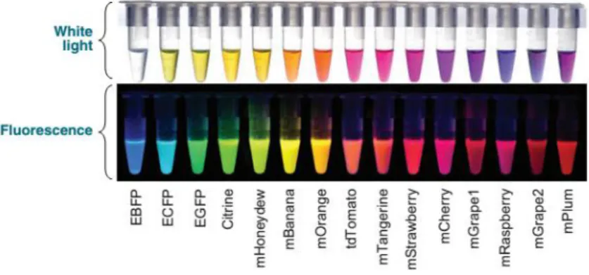
Advances in fluorescence probes and microscopy technologies have provided valuable approaches that present advantages and complement traditional western and immunostaining assays. Since the discovery of green fluorescent protein (GFP) (Prasher et al.,. 1992; Chalfie et al., 1994;; Tsien, 1998), a variety of fluorescent proteins (FPs) with different colours has been developed. Limitations of single-coloured FPs become apparent when the aim is to understand spatiotemporal aspects of interactions between similar organelles and local and often transient alterations in the organization of dynamic subcellular elements like the cytoskeleton and endomembranes. Nowadays fluorescent proteins covering the entire visible spectrum (Fig. 1.5) are considered essential tools for studying gene activity, protein localization, and subcellular interactions. In the recent years, a diversity of optical highlighters namely, photoactivatable, photoswitchable, and photoconvertible fluorescent proteins, have become available. These proteins react to specific wavelengths and undergo structural changes that result in changing from a dark to a bright fluorescent state (photoactivatable FPs) or cause a shift in their fluorescence emission wavelength (photoconvertible FPs) (revised in Shanner et al., 2007).
Figure 1.5.: Diversity of the different fluorescent proteins available for cloning. The top panel shows the FPs under
white light and the bottom panel demonstrates the fluorescence of these FPs. (Adapted from Wang et al., 2008).
1.5.1 Fluorescent proteins: Green fluorescent protein and mCherry
GFP complex was first isolated from the jellyfish Aequorea Victoria. The fusion of wild-type GFP sequence with genes encoding target molecules allows the tracking of subcellular localisations and dynamic motions by fluorescence microscopy. Recently it has been demonstrated new fluorescent protein-based technologies with this protein reinforcing that GFP is a valuable tool for in vivo studies (revised by Sparkes and Branzzini, 2012).
17 Several proteins have been purified from Anthozoa species. Among them, the red fluorescent protein (RFP) was of particular interest. A large amount of effort has been devoted to producing a monomeric RFP such as mCherry (Shanner et al., 2004). mCherry has excellent photostability, fast maturation rate and high resistance to pH variations.
Despite highlighting their target clearly the green and red fluorescent probes are limited in their applications since their colours cannot be switched on or altered as and when required.
1.5.2 Photoconvertible Proteins
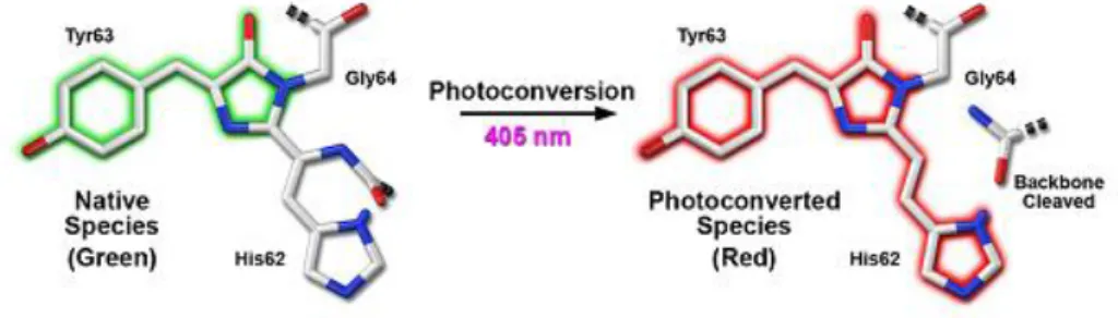
Improved classes of chromophores that are photoswitchable or photoconvertible, capable of pronounced light-induced spectral changes, have enabled users to study the subcellular dynamics in vivo. More recent optical highlighters have chromophores that can be activated either to initiate fluorescence emission from a quiescent state (photoactivation) or to be optically converted from one fluorescence emission bandwidth to another (photoconversion) through a cleavage in peptide backbone represented in the figure 1.5. These tags allow specific tracking of one subcellular structure (organelle or cell subdomain) within a differentially labelled population that used to be a methodological challenge for the cell biologist. Like green fluorescent protein (GFP), monomeric EosFP (mEos) and Kaede are bright green in colour but are efficiently photoconverted into a red fluorescent form using a violet-blue excitation. Moreover mEos and Kaede are fully compatible with cyan fluorescent protein, GFP, yellow fluorescent protein, and red fluorescent protein for use in simultaneous, multicolor labeling schemes. In this section we focus in these two photoconvertible proteins that were optimized in our laboratory for cardosins expression studies, Kaede and mEos, that have different applications due to their different properties.
Figure 1.6.: Green-to-red photoconversion mechanism for Kaede and mEos that occurs when the FP is illuminated with ultraviolet or violet radiation to induce cleavage between the amide nitrogen and carbon atoms in the His62 residue leading to subsequent formation of a conjugated dual imidazole ring system. (Adapted from Shanner et al., 2007)
18 The tetrameric Kaede
The Kaede gene codes for a tetrameric protein found in the stony coral Trachyphyllia geoffroyi. Emits green fluorescence and irreversibly shifts to red fluorescence following irradiation with UV or violet light (Ando et al., 2002). The emission spectra of Kaede before and after photoconversion are distinct from each other which enable simultaneous dual-colour labelling. Kaede has been reported as an excellent subcellular marker for endomembrane compartments such as Golgi bodies (Brown et al., 2010). In this work Kaede proved to be a great reporter to track the dynamic behaviour of designated subpopulations of Golgi within living cells, while visualizing the de novo formation of proteins and structures (Brown et al., 2010). Brown and co-workers demonstrated the applicability of Kaede to explore the secretory pathway in plant cells. The tetrameric structure of Kaede represents advantageous properties like its robustness, the stability of its green and red forms, its high contrast (high absorptivity and distinctness of its forms), and its sensitivity to violet light (Brown et al., 2010). It was suggested that this three-dimensional nature possibly retains protein fusions which induced artefacts in Golgi compartments (Brown et al., 2010).
mEos: a powerful addition to study trafficking dynamic events
EosFP, a homolog of Kaede derived from Lobophyllia hemprichii, has been engineered to a monomeric form without loss in fluorescence and photoconversion properties (Wiedenmann et al., 2004). In its unconverted form, mEos displays bright green fluorescence that, upon illumination with an approximately 390- to 405-nm waveband, changes irreversibly to red fluorescence (emission maximum of 581 nm). Monomeric fluorescent proteins of different colours are widely used to study behaviour and targeting of proteins in living cells. mEos is especially useful for application in plants as it provides the ability to differentially colour and track a single organelle within a population, as well as follow endomembrane and cytoskeletal dynamics over time (Mathur et al., 2010). An important property of monomeric EosFP (mEosFP) is the high stability and irreversibility of its red fluorescent photoconverted form. Thus, if green fluorescence of a target organelle or cell increases and reappears after photoconversion it can be attributed largely to newly synthesised green fluorescent form (recovery after photoconversion).
19 2 Materials and Methods
In order to study cardosins trafficking and their respective targeting signals, classical molecular biology tools were employed to obtain fusions with fluorescent proteins. These constructs were used to transform Arabidopsis thaliana using Agrobacterium tumefaciens mediated methods, either to achieve a transient expression or to obtain stably transformed plants expressing cardosins. Several truncated and mutated versions (already available in our lab) were used and tested for the first time in A. thaliana through transient expression. The same constructs and the ones produced during this work (represented in the figure 2.2) were used to obtain transgenic A. thaliana plants. Cardosins expression was evaluated by confocal microscopy and confirmed by RT-PCR analyses.
2.1 mEos-based constructs
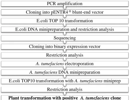
The methodology to obtain mEos constructions and subsequent screening is showed schematically in figure 2.1 and will be detailed in this chapter.
Figure 2.1.: Flowchart representation of the steps performed for obtaining, cloning and screening the mEos constructs analyzed during the work.
Plant transformation with positive A. tumefaciens clone Restriction analyis
E.coli TOP10 transformation with A. tumefaciens miniprep A. tumefaciens DNA minipreparation
A. tumefaciens electroporation Restriction analysis
Cloning into binary expression vector Sequencing
E.coli DNA minipreparation and restriction analysis E.coli TOP 10 transformation
Cloning into pENTR4 ® blunt-end vector
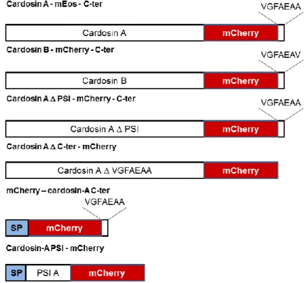
20 Figure 2.2.: Schematic representation of mEos-based constructs engineered during this work and detailed in this section. Cardosin A and B fused to mEos fluorescent protein with cardosins terminal sequence fused to the C-terminus of mEos and cardosin-A and cardosin-B PSI fused to the N-terminal of mEos and mCherry were obtained.
2.1.1 Construction of Cardosins-mEos fusions
The sequence of unmodified cardosin A (Duarte et al., 2008) and B (Soares da Costa et al., 2010) were fused to the monomeric photoconvertible fluorescent protein mEos (Wiedenmann et al., 2004) and were already available in our laboratory from a previous work (Vieira, data). The expression of these versions in N. tabacum leaf epidermis cells showed a pattern suggesting that cardosins were retained in the early endomembrane compartments. Similarly to what happens with mCherry fusions we propose that the cardosins’ C-terminal region may not be exposed to vacuolar receptors due to the conformation of the fusions with mEos that would not allow the proper folding of the protein. In order to obtain chimeric proteins, a C-terminal region was introduced in the 3’ end of the chimeric fusions by amplification with a set of primers (Table 2-1).
21 Table 2-1: Primers used in the amplification of Cardosins-mEos-C-terminal fusions
Primer name Sequence Description
RevEOS-CterA_Sac TTGAGCTCTTAAGC
TGCTTCTGCAAATCCAACTCGTCT GGCATTGTCAGGCAATCCAGAA
Introduces a cardosin A C-terminal region at the 3’ end of the mEos sequence (green), alters the STOP codon of cardosin sequence (red) and introduces a Sac I recognition site (yellow) at the 3’ end of the fusion sequence
A 5’ Xba TCTAGAGCCGCCAC
CATGGGTACCT
Introduces a Xba I restriction site (yellow) at the 5’end of Cardosin A sequence
RevEOS-CterB_Sac TTGAGCTC TTAAAC
TGCTTCTGCAAATCCAACTCGTCT GGCATTGTCAGGCAATCCAGAA
Introduces a cardosin B C-terminal region at the 3’ end of the mEos sequence (green), alters the STOP codon of cardosin sequence (red) and introduces a Sac I recognition site (yellow) at the 3’ end of the fusion sequence
B F KZ Xba 5’- CATCTAGACTCGAG CCACCATGGGAACCCC AATCAAAGCAAACG-3’
Introduces a Xba I restriction site (yellow) at the 5’end of Cardosin B sequence
PCR fragments were cloned into pENTR4® (Invitrogen) digested with Hinc II restriction enzyme that permits the blunt-ended cloning. Positive clones were selected using Xba I and SalI restriction enzymes and sent for sequencing using the universal primer pENTattL2rev (Eurofins MWG Operon, http://www.eurofinsdna.com/home.html) and the listed primers in table 2-2.
22 Table 2-2: Primers sent to Eurofins MWG Operon for the sequencing of pENTR4-Cardosins-mEos-C-terminal constructs
Primer name Sequence Description
Card A 5’ Xba TCTAGAGCCGCCACCATGGGTA CCTCAATCAAA
Primer designed for the 5’ end of Cardosin A heavy chain.
A15F GGTGCTGCAGGTGGTACTTCAT CTGAAGAATTAC
Primer designed for the 5’ end of Cardosin A light chain.
Card B1 AAAACTCGAGCCACCATGGGAA CCCCAATCAAAGCAAGCC
Primer designed for the 5’ end of Cardosin B heavy chain.
B15F GCTGCAGGTGGTTCGATGGTAG ACTGCAAT
Primer designed for the 5’ end of Cardosin B light chain
Positive sequenced clones were selected and used for cloning in the Gateway-compatible cloning vectors designed for expression in Arabidopsis. Cardosin B construct was subcloned into pFAST-G02 (Shimada et al., 2010) and pMDC83 (Curtis et al., 2003) adapted for classical cloning was used for Cardosin A construct. Positive cardosin B-mEos-C-terminal clones were selected using the Eco RI restriction enzyme. Cardosin A-mEos-C-terminal clones were screened with Sal I and Sac I enzymes.
2.1.2 Fusion of Cardosin-A PSI and Cardosin-B PSI to mEos
SP-PSI A-mEos fusion
The construction SP-PSIA fused to mCherry fluorescent protein (Shanner et al., 2004) cloned into pVKH18-En6- was already available in our laboratory. The restriction enzymes Xba I and Sac I were used to excise SP-PSIA-mCherry insert. The reaction product was analysed by agarose gel electrophoresis and the fragment of interest was excised from the gel and purified. The purified DNA was used for cloning into pMDC83 for expression in Arabidopsis. The ligation product was used for TOP10 E.coli transformation and the clones were screened using Xba I and Sac I enzymes.
A positive clone was selected and the DNA extracted from this clone was partially digested for 15 minutes with Sal I and Sac I restriction enzymes to avoid excision of the PSI sequence but