Vol.50, Special Number : pp. 29-35, September 2007
ISSN 1516-8913 Printed in Brazil BRAZILIAN ARCHIVES OF BIOLOGY AND TECHNOLOGY
A N I N T E R N A T I O N A L J O U R N A L
Common Causes of False Positive F
18FDG PET/CT Scans in
Oncology
Kevin R. Carter* and Eduard Kotlyarov
Michigan State University, POH Medical Center, 50 N. Perry, kcarter99@hotmail.com; Pontiac 48342 - Michigan - USA
ABSTRACT
PET/CT is a common imaging modality used in the evaluation of oncology patients. While being extremely sensitive to identifying sights of malignancy F18FDG is very non-specific. We attempted to provide a brief review of some of the more common processes that a nuclear radiology physician may encounter in daily clinical practice that could result in a false positive diagnosis with F18FDG PET/CT. A fundamental understanding of the limitations of this technology by the interpreting physician is necessary to avoid making inaccurate diagnosis and potentially limiting important treatments for our patients.
Key words: PET/CT, False Positives
*
Author for correspondence
INTRODUCTION
Radiologic imaging provides an important component to both the diagnosis and management of oncology patients. Through the accurate noninvasive visualization of pathologic processes, there is a resulting decrease in the overall morbidity and mortality. Traditionally, Computerized Tomography (CT) has been utilized to evaluate anatomical pathology. This information was visually combined with the functional information of Positron Emission Tomography (PET) with F18 fluorodioxyglucose (FDG). Current technology allows for simultaneous acquisition of this information which allows precise localization and characterization of pathological processes. Utilization of PET/CT has been shown to be beneficial in altering the management of patients compared to imaging with just CT or PET alone (Mavi et al., 2005). PET/CT is currently utilized in the diagnosis and
staging of multiple malignancies (Carter and Kotlyarov, 2005). It is approved in the United States for the characterization of solitary pulmonary nodules, the diagnosis-staging-restaging of colon carcinoma, lymphoma, melanoma, esophageal carcinoma, and head and neck cancers. (See Fig. 1) It is also useful to evaluate the efficacy of chemotherapy and/or radiation therapy following treatment (MNCDM, 2007). (See Fig. 2)
space in substantial amounts, essentially trapping the FDG (Kostakoglu et al., 2003).
myocardium, gastrointestinal system, urinary system, thyroid gland, and gonadal issues).
Figure 1 - This 56 year old male presented with an abnormal pre-employment screening chest x-ray. The PET/CT was performed to determine if an abnormality present in the lung was malignant or benign. Whole body maximum intensity projection (MIP) image (A) shows a focal area of FDG uptake in the right lower lobe (arrow). Axial PET image at the level of the lesion (B) showed the approximate location of the area of increased activity (arrow), but the fused PET/CT image (C) confirmed that this lesion was actually within the lung. This lesion underwent biopsy and proved to be well differentiated adenocarcinoma
Figure 2 - This 47 year old female initially presented for a lymphoma staging exam in March 2007 (A) Whole body MIP image, demonstrated marked uptake of FDG within the lymph nodes of the mediastinum and right supraclavicular region (arrow). Following chemotherapy, whole body MIP image obtained July 2007, (B) there has been a dramatic reduction in the uptake of FDG within the mediastinum and supraclavicular region, but there is increased uptake within the bone marrow that is related to increased hematopoietic activity as a response to chemotherapy
A
B
C
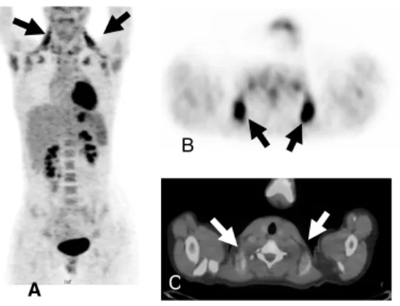
Figure 3 - This 6 year old child underwent PET/CT for evaluation of right supraclavicular lymphadenopathy. The MIP image (A and B) shows intense uptake of FDG in the bilateral supraclavicular regions that would be suspicious for disease, but when correlated with the fused image (C), this area of increased uptake is in the region of fat and is consistent with brown fat. The CT portion of the exam helped exclude our original diagnosis of lymphoma that was obtained from interpreting only the PET images
DISCUSSION
FDG is not only a cancer specific imaging agent, false positive results may be observed with benign diseases. False positive results are commonly observed in areas of active inflammation or infection (Gupta et al., 20000), with a reported false positive rate of 13% and false negative rate of 9% (Alavi et al., 2002). Inflammatory cells (neutrophils and activated macrophages) at the sites of inflammation or infection will show increased FDG accumulation (Alavi et al., 2002). These false-positive areas of metabolic activity have the potential for significant morbidity and mortality if not accurately recognized.
Brown adipose tissue has been reported to be observed in 2-4% of patients and is especially common to be observed in women and children during cold weather months. This tissue is responsible for cold induced and diet induced thermogenesis. Mitochondria in brown adipose tissue exclusively express the thermogenic protein, and F18 FDG uptake in this hypermetabloic brown fat can occur (Kostakoglu et al., 2003). FDG is normally taken up in brown adipose tissue in the neck, paraspinal location as well as in the pararenal space (See Fig. 3).
Multiple nonspecific infectious/ inflammatory processes can cause increased FDG uptake and
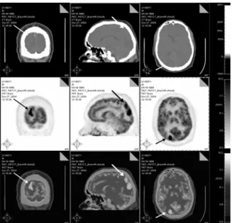
result in a false positive diagnosis for malignancy on PET/CT. Several cases have been reported including abscesses (brain, abdominal, renal, tubo-ovarian), osteomyelitis, sinusitis, myositis, thryoridits, matitis, esophagitis and gastritis (Alavi et al., 2002). Other described etiologies include pneumonia from multiple causes (Bunyaviroch and Coleman, 2005). While there are numerous etiologic causes for infection, tuberculosis and the fungal infections (Cryptococcosis, Histoplasmosis, Coccidioidomycosis, Blastomycosis, and Aspergillosis) are most commonly described as source of false positive results with PET/CT examinations (Bunyaviroch and Coleman, 2005). Tuberculosis is one of the oldest diseases to affect humans. It is caused by bacteria belonging to the mycobacterium tuberculom complex (Raviglione and O’Brien, 2005). It usually affects the lungs, but in one third of cases, other organs are involved. Extrapulmonary sites may also be involved including: regional and distant lymph nodes, pulmonary pleura, genitourinary system, gastrointestinal system, skeletal system, pericardium, and the central nervous system (Raviglione and O’Brien, 2005). All of these areas of infection/inflammation will demonstrate intense uptake of FDG and unless correlated with the patient’s clinical history and pathologic biopsy will result in an incorrect diagnosis. (See Fig. 4). A
B
Figure 4 - Coronal, Sagital, and axial PET/CT images of the brain that shows a focal area of increased metabolism within the Right occipital lobe (arrow). This area correlated with the enhancing mass that was observed on an MRI (not shown) and was felt to represent a primary brain neoplasm (astrocytoma). Biopsy revealed that this lesion was actually a walled off abscess resulting from infection with M. Tuberculosis
Many noninfectious inflammatory granulomatous processes can also lead to localized and disseminated inflammatory processes throughout the body that may result in focal areas of increased metabolism on PET/CT examinations resulting in a false positive diagnosis. While there may be multiple etiologies, those most commonly encountered includes: sarcoidosis, athrosclerosis, and pneumoconiosis (Shreve et al., 1999).
Sarcoidosis is a chronic multisystem noncaseating granulomatous disease. It is characterized by accumulation of T lymphocytes and mononuclear phagocytes, noncaseating epithelioid granulomas, and derangements of the normal tissue architecture in affected organs. The etiology is unknown, but is though to be caused by an exaggerated cellular immune response. Several groups have reported FDG uptake by sarcoid granulomas (Shreve et al., 1999; Brudin et al., 1994; Lewis and Salama, 1994; Yasuda et al., 1996) due to the inflammatory cell infiltrates composed of lymphocytes, macrophages, and epithelioid cells. Sarcoidosis may show multiorgan involvement at PET/CT examination. Respiratory tract involvement will occur at some time period in the course of almost all cases of sarcoidosis, with an interstitial lung presentation that involves the alveoli, blood vessels, and bronchioles (Shreve et al., 1999). Intrathoracic and peripheral lymphadenopathy
with increased FDG accumulation is also commonly observed on PET/CT (Shreve et al., 1999). Approximately 5-10% of patients with sarcoidosis will manifest cardiac involvement with reduced left ventricular function, valvular abnormalities, pericardial effusions, and ventricular aneurysms (Brudin et al., 1994). Other areas of involvement include the skin, ears, and central nervous system (Shreve et al., 1999). Involvement of the kidneys and liver is also known, with almost 40-70% of patients having granulomatous involvement of the liver at biopsy (Shreve et al., 1999). Any of these sites of active involvement will show accumulation of FDG on PET/CT and this accumulation as long as not mistaken for malignancy may be useful for assessing systemic involvement and the response to therapy (Alavi et al., 2002). In cases of suspected sarcoid based upon PET/CT findings and clinical history; correlation with bronchoscopy and bronchoalveolar lavage, or biopsy of an accessible lymph node may is necessary to differentiate this entity from metastasis, military infection, or lymphoma (See Fig. 5).
asbestosis, and berylliosis (Alavi et al., 2002). These diseases tend to be progressive continuing even after the inhalation of the dust has ceased. The inhalation of these dust particles result in activation of marcrophages that result in secretion of cytokines which mediate an inflammatory reaction that includes fibroblast proliferation and collagen deposition (Yasuda et al., 1996). Numerous reports have described FDG uptake in pneumoconiosis, this is likely related to the activation of the inflammatory cells as well as the fibroblasts (Yasuda et al., 1996). Correlation with the CT portion of the examination is necessary to evaluate for changes in the lungs which would be indicative of a fibroitic process.
Known to be a major cause of coronary heart disease, stroke, and peripheral vascular disorders, atherosclerosis mainly involves the aorta and arterial circulation of the heart, brain, kidneys, and extremities. Histologically, inflammation is known to occur early in the development of atherosclerosis (Newman et al., 1997). Uptake of FDG within atherosclerotic plaques in multiple arteries, including the aorta, iliac arteries, and proximal femoral arteries has been reported (Hirose et al., 1994; Libby et al., 2002), and correlation with the CT portion of the examination should be made to confirm that these areas of increased metabolism reside within a vessel (See Fig. 6).
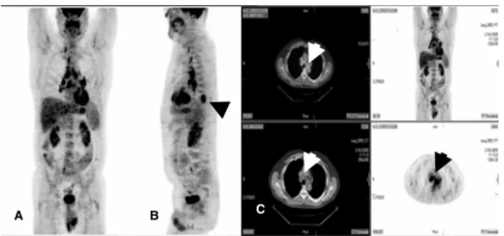
Figure 5 - Whole Body PET/CT (A and B) was performed on this 62 year old male to stage his known lung adenocarcinoma involving the left lower lobe (arrow in B). In addition to uptake within his know left lower lobe neoplasm, there are diffuse areas of FDG accumulation within the bilateral hilar regions and multiple lymph nodes (arrows in C) within the mediastinum. The patient was initially diagnosed with diffuse metastatic disease. After a full review of his clinical history and biopsy, the diffuse hilar and mediastinal uptake is consistent with the patients known diagnosis of sarcoidosis, not advanced metastatic carcinoma.
Numerous iatrogenic may result in areas of false positive increased metabolic activity on FDG PET/CT exams. This includes increased metabolically activity resulting from injections such as diffuse muscular uptake following administration of insulin prior to the injection of FDG (Bunyaviroch and Coleman, 2005). Following the administration of colony-stimulating factors, diffuse increased bone uptake has also been described (Yao et al., 1995). Post surgical changes will result in areas of increased FDG uptake including areas of prior biopsies, sites of catheter insertions, and sites of other drainage tube insertions (El-Haddad et. al, 2004). (See Fig. 7) Some of the most common iatrogenic causes of uptake on PET/CT include the response from
radiation with development of pneumonitis/fibrosis, as well as talc pleurodesis (Shreve et al., 1999).
An awareness of prior treatments and interventions is necessary prior to interpreting any patients PET/CT examination in order to avoid making an interpretation error and potentially overestimating the amount of disease.
Benign tumors that result in intense FDG accumulation on PET/CT examination have been described, and these should be recognized at potential causes for a false positive diagnosis. These tumors include: fibrous mesothelioma, schwannoma, aggressive neurofibromas and enchondromas (Shreve et al, 1999).
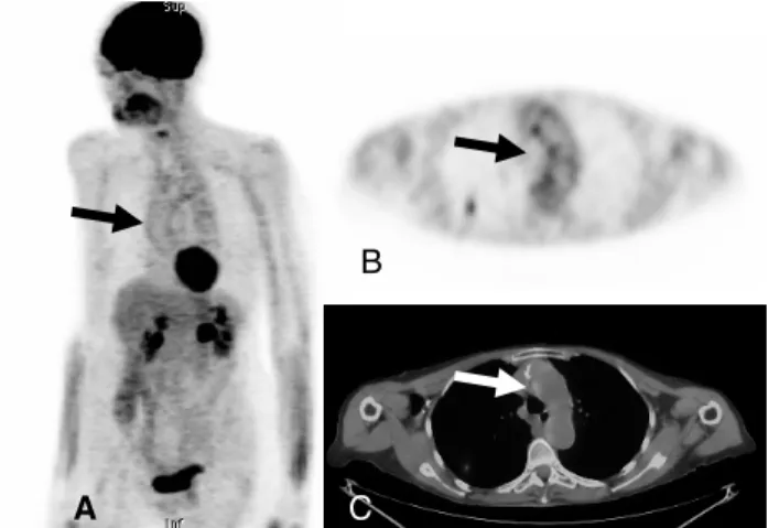
Figure 6 - This 72 year old female presented for evaluation of a right upper lobe mass. Whole body MIP image (A) shows diffuse curvilinear uptake in the mediastinal region. Axial PET image (B) confirmed this finding which was felt to represent the aorta. This was confirmed on the axial fused PET/CT image (C) and is consistent with diffuse atherosclerotic disease.
Correlation with the patient’s history and other imaging modalities may be necessary to suggest the correct diagnosis. Most importantly, long term stability will be seen in all of these lesions ensuring that they are benign processes. These lesions are usually misdiagnosed as malignant processes and will undergo biopsy or resection (Shreve et al., 1999; Libby et al., 2002; Yao et al., 1995).
Well-known focal false positive artifacts are metallic or otherwise dense foreign bodies on the surface of the body or implanted (pace-maker, stimulators, leads, prosthetics, bullet fragments, brachotherapy, etc) bodies. This false positive artifact is only seen on Attenuation Correction (AC) scans and is not seen on Non-Attenuation Correction (NAC) images. It is imperative to analyze both AC and NAC images routinely to avoid these false positive artifacts. Additionally, reviewing NAC images increases study sensitivity by about 13-15%.
CONCLUSION
Physicians interpreting PET/CT examinations must understand that accumulation of FDG only indicates enhanced cellular metabolism, irrespective of the nature of the cells. Areas of activated macrophages as well as metabolically active carcinomas will all accumulate FDG. Due to the limited specificity of FDG-PET/CT in
excluding malignancy, FDG positive lymph nodes and metabolically active masses require histologic assessment to avoid incorrect diagnosis and potentially limiting treatment options for patients. The absences of FDG accumulation in lesions and lymph nodes is highly predictive for the absence of active disease, but false negative results can also be observed in malignancies with low metabolic activities including: prostate carcinoma, renal carcinoma, carcinoid, brochoalveolar cell carcinoma (Kostakoglu et al., 2003). It is essential to have an understanding of the causes of both false positive and false negative results when interpreting PET/CT to avoid making inaccurate diagnoses. Overall the FDG PET/CT is a powerful new diagnostic tool that has made a significant contribution to the diagnosis and management of oncology patients.
RESUMO
PET/CT é uma modalidade de imagem usada para avaliação de pacientes oncológicos. Embora seja extremamente sensível para identificar sinais de
malignidade, o F18FDG não é ainda muito
específico. Nós buscamos mostrar uma breve revisão de alguns dos processos mais comuns que um médico nuclear pode encontrar na prática clínica diária que poderia resultar em um diagnóstico falso positivo com a F18FDG PET/CT. Uma melhor compreensão das limitações dessa
A
B
tecnologia é necessária para que a interpretação pelo médico não possibiite diagnósticos imprecisos, que potencialmente podem limitar os tratamentos dos pacientes.
REFERENCES
Alavi, A., Gupta, N., Alberini, J. L., Hickeson, M., Adam, L. E., Bhargava, P., Zhuang, H. (2002), Positron emission tomography Imaging in nonmalignant thoracic disorders. Semin. Nucl. Med.,
32, 293-321.
Bunyaviroch, T., Coleman, E. (2005), PET evaluation of Lung Cancer. J. Nucl. Med., 47, 451-469.
Brudin, L. H., Valind, S. O., Rhodes, C. G., Pantin, C. F., Sweatman, M., Jones, T., Hughes, J. M. (1994), Fluorine-18 deoxyglucose uptake in sarcoidosis measured with positron emission tomography. Eur. J. Nucl. Med., 21, 297-305.
Carter, K., Kotlyarov, E. (2005), PET and PET/CT imaging for the earliest detection and treatment of colorectal carcinoma. Braz. Arch. Biol. Technol., 48
(suppl.), 169-174.
El-Haddad, G., Zhuang, H., Gupta, N., Alavi, A. (2004), Evolving role of positron emission tomography in the management of patients with inflammatory and other benign disorders. Sem. Nucl. Med., 34, 313-329.
Gupta, N. C., Graeber, G. M., Bishop, H. A. (2000), Comparative efficacy of positron emission tomography with fluorodeoxyglucose in evaluation of small (<1cm), intermediate (1 to 3 cm), and large (>3 cm) lymph node lesions. Chest, 117, 773-778. Hirose, Y., Ishida, Y., Hayashida, K., Maneo, M.,
Takamiya, M., Ohmori, F., Miyatake, K., Uehara, T., Nishimura, T., Tachibana, T. (1994), Myocardial involvement in patients with sarcoidosis: an analysis of 75 patients. Clin. Nucl. Med.,19, 522-526.
Kostakoglu, L., Agress, H., Goldsmith, S. J. (2003), Clinical role of FDG PET in evaluation of cancer patients. Radiographics, 23, 315-340.
Lewis, P. J., Salama, A. (1994), Uptake of fluorine-18-fluorodeoxyglucose in sarcoidosis. J. Nucl. Med., 35, 1647-1649.
Libby, P., Ridker, P., Maseri, A. (2002), Inflammation and atherscelerosis. Circulation, 105, 1135-1143. Mavi, A., Lakhani, P., Zhuang, H., Gupta, N. C., Alavi,
A. (2005), Fluorodeoxyglucose-PET in characterizing solitary pulmonary nodules, assessing pleural diseases, and the initial staging, restaging, therapy planning, and monitoring response of lung cancer,
Radiol. Clin. North Am., 43, 1-21.
Medicare National Coverage Determinations Manual, Pub. 100-03, Part 4, Section220.6, accessible online athttp://cms.hhs.gov/manuals/103_cov_determ/ncd10 3c1_Part4.pdf.
Newman, L. S., Rose, C. S., Maier, L. A. (1997), Sarcoidosis. New Engl. J. Med., 336, 1224-1234. Raviglione, M. C., O’Brien, R. J. (2005), Tuberculosis
In: Harrison’s Principles of Internal Medicine. 16th Ed, Kasper, D. L., Braunwald, E., Fauci, A. S., McGraw-Hill New York, NY pp.953-966.
Shreve, P., Anzai, Y., Wahl, R. L. (1999), Pitfalls in oncolgic diagnosis with FDG PET imaging: physiologic and benign variants. Radiographics,
19,61-77.
Yao, W., Hoh, C., Hawkins, R. A., Glaspy, J. A., Weil, J. A., Lee, S. J., Maddahi, J., Phelps, M. E. (1995), Quantitative PET imaging of bone marrow glucose metabolic response to hematopoietic cytokines. J. Nucl. Med, 36, 794-799.
Yasuda, S., Shohtsu, A., Ide, M., Takagi, S., Ogawa, J., Mitomi, T., Suzuki, Y. (1996), High fluorine-18 labeled deoxyglucose uptake in sarcoidosis. Clin. Nucl. Med., 21, 983-984.