Received for publication 19/08/2015 - Accepted for publication 07/10/2015
Sarcoidose ocular: a nossa realidade
nos últimos seis anos
Ocular sarcoidosis: our reality
for the past six years
Inês Coutinho
1, Ana Bastos Furtado
2, Cristina Santos
1, Susana Pina
1, Maria Lisboa
1, Isabel Ferreira
2, Bruno Grima
2,
Manuela Bernardo
1R
ESUMOIntrodução: Na sarcoidose, as manifestações oculares são comuns e podem constituir a manifestação inicial da doença ou mesmo a única. O objetivo deste trabalho foi analisar os parâmetros demográficos, manifestações clínicas, terapêutica e principais complicações oculares em doentes com sarcoidose ocular. Métodos: Estudo descritivo e retrospectivo que incluiu doentes com o diagnóstico de sarcoidose ocular, observados nas consultas de Inflamação Ocular e de Doenças Auto-Imunes do Hospital Prof. Doutor Fernando Fonseca, no período entre 2009 e 2015. Resultados: Foram identificados 11 doentes com o diagnóstico de sarcoidose ocular, com predomínio do sexo feminino (54,5%) e caucasianos. A média da idade ao diagnóstico foi de 45±14 anos. A sarcoidose manifestou-se de forma exclusivamente ocular em 36% dos casos. O envolvimento ocular foi a manifestação inicial em 90,9% dos casos. Identificaram-se 9 casos de uveíte, 1 de esclerite anterior nodular e 1 de queratite intersticial. O tratamento com corticoterapia tópica foi realizado em 100% dos casos, sendo o tratamento único em apenas 1 doente. Nos restantes, foi necessário associar corticoterapia oral. Em 4 desses doentes, pela gravidade da doença e atingimento binocular, utilizou-se também corticoterapia pulsada endovenosa. O tratamento adjuvante imunossupressor mais frequentemente utilizado foi o metotrexato (45%). Um doente necessitou de terapia biológica com infliximabe para controle da doença. Conclusão: A manifestação ocular mais comum foi a uveíte, com predomínio da panuveíte. O tratamento mais utilizado e com maior taxa de controle da doença foi a corticoterapia sistêmica em associação com o metotrexato.
Descritores: Sarcoidose; Sarcoidose ocular; Corticoterapia/uso terapêutico; Metotrexato/uso terapêutico; Infliximabe/uso terapêutico
A
BSTRACTPurpose: Insarcoidosis, ocular manifestations are common and can be the initial or even the only clinical manifestation. The aim of this study was to analyze the demographic parameters, clinical manifestations, treatment and the major ocular complications in patients with ocular sarcoidosis. Methods: We conducted a descriptive and retrospective study that included patients with the diagnosis of ocular sarcoidosis, followed by inflammatory ophthalmology and immune-mediated disease consults at the Prof. Doutor Fernando Fonseca Hospital, between 2009 and 2015. Results: Eleven patients with the diagnosis of ocular sarcoidosis were identified, with a predominance of females (54,5%) and Caucasians. The average age at diagnosis was 45 ± 14 years. Sarcoidosis was exclusively ocular in 36%. The first manifestation of sarcoidosis was eye disease in 90.9 % of cases. Nine cases of uveitis, one of nodular scleritis and one of interstitial keratitis were observed. Topical corticoid treatment was applied in 100% of cases, with only one achieving remission of the disease. Oral corticoid treatment was necessary in 10 cases, four of which needed a high dose methylprednisolone induction. Methotrexate was the adjunctive immunosuppressive treatment of choice in 45% of cases. There was one refractory case for conventional immunosuppressive therapy, having achieved remission with biologic agent infliximab.Conclusion: Uveitis was the commonest ocular manifestation, and there was a predominance of panuveitis. Systemic corticoid and methotrexate were the most used immunosuppressive treatments for maintaining the controlled stated of the disease.
Keywords: Sarcoidosis; Ocular sarcoidosis, Corticosteroid/therapeutic use; Methotrexate/therapeutic use; Infliximab/therapeutic use
The authors declare no conflicts of interests.
I
NTRODUCTIONS
arcoidosis is a systemic inflammatory disease characterized by the formation of noncaseating granulomas in the organs and tissues involved. The pathophysiological mechanism is not fully understood yet, and is the result of a complex immune process mediated by Th1 and Th17 cell response. Response patterns were identified in association to genetic and environmental factors, whether in the presentation form of antigen (determined by MHC2 alleles) or in the form how the immune response is developed and maintained [regulatory T cells (Tregs)].(1,2) The disparity observed among individuals withregard to the clinical course of the disease can be explained by the different ability to develop and control the immune response. Sarcoidosis can be self-limiting, without the need of therapeutic intervention, or can become persistent and develop to chronicity, requiring suppression by pharmacological means.
Epidemiologically, it is more common in young female adults aged from 20 to 40 years, and in European women after the age of 50, where a second peak of incidence is described1. It can affect all
ethnic groups, although it is reported with greater frequency in individuals from Northern Europe and in the black race.(1,2)
Sarcoidosis can involve any organ, the lung being the main organ affected (90% of cases). The skin and eye attainment are common with an estimated prevalence of 20 to 35% and 10 to 60% respectively.(3-5)
Sarcoidosis can be the initial manifestation of ocular disease or even the only one.(3,6) It can affect all structures of the eyeball
and attachments, but classically the bilateral granulomatous an-terior uveitis is the most common ocular manifestation.
Diagnosis is made by a detailed evaluation of all clinical, imaging, histological, cytological information and exclusion of other causes of granulomatous disease such as tuberculosis.(1,7)
Obtaining noncaseating granulomas in the histological sample confirms the diagnosis. However, it is not always possible to have a biopsy of the affected tissues, in particular in the patient with isolated uveitis. So, given the wide variety of systemic and ocular manifestations, the International Workshop on Ocular Sarcoidosis (IWOS) proposed 7 clinical signs and 5 laboratory and imaging results in order to assist and support the diagnosis of ocular sarcoidosis (Table 1).(8-11)
According to the criteria proposed by IWOS, a diagnosis of ocular sarcoidosis can range from possible to definitive (Table 2), after the exclusion of other causes of uveitis, especially tuberculosis, syphilis, toxoplasmosis and lymphoma. A global and detailed evaluation of the patient by experienced clinicians is therefore essential in systemic diseases such as sarcoidosis.(12)
The treatment of sarcoidosis, particularly ocular sarcoidosis, aims to halt the inflammatory process and is guided by the severity of the disease presentation, i.e., more intense inflammatory activity with loss or risk of loss of vision requires a more aggressive initial immunosuppressive approach. The treatment may be topical in the uveitis with previous location or associated to systemic therapy in cases of intermediate ou poste-rior uveitis, or if refractory to topical therapy. The systemic treatment includes classic immunosupressants drugs such as corticosteroids, azathioprine, methotrexate and Cyclosporine.(13)
Methotrexate is the most widely used corticosteroid-sparing drug in the series reported(14,15), with positive results in chronic uveitis
by sarcoidosis. In cases refractory to classic immunosuppressive therapy, biological therapy with the monoclonal antibody anti-TNF á infliximab has shown very positive results. (16, 17)
M
ETHODSA descriptive and retrospective study included 11 patients with diagnosis of sarcoidosis observed in the appointments of Eye Inflammation and Auto-Immune Diseases at the Hospital Prof. Doctor Fernando Fonseca, Portugal, between June 2009 and January 2015.
R
ESULTSDemographic characteristics
Of the 11 individuals studied with ocular sarcoidosis, 6 women (54.5%) and 5 men (45.5%) were identified.
Table 1
Clinical signs, laboratory tests and imaging suggestive of the diagnosis of ocular sarcoidosis
Ocular clinical signs
1. Granulomatous keratic precipitates and/or iris nodules (Koeppe/Busacca)
2. Nodules on trabecular meshwork and/or peripheral anterior synechiae in tent form
3. Vitritis, snowballs and vitreous infiltrated in “string of pearls” 4. Active or atrophic peripheral chorioretinal lesions (Dalen
Fuchs nodules)
5. Nodular or segmental periphlebitis (wax drop) and/or retinal macroaneurysm
6. Granulomas of the optic disc and /or solitary choroidal 7. Bilateralism
Investigational tests
1. Negative tuberculin test
2. High serum values of ACE e/ou Lysozyme 3. Chest x-rays revealing bilateral hilar adenopathy 4. High liver enzymes (AST, ALT, alkaline phosphatase) 5. Chest CT with changes in patient with negative chest x-ray
Table 2
Diagnostic criteria of ocular sarcoidosis
Diagnostic criteria
1. DEFINITIVE Confirmation by biopsy and ocular sarcoidosis compatible uveitis
2. PRESUMED Biopsy not doneBilateral hilar ocular sarcoidosis adenopathy + compatible uveitis 3. PROBABLE Biopsy not performed and absence ocular sarcoidosis of bilateral hilar adenopathy
3 Suggestive intraocular signs + 2 positive investigational tests 4. POSSIBLE Biopsy negative 4 Suggestive ocular sarcoidosis intraocular signs + 2 positive
Figure 1. Ocular manifestations of sarcoidosis
Figure 2. Uveitis type
The average age at date of diagnosis was 45 ± 14 years, most patients (90.9%) being caucasians.
Diagnosis
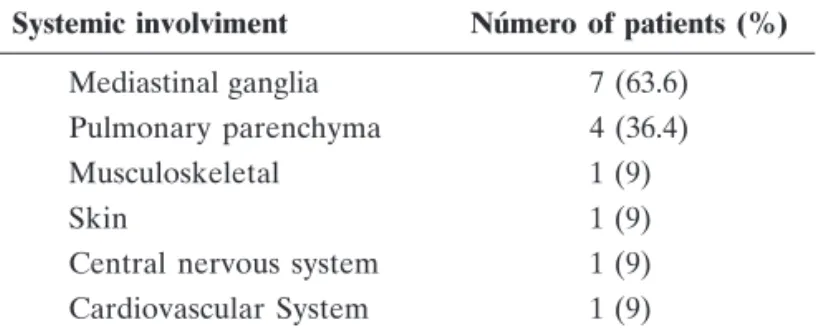
The eye disease was the first clinical manifestation in 90.9% of the sample (n = 10), leading to research and diagnosis of sarcoidosis. Sarcoidosis is expressed exclusively in the ocular form in 36% (n = 4) of the cases and 63.6% (n = 7) systemically with ganglion predominance of mediastinal location of 63.6% (n = 7) and pulmonary parenchyma 36.4% (n = 4) (Table 1). The average time between the first ocular manifestation and the systemic ones was 9.8 months [0-36M]. In addition to clinical observation, additional diagnostic test performed which assisted the most the diagnosis was chest CT. It was possible to confirm the diagnosis with the histologic sampling containing noncaseating granulomas in 45% (n=5) of cases.
According to the diagnostic criteria of the International Workshop on Ocular Sarcoidosis (IWOS), the cases of Uveitis (n=9) were classified as definite (n=5), presumed (n=1), and probable (n=3) ocular sarcoidosis.
Characterization of ocular involvement
The bilateral ocular involvement expressed in 54.5% of the sample.
Uveitis was the most frequent ocular manifestation with 81% of prevalence (n = 9), followed by 1 case of anterior scleritis and 1 of interstitial keratitis (Figure 1).
Of the 9 patients with uveitis, 2 had anterior uveitis and 7 panuveitis, not being observed isolated posterior involvement (Figure 2).
Table 1
Location of systemic involvement
Systemic involviment Número of patients (%)
Mediastinal ganglia 7 (63.6) Pulmonary parenchyma 4 (36.4) Musculoskeletal 1 (9)
Skin 1 (9)
Central nervous system 1 (9) Cardiovascular System 1 (9)
Figure 3: Keratic precipitates in “mutton fat”
Figure 4: Posterior Synechia
Ocular inflammation was presented as granulomatous uveitis in 4 patients (Figure 3) and not granulomatous in 5 patients. This data emphasizes that ocular sarcoidosis can also manifest in the form of non-granulomatous anterior uveitis.
The formation of posterior synechiae was frequent (Figu-re 4), not being documented nodules in the iris (Koeppe or Busacca).
Figure 5: Segmental papillitis and periphlebitis (retinography and angiography)
Figure 6: Isolated choroidal granuloma
The impairment of the episclera and sclera with episcleritis and scleritis are frequent demonstrations, while the corneal involvement is rare. In our analysis, a case of nodular anterior scleritis (Figure 7) and another of interstitial keratitis (Figure 8) were identified.
Complications and visual acuities
The ocular prognosis is relatively benign, and some patients may develop severe ocular complications with low visual acuity. Ocular complications have been documented in 5 of the 11 patients who included posterior synechiae (n=3), cataract (n=2), ocular hypertension (n=1), glaucoma (n=1) and macular edema (n=1).
Figure 7: Nodular anterior scleritis
Figure 8: Interstitial keratitis with corneal neovascularization
The visual acuities (VA) of the 39 patients ranged from 6/ 10 to 10/10, except for one patient in which the VA was 1/10 due to developing nuclear cataract with posterior synechiae to 360º, and who is waiting for surgery.
Therapy
Patients with a diagnosis of ocular sarcoidosis were referred to the appointment of the Autoimmune Diseases of the IV Medicine Service for the study of systemic involvement and, when necessary, start the appropriate therapy.
Topical corticosteroids have been used in all patients (100%), and the disease was controled in one case. In the remaining patients (n=10), it was necessary to establish a systemic immunosuppressive therapy.
Given the severity of the ocular presentation, 4 patients (36%) were treated with intravenous methylprednisolone in high dose (1g/day), followed by oral prednisolone (1mg/kg/day), and subsequent adjuvant immunosuppressive therapy with methotrexate (10mg/week). One of these patients (patient No. 5
-Table 2) kept refractory disease, and the association to other classic immunosuppressants, azathioprine and cyclosporine was tempted without effectiveness and with the need for systemic corticosteroid therapy in very high doses. So, given the severity of the clinical profile the treatment with anti-TNF á (infliximab in association with methotrexate) was started, with a good and safe response.
Of the remaining patients in need of systemic therapy, 3 held only oral corticosteroid therapy and 1 oral corticosteroid therapy in association with methotrexate, with control of the disease.
D
ISCUSSIONThe main ocular manifestation of sarcoidosis was uveitis, and 90.9% of the patients the ocular disease was the first clinical manifestation, which supports the ocular impairment as an early manifestation of the disease.(17)
Ocular involvement in sarcoidosis varies with race and age, with the involvement of the posterior segment being more common in caucasians and of the anterior segment in the black race(18), which explains in part the large posterior involvement
found in the sample studied.
Although not occuring in our study, the involvement of the conjunctiva and iris with granulomas, and the inflammation of the lacrimal gland may occur, affecting an increase thereof and secondarily sicca keratoconjunctivitis, as well as the involvement of the orbit and the extraocular muscles, sometimes simulating a condition of thyroid orbitopathy or non-specific inflammation of the orbit. (3,6,19)
The topical and/or oral corticosteroid therapy is the immunosuppressive treatment of choice, but in refractory cases or in need of savers of oral corticosteroids the methotrexate was the most widely used alternative and with a good rate of disease control.
C
ONCLUSIONOcular involvement is a common and early manifestation of sarcoidosis.
N Age at Sex Clinic Class uveitis MCDTs diagnosis Initial Organ involved IWOS
(years) manifestation Chest CT Bx Eye Lung Ganglia NS MSK CV Skin GI
1 48 F Ocular Ant. uveitis Ø Y Ø Y Ø Ø Ø Probable Y Ø
2 55 F Ocular Panuveitis Y Y Y Ø Ø Y Ø Definitive Y Y 3 28 M Ocular Panuveitis Ø Ø Ø Ø Ø Ø Ø Presumed Y Ø
4 71 M Ocular Panuveitis S Y Ø Ø Y Ø Ø Definitive Y Y 5 31 M Ocular Instersticial Ø Ø Ø Ø Ø Ø Ø Ø Y Ø keratitis
6 53 F Ocular Panuveitis Y Y Ø Ø Ø Ø Ø Definitive Y Y 7 45 F Systemic Panuveitis Ø Y Ø Ø Ø Ø Ø Definitive Y Y 8 29 M Ocular Ant. Scleritis Ø Ø Ø Ø Ø Ø Ø Ø Y Ø
9 30 M Ocular Panuveitis Y Y Ø Ø Ø Ø Ø Probable Y Ø
10 45 F Ocular Ant. uveitis Ø Y Ø Ø Ø Ø Ø Definitive Y Y 11 61 F Ocular Panuveitis Ø Ø Ø Ø Ø Ø Ø Probable Y Ø
N Terapy Status of the disease
Topic Systemic
MPDN PDN MTX AZAT CICLOSP Anti-TNF – INF
1 Y Ø Ø Ø Ø Ø Ø Controled
2 Y Y Y Y Ø Ø Ø Controled
3 Y Y Y Y Ø Ø Ø Controled
4 Y Ø Y Ø Ø Ø Ø Controled
5 Y Y Y Y Y Y Y Controled
6 Y Ø Y Ø Ø Ø Ø Controled
7 Y Ø Y Ø Ø Ø Ø Controled
8 Y Y Y Y Ø Ø Ø Controled
9 Y Ø Y Ø Ø Ø Ø Controled
10 Y Ø Y Y Ø Ø Ø Controled
11 Y Ø Y Ø Ø Ø Ø Controled
Caption: N. patient; Y = yes; Ø = no; MDPN methylprednisolone; PDN prednisolone; MTX methotrexate, AZAT azathioprine; CICLOSP cyclosporin, INF infliximab
Table 2
Characteristics analysed in each case
stage of the disease. The clinical development of the disease can be one of the most important data for the diagnosis and subsequent therapeutic control. Awareness of the difficulty in diagnosis goes along with the treatment. Ocular inflammatory disease may have significant implications on visual acuity, and may lead to the development of potentially irreversible lesions. At the same time, the choice of immunosuppressive therapy is difficult and not without harmful side effects, both locally and systemically.
The multiplicity of forms of presentation without pathognomonic signs together with the fact of organ involvement does not necessarily cause symptoms makes it challenging to
diagnose and give therapeutic guidance for these patients, requiring interdisciplinary work between physicians.
A
CKNOWLEDGEMENTSTo the IV Medicine Service of Hospital Prof. Doutor Fernando Fonseca
R
EFERENCES2. Broos CE, van Nimwegen M, Hoogsteden HC, Hendriks RW, Kool M, van den Blink B.Granuloma formation in pulmonary sarcoidosis. Front Immunol. 2013;4:437.Review.
3. Rothova A. Ocular involvement in sarcoidosis. Br J Ophthalmol. 2000;84(1):110-6. Review.
4. Jamilloux Y, Kodjikian L, Broussolle C, Sève P. Sarcoidosis and uveitis. Autoimmun Rev. 2014;13(8):840-9.
5. Oréfice F, Santos D, Oréfice J. Uveítes. 2a ed. Rio de Janeiro: Cultura Médica: Guanabara Koogan; 2011.
6. Bustelo M, Garcia S. Tratamiento de la sarcoidosis ocular. Rev Esp Inv Oftal. 2012; 2(4):273-8.
7. Heinle R, Chang C. Diagnostic criteria for sarcoidosis. Autoimmun Rev. 2014;13(4-5):383-7. Review.
8. Papadia M, Herbort CP, Mochizuki M. Diagnosis of ocular sarcoi-dosis. Ocul Immunol Inflamm. 2010;18(6):432-41.
9. Gil J. Sarcoidose ocular. [dissertação de mestrado] Coimbra: Faculdade Medicina Universidade de Coimbra; 2011.
10. Herbort CP, Rao NA, Mochizuki M; members of Scientific Com-mittee of First International Workshop on Ocular Sarcoidosis. International criteria for the diagnosis of ocular sarcoidosis: re-sults of the first International Workshop On Ocular Sarcoidosis (IWOS). Ocul Immunol Inflamm. 2009;17(3):160-9.
11. Takase H, Shimizu K, Yamada Y, Hanada A, Takahashi H, Mochizuki M. Validation of international criteria for the diagno-sis of ocular sarcoidodiagno-sis proposed by thefirst international work-shop on ocular sarcoidosis. Jpn J Ophthalmol. 2010;54(6):529-36.
Corresponding author:
Inês Coutinho
E-mail: [email protected]
12. Baughman RP, Lower EE, Kaufman AH. Ocular sarcoidosis. Semin Respir Crit Care Med. 2010;31(4):452-62.Review.
13. Smith JR, Rosenbaum JT. Management of uveitis: a rheumatologic perspective.Arthritis Rheum. 2002;46(2):309-18. Review. 14. Dev S, McCallum RM, Jaffe GJ. Methotrexate treatment for
sar-coid-associated panuveitis. Ophthalmology. 1999;106(1):111-8. 15. Baughman RP, Lower EE, Ingledue R, Kaufman AH.
Manage-ment of ocular sarcoidosis. Sarcoidosis Vasc Diffuse Lung Dis. 2012;29(1):26-33.
16. Cottin V. Update on bioagent therapy in sarcoidosis. F1000 Med Rep. 2010;2.pii:13.
17. Rao DA, Dellaripa PF. Extrapulmonary manifestations of sarcoi-dosis. Rheum Dis Clin North Am. 2013;39(2):277-97. Review. 18. Bodaghi B, Touitou V, Fardeau C, Chapelon C, LeHoang P. Ocular
sarcoidosis. Presse Med. 2012;41(6 Pt 2):e349-54