17 artigo 477
CASE REPORT
1- General Surgeons. Oncological Surgery Residents of the Hospital Erasto Gaertner – Uberaba, Curitiba, PR, Brazil. 2- Orthopedists of the Oncology Orthopedics Service Hospital Erasto Gaertner – Uberaba, Curitiba, PR, Brazil. Work carried out at the Hospital Erasto Gaertner – Curitiba, PR.
Correspondence: R. Pe. Julio Saavedra, 74, casa 4 Uberaba, Curitiba – PR. CEP: 81570-180 - E-mail: [email protected] Received for publication: 01/30/2011, accepted for publication: 07/13/2011
RECONSTRUCTION FOLLOWING EXTENSIVE TUMOR RESECTION OF
THE PELVIC AND SCAPULAR GIRDLE: A REPORT OF TWO CASES
Juliana Corrêa Dallagnol1, Rosyane Rena de Freitas1, André Luiz Soares Crivellaro1, Glauco José Pauka Mello2, Mário Armani Neto2, Geraldo de Freitas Filho2
The authors declare that there was no conflict of interest in conducting this work
This article is available online in Portuguese and English at the websites: www.rbo.org.br and www.scielo.br/rbort ABSTRACT
Radical surgeries for treatment of scapular and pelvic girdle tumors (hemipelvectomy and interscapulothoracic amputation) are generally extended procedures, with large areas of local tissue loss after tumor resection. The use of a flap that includes all the anterior and posterior thigh musculature after femur dissection, pedicled in the super-ficial femoral vessels, has been described was only once in the medical literature, and there have been no reports on
a similar flap using the whole anterior and posterior mus-culature of the arm after humerus dissection, pedicled in the subclavian vessels, for reconstruction after interscapu-lothoracic amputation. Here, we describe two cases – one hemipelvectomy and one interscapulothoracicl amputation - using these two the flaps to close the defect.
Keywords - Surgical Flaps;Pelvic Neoplasms;
Scapu-la; Hemipelvectomy;Bone Neoplasms; Disarticulation
INTRODUCTION
Radical surgery to treat tumors of the pelvic and scapular belts (hemipelvectomy and interscapular-thoracic disarticulation) generally consists of exten-sive procedures with large areas of local substance loss after the tumor resection.
Hemipelvectomy is normally indicated for treating sarcomas of the gluteal region and the proximal por-tion of the thigh, as well as for pelvic bone tumors that extend posteriorly(1,2).
Interscapular-thoracic amputation (ISTA) consists of ablation of the upper limb together with the scapula and clavicle, partially or totally. It is indicated for resection of primary or metastatic tumors that invade the axillary vessel-nerve bundle.
There are many technical variations for reconstruc-tions after these resecreconstruc-tions, but the skin closure is limi-ted to the size of the lesion(3). Use of a flap composed
of all of the anterior and posterior musculature of the thigh and pedicled with the superficial femoral vessels, after dissection of the femur, has only been described
once in the literature(2). There have not been any reports
on similar flaps using all the anterior and posterior musculature of the arm after dissection of the humerus, pedicled with the subclavian vessels for reconstruction after interscapular-thoracic disarticulation.
We describe two cases – one of hemipelvectomy and the other of interscapular-thoracic disarticulation – using these two flaps to close the defects created after the resection.
REPORT ON CASE 1
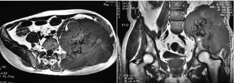
Figure 1 – Magnetic resonance imaging showing extensive expansive lesion on the left iliac wing with muscle and nerve invasion.
Figure 2 – Malignant round-cell neoplasm on slide with hematoxylin-eosin and with the immunohistochemical marker vimentin, compatible with mesenchymal chondrosarcoma.
Computed tomography on the pelvis showed an expansive bone lesion, with thinning of the cortical bone anteriorly and lytic cortical bone in the upper part of the wing of the left iliac bone, associated with increased volume of soft tissue adjacent to the lesion. Magnetic resonance imaging showed an extensive expansive lesion on the iliac wing, with clear invasion of the gluteal and left paravertebral musculature, and compromising of the sciatic nerve. The staging exami-nations did not show any distant metastases. Figure 1 shows the magnetic resonance imaging in axial and coronal slices, illustrating the extent of the lesion.
An incisional biopsy showed an undifferentiated malignant round-cell neoplasm. The immunohisto-chemical test was compatible with mesenchymal chondrosarcoma, as illustrated in Figure 2.
Since the physical examination and magnetic reso-nance imaging indicated invasion of the sciatic nerve
Figure 3 – Illustration of the flap.
Source: Mnaymneh W, Temple W. Modified hemipelvectomy utilizing a long vascular myocutane-ous thigh flap. Case report. J Bone Joint Surg Am. 1980;62(6):1013-5.
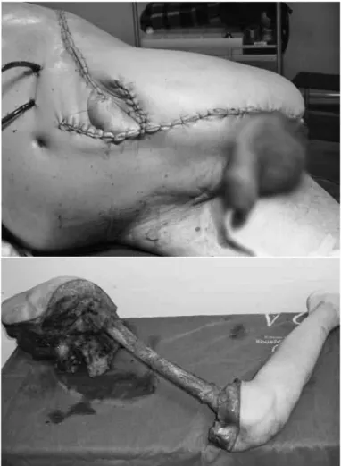
Figure 4 – Final result after resection and the surgical specimen.
means of the superficial and deep femoral arteries. After dissection of the flap, we rotated it posteriorly to cover the area of substance loss in the left hemi-pelvis. Figure 4 shows the surgical specimen and the final result after resection of the lesion.
After the resection, the patient evolved well, re-maining in the intensive care unit for two days and in the ward for 10 days. During the hospital stay, he evolved with a decubitus ulcer over the right iliac crest and in the sacral region close to the flap, with partial dehiscence of the edge of the wound in this region. Debridement was required, and complete res-olution was achieved after two months. During the first month of follow-up, the patient presented fever, and hyperemia and fluctuance under the flap were observed. Ultrasonography on the region showed an abscess under the flap, which was drained percutane-ously, guided by ultrasound, with resolution of the condition. There was good integration of the flap and the patient adapted well to using the pad for sitting down. No prosthesis was adapted for the case and the patient was referred for adjuvant chemotherapy and radiotherapy. Fourteen months after the resection, he presented local recurrence that was not resectable and was treated with palliative chemotherapy. During the chemotherapy, he evolved with febrile neutropenia and urinary sepsis, leading to death.
REPORT ON CASE 2
The patient was a 51-year-old divorced white man who was admitted to our service in February 2009 with a complaint of “a nodule in the right axilla”, which had been growing progressively for approxi-mately two months, associated with weight loss of 2
kg over six months. On examination, he presented an extensive hardened lesion occupying the entire right axillary region, measuring 26 x 19 x 17 cm. There were signs of collateral circulation; the scapular-hu-meral joint was displaced upwards and no palpable lymphadenopathy was observed. An incisional biopsy showed undifferentiated pleomorphic malignant neo-plasia with giant cells. Immunohistochemical evalu-ation showed that the condition was compatible with leiomyosarcoma with osteoclastic giant cells.
Thoracic tomography showed an expansive pro-cess in the soft-tissue region of the right axillary re-gion, measuring 175 mm along its longest axis, in-volving the scapula and presenting a contact surface with the clavicle, scapula and right axillary vessel-nerve bundle. The staging examinations did not show any distant metastases. Figure 5 shows the initial ap-pearance of the lesion and the corresponding imaging examination.
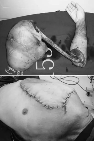
Figure 6 – Surgical specimen and final result after the resection.
Figure 5 – Appearance at the initial physical examination on the patient and the corresponding imaging examination.
adhere to the thoracic wall, but there was extensive ulceration of the overlying skin, associated with inva-sion of the subclavian vein and brachial plexus, while the subclavian artery was free. There were enlarged lymph nodes in the right axilla.
The patient underwent interscapular-thoracic dis-articulation. The reconstruction was done by means
of a posterior median incision along the axis of the right upper arm, taking a myocutaneous flap contai-ning the skin and all the anterior and posterior muscle tissue from the region of the right upper arm. The flap was isolated from the humerus and was dissected as far as the base of the clavicle, while maintaining the vascularization by means of the subclavian-axillary-brachial artery. After dissection of the flap, we rotated it posteriorly in order to cover the area of substance loss from the thoracic wall. Figure 6 shows the surgi-cal specimen and the final result after the resection.
The anatomopathological examination on the sur-gical specimen showed that this was a case of high-grade pleomorphic sarcoma, rich in giant cells, mea-suring 12 x 8.5 x 7.8 cm, with free surgical margins, as illustrated in Figure 7.
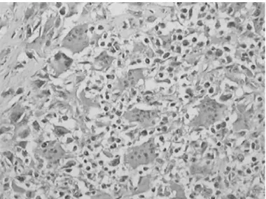
Figure 7 – Malignant giant-cell neoplasia, compatible with pleo-morphic sarcoma.
There was no complication relating to the flap, which integrated perfectly, without infection, dehiscence or necrosis. No prosthesis was used, and the patient did not receive any adjuvant therapy. He is now being followed up every six months, without any signs of disease recurrence.
DISCUSSION:
Tumors of the scapular and pelvic belt of small dimensions can be resected and reconstructed using local skin flaps(4-6). However, locally advanced tumors
of large dimensions require skin or myocutaneous flaps that can cover the area of substance loss(7- 9).
In conventional hemipelvectomy, the substance loss is repaired by means of a myocutaneous flap from the gluteus maximus muscle(10). Bowden and
Booher reviewed the treatment for sarcomas of the gluteal region and described a hemipelvectomy pro-cedure in which the gluteal myocutaneous flap could be partially resected with the lesion in cases in which the tumor invaded the gluteus muscle. To cover the posterior defect, the external iliac vessels and a small portion of the superficial femoral vessels were pre-served to nourish an anterior skin flap(1). Frey et al(11)
described using an anterior myocutaneous flap from the thigh. Sugarbaker and Chretien(12) gave a detailed
description of a hemipelvectomy procedure in which a large posterior defect was closed using an antero-lateral myocutaneous flap from the thigh, composed of femoral quadriceps muscles and nourished through the superficial femoral vessels.
Anatomical studies have shown that anterolateral flaps from the thigh are irrigated by myocutaneous
perforating vessels in more than 80% of the cases(13).
Today, such flaps have a very wide range of indi-cations such as in reconstructions of the head and neck, trunk and limb extremities(14-18), especially
because of the anatomical and safety characteristics of the flap and because of the minimal morbidity in the donor area.
Although the skin area of anterolateral thigh flaps is large, such that areas of 25 cm in length by 18 cm in width can survive with only one perforating vessel(13),
we decided to use a modification of the anterolateral myocutaneous thigh flap in the case presented here. In this, we used all of the musculature of the thigh after dissection of the femur, thus preserving the entire vascular bundle of the femoral artery and providing a tissue pad of greater volume over the sacrum, in order to cover the extensive area of substance loss. This would also have the weight-bearing capacity required for an upper prosthesis, when indicated. Although this type of flap modification is an option among the various flaps used for reconstruction subsequent to external hemipelvectomy, it has only been described once in the literature(2).
With regard to interscapular-thoracic disarticula-tion, large-dimension locally advanced tumors can be resected en bloc with ribs (from the first to the ninth), with or without associated pneumonectomy. In such cases, it is important to use skin or myocutaneous flaps(7-9). In cases in which the rib cage is preserved,
an open area can be left for secondary granulation and subsequent repair using a free skin graft from the fo-rearm or another region(1). When there is enough skin,
local flaps can be used(3,5,6), or a myocutaneous flap
from the deltoid(4). Other proposals for reconstructing
these substance losses include use of the pectoralis minor muscle, rotation of the greater omentum, and muscle flaps based on the latissimus dorsi with a ho-molateral pedicle(19-22). These flaps may be limited to
the extent of the substance loss and, in some cases, to the needs of the rib cage. In these cases, flaps with abdominal strips have a defined role(22). Flaps with the
homolateral latissimus dorsi can be used in recons-tructing moderate to large skin losses(23), in which the
main thoracodorsal pedicle is a branch of the subsca-pular artery coming from the axillary artery(24). Use
Mnaymneh and Temple apud Vieira et al(25) for ISTA.
In the case presented here, to reconstruct the ex-tensive area of substance loss after the interscapular-thoracic amputation, we chose to use a flap consisting of all of the anterior and posterior musculature of the upper arm after dissection of the humerus, thereby maintaining vascularization through the subclavian artery. With this flap, we provided the coverage needed
for the exposed region, by means of a flap for which the pedicle included a large-caliber vessel. This sustai-ned a substantial area of underlying soft tissue, thereby minimizing the depression in the thoracic wall that is created through substance loss.
Because these modified flaps are large, extensive unreconstructed open areas can easily be closed using conventional pedicled flaps.
REFERENCES
1. Vieira LJ, Vieira JP, Oliveira AF, Freitas RR. Hemipelvectomia com reconstrução por retalho miocutâneo anterior de coxa: relato de caso e descrição da técnica cirúrgica. Rev Bras Cancerol. 2004;50(4):301-5.
2. Mnaymneh W, Temple W. Modified hemipelvectomy utilizing a long vascular myocutaneous thigh flap. Case report. J Bone Joint Surg Am. 1980;62(6):1013-5. 3. Capanna R, Manfrini M, Briccoli A, Gherlinzoni F, Lauri G, Caldora P. Latissimus dorsi pedicled flap applications in shoulder and chest wall reconstructions after extracompartimental sarcoma resections. Tumori. 1995;81(1):56-62. 4. Gitelis S, Bertoni F, Picci P, Campanacci M. Chondrosarcoma of bone.
The experience at the Istituto Ortopedico Rizzoli. J Bone Joint Surg Am. 1981;63(8):1248-57.
5. Delay E, Bobin JY, Rivoire M, Franc C. [Full thickness reconstruction of the anterior chest wall with osteomusculocutaneous flap of the latissimus dorsi muscle]. Ann Chir Plast Esthet. 1994;39(2):204-10.
6. Kuhn JA, Wagman LD, Lorant JA, Grannis FW, Dunst M, Dougherty WR, et al. Radical forequarter amputation with hemithoracectomy and free exten-ded forearm flap: technical and physiologic considerations. Ann Surg Oncol. 1994;1(4):353-9.
7. Fianchini A, Bertani A, Greco F, Brunelli A, Muti M. Transthoracic forequarter amputation and left pneumonectomy. Ann Thorac Surg. 1996;62(6):1841-3. 8. Gentil FC. Indicações, técnica, tática cirúrgica e resultados da amputação
inter-escápulo-torácica no tratamento das neoplasias malignas [tese livre docência]. Campinas, SP: Faculdade de Ciências Médicas da Pontifícia Universidade Católica de Campinas; 1978.
9. Lassen M, Krag C, Nielsen IM. The latissimus dorsi flap. An overview. Scand J Plast Reconstr Surg. 1985;19(1):41-51.
10. Sugarbacker P, Henshaw R, Malawer M. Hemipelvectomia de retalho anterior. In: Malawer MM, Sugarbacker PH, Lopes A. Atlas de cirurgia para sarcomas ósseos e de partes moles. São Paulo: Lemar; 2003.
11. Frey C, Matthews LS, Benjamin H, Fidler WJ. A new technique for hemipelvec-tomy. Surg Gynecol Obstet. 1976;143(5):753-6.
12. Sugarbaker PH, Chretien PA. Hemipelvectomy for buttock tumors utilizing na anterior myocutaneous flap of quadriceps femoris muscle. Ann Surg. 1983;197(1):106-15.
13. Wei FC, Jain V, Celik N, Chen HC, Chuang DC, Lin CH. Have we found an
ideal soft-tissue flap? An experience with 672 anterolateral thigh flaps. Plast Reconstr Surg. 2002;109(7):2219-26.
14. Koshima I, Fukuda H, Yamamoto H, Moriguchi T, Soeda S, Ohta S. Free antero-lateral thigh flaps for reconstruction of head and neck defects. Plast Reconstr Surg. 1993;92(3):421-8.
15. Kimura N, Satoh K, Hasumi T, Ostuka T. Clinical application of the free thin anterolateral thigh flap in 31 consecutive patients. Plast Reconstr Surg. 2001;108(5):1197-208.
16. Hsieh CH, Yang CC, Kuo YR, Tsai HH, Jeng SF. Free anterolateral thigh adi-pofascial perforator flap. Plast Reconstr Surg. 2003;112(4):976-82.
17. Adani R, Tarallo L, Marcoccio I, Cipriani R, Gelati C, Innocenti M. Hand reconstruction using the thin anterolateral thigh flap. Plast Reconstr Surg. 2005;116(2):467-73.
18. Yang WG, Chiang YC, Wei FC, Feng GM, Chen KT. Thin anterolateral thigh per-forator flap using a modified perper-forator microdissection technique and its clini-cal application for foot resurfacing. Plast Reconstr Surg. 2006;117(3):1004-8. 19. Levine EA, Warso MA, McCoy DM, Das Gupta TK. Forequarter amputation for
soft tissue tumors. Am Surg. 1994;60(5):367-70.
20. Mansour KA, Powell RW. Modified technique for radical transmediastinal forequarter amputation and chest wall resection. J Thorac Cardiovasc Surg. 1978;76(3):358-63.
21. Nielsen IM, Lassen M, Gregersen BN, Krag C. Experience with the latissimus dorsi flap. Scand J Plast Reconstr Surg. 1985;19(1):53-63.
22. Petty PM, Terkonda SP, Shives TC. Reconstruction of soft-tissue defects. In: Simon MA, Springfield D. Surgery for bone and soft-tissue tumors. Philadelphia: Lippincott-Raven, 1998;585-96
23. Stafford ES, Williams GR Jr. Radical transthoracic forequarter amputation. Ann Surg. 1958;148(4):699-703.
24. Yamamoto Y, Sugihara T, Kawashima K, Qi F. An anatomic study of the latis-simus dorsi-rib flap: an extension of the subscapular combined flap. Plast Reconstr Surg. 1996;98(5):811-6.