LANG MG ETAL.
298 REV ASSOC MED BRAS 2014; 60(4):298-301
IMAGE IN MEDICINE
Mycotic aneurysm as a complication of infective endocarditis – a
case report
A
NEURISMA MICÓTICO COMO COMPLICAÇÃO DE ENDOCARDITE INFECCIOSA–
RELATO DE CASOMARIA GABRIELA LANG1*, MARIELLE LANG2, RAFAEL RONSONI3
1 Resident physician of Clinical Medicine at the Hospital Municipal São José, Joinville, SC, Brazil.
2 Resident physician of Clinical Medicine at the Hospital Universitário da Região Norte do Paraná - State University of Londrina. Londrina, PR, Brazil. 3 Assistant physician to the Clinical Medicine service at the Hospital Municipal São José - Joinville, SC, Brazil.
Study conducted at the Hospital Municipal São José, Joinville, SC.
Approved by the Research Ethics Committee at the Hospital Municipal São José on 09/19/2013.
*Correspondence:
Address: Rua Saí, 44, apt 204; CEP 89202-170; B. Anita Garibaldi; Joinville/SC - Brazil
Phone: +55 47 3441-6666 / 9600-1172 [email protected]
http://dx.doi.org/10.1590/1806-9282.60.04.005
Conflict of interest: none
I
NTRODUCTIONThe first mention of the clinical aspects of infectious endocarditis (IE) comes from French physician Jean François Fernel in the 16th century.1 However,
appropri-ate treatment only started being implemented more than a century later, when the presence of microorgan-isms as the cause of the disease was identified.2 IE
nor-mally affects the native valves as a primary site, being the mitral valve the most common.3 In the last thirty
years there have been important advancements in the diagnostic and therapeutic methods for the disease, which despite the low incidence in the population in general, still presents a poor prognosis and high mor-tality rate.4,5,6 IE complications appear in 60% of patients,
being mycotic aneurysm one of the most serious. Caused by weakening of the vessel walls, it is clinically recog-nized in 3-10% of IE cases.7 The most dramatic event is
subarachnoid hemorrhage, which usually occurs in the stage of bacteremia.6
C
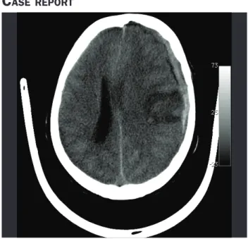
ASE REPORTFIGURE 1 Computerized tomography of the cranium showing
acute frontoparietal subdural hematoma to the left with a subjacent frontoparietal focus of contusion
MYCOTICANEURYSMASACOMPLICATIONOFINFECTIVEENDOCARDITIS – ACASEREPORT
REV ASSOC MED BRAS 2014; 60(4):298-301 299
FIGURE 2 Angiography showing aneurysm formation, measuring
about 8 × 9mm on topographic examination of cortical branch of the middle cerebral artery associated with parietal irregularity of the vessel, as indicated by the white arrow.
Source: HMSJ neuroradiology service.
FIGURE 3 Postembolization arteriogram with coils, as indicated
by the white arrow. Source: HMSJ neuroradiology service.
Female patient, 45 years old, born in São Jorge d’Oeste, state of Paraná, Brazil, and originating from Joinville, San-ta CaSan-tarina, white, married, building janitor. She was ad-mitted to the Hospital Municipal São José (HMSJ) for in-vestigation of epigastric pain associated with holocrani-al headache, intermittent and progressive, with 1 month of evolution and significant deterioration in the last 2 days. No further complaints.
Hypertensive and hypothyroid patient, treated with captopril 25 mg T.I.D. and levothyroxine 50 mcg/day. Un-derwent thyroidectomy 8 months before admission for adenomatous goiter. Patient denies allergies and previ-ous hospitalization.
On physical examination, she showed good general condition, pale + / 4 +, hydrated, eupneic, tachycardia (HR: 110), afebrile, acyanotic, anicteric, lucid and orient-ed in time and space. In the lung auscultation, vesicular murmur present and symmetrical, with no adventitious sounds. In the cardiovascular examination, presence of systolic murmur from regurgitation, pancardiac 4+/6+ radiating to the left armpit and more audible in the mi-tral zone, with tremor present, ictus cordis of normal size,
palpable and visible. The presence of lesions compatible with Janeway lesions on the right fifth toe was verified. No signs of meningeal irritation and no focal deficits.
Additional tests were requested: cranial tomography that showed no presence of acute lesions; CSF without changes in cellularity, protein, glucose and culture-neg-ative; negative blood culture; anti-HCV non-reactive, an-ti-HIV nonreactive and HBsAg reactive serologies; echo-cardiogram demonstrating redundant mitral valve with intense systolic posterior movement and nodule image on atrial side of the anterior leaflet suggesting vegetation. Given the findings, IE diagnosis was established based on the modified Duke criteria (two major and one mi-nor).8 Treatment started with 60 mg of gentamicin T.I.D.
and 1g of vancomycin B.I.D. after hospital admission. Transesophageal ultrasound performed on the twenty-seventh day of hospitalization showed a 40% reduction of mitral valve vegetation, with antibiotic treatment re-placed by 2 g oxacillin Q.I.D.
frontoparie-LANG MG ETAL.
300 REV ASSOC MED BRAS 2014; 60(4):298-301
tal focus of contusion. The patient was sent for an emer-gency decompressive hemicraniectomy. The next day the patient was submitted to angiography, which revealed an aneurysm measuring about 8 × 9mm on topography of the cortical branch of the left middle cerebral artery asso-ciated with parietal irregularity of the vessel – image com-patible with a ruptured mycotic aneurysm, which was em-bolized with coils. After the procedure, the patient was referred to the Intensive Care Unit at the hospital.
On the sixty-seventh day of hospitalization orotra-cheal extubation was performed, and after three days the patient was then referred to clinical medicine ward. The patient initially remained aphasic, but presented progres-sive improvement with multi-professional rehabilitation. The patient was discharged on the ninety-fifth day of hos-pitalization with moderate disability (grade 3 on the mod-ified Rankin scale),9 after completing 42 days of treatment
with the latest antimicrobial scheme. They were referred for monitoring by hepatitis and cardiology outpatient clinics (with valvuloplasty scheduled). The patient under-went skull reconstruction 60 days after discharge, with no adverse events.
D
ISCUSSIONThe case reported is consistent with the reality of IE in medical practice in various aspects. The patient, aged 47, and with native valves is categorized under the new epi-demiological profile of the disease, which was previously reserved for patients with compromised valves and young adults. However, the entry point that could have caused bacteremia was not identified, as is possible in the majo-rity of cases.10
The patient sought the health service with specific complaints, presenting a physical exam with various signs of cardiovascular impairment. This paradox is not fre-quent, but possible, considering the presence of underly-ing heart disease and the characteristics of the patient.6
The literature demonstrates that although approximate-ly 90% of patients present fever and 80% have heart mur-murs, the diagnosis is difficult.10 In the majority of cases,
IE is only diagnosed accurately around one month after hospital admission, with transesophageal ultrasound as an indispensable exam for it presents sensitivity over 90%. In the present case, the exam was capable of showing mi-tral valve vegetation, at the location most commonly af-fected by IE.11,12 Delays of weeks or months in
antimicro-bial treatment increase the chance of the occurrence of embolic or immunological events.6
In relation to the blood culture, the case presented differs from European studies where 2.5 to 31% of cases
demonstrate negativity, but is very similar to that found in a Senegalese study conducted from 2005 to 2011, in which 60% of blood cultures were negative.4,12,13
After almost two months of clinical evolution the pa-tient presented a mycotic aneurysm, as a consequence of displacement of septic emboli from valvular vegetation to the arterial vasa vasorum, disseminating the infection
to the inner layer and wall of the vessel.5,13 This
compli-cation is rare, occurring in around 2-4% of cases, and as reported, more than half (57.4%) are located in the distal branches of the medium cerebral artery.7,13,14,15
The cerebral hemorrhage presented by the patient can be justified by the fact that mycotic aneurysms gen-erally present a fine and fragile wall.7 A multicenter study
conducted in France showed that 65-80% of mycotic an-eurysms are asymptomatic, diagnosed only by imaging exams. In the case of rupture, however – as in the case pre-sented here, in which the patient experienced convulsions and changes to her level of consciousness –, mortality reaches 80%.5,10,11,14,16 In contrast with ruptured aneurysms,
unruptured ones have a much lower mortality rate (30%) and treatment is generally based on antibiotics and pa-tient monitoring.5,14
In addition to the difficulty in diagnosis, the treat-ment of this complication does not yet have defined pro-tocols, which makes conduct individualized and based on the clinical symptoms of the patient, as well as on the cost-benefit analysis of the treatment strategies.10 The
majority of the Guidelines advise waiting 6 to 8 weeks for performing cardiac surgery on patients with a hemor-rhagic event secondary to IE.15 Even if single antibiotic
treatment is responsible for curing the majority of cases, not conducting surgery could lead to rupture of the an-eurysm and catastrophic consequences.5
The long period of hospitalization of the patient (near-ly 100 days) may be explained by the occurrence of cere-bral hemorrhage, corroborated by the study conducted in northeastern Italy, which demonstrates an increase in the average hospitalization from 23 to 35 days in the pres-ence of complications associated with IE.17 Despite this,
like the majority of survivors of cerebrovascular events secondary to IE evaluated by the study by Yeates, the pa-tient recovered well from her neurological impairments.15
R
EFERENCES1. Millar BC, Moore JE. Emerging issues in infective endocarditis. Emerg Infect Dis. 2004;10:1110-6.
2. Contrepois A. Towards a history of infective endocarditis. Med Hist. 1996;40:25-54.
MYCOTICANEURYSMASACOMPLICATIONOFINFECTIVEENDOCARDITIS – ACASEREPORT
REV ASSOC MED BRAS 2014; 60(4):298-301 301
4. Leone S, Ravasio V, Durante-Mangoni E, Crapis M, Carosi G, Scotton PG, et al. Epidemiology, characteristics, and outcome of infective endocarditis in Italy: Italian study on endocarditis. Infection. 2012;40:527-35. 5. Bayer AS, Bolger AF, Taubert KA, Wilson W, Steckelberg J, Karchmer AW,
et al. Diagnosis and management of infective endocarditis and its complications. Circulation. 1998;98:2936-48.
6. Suknjaja V, Popovic N, Božic S, Sakalaš L, Milojkovic J, Hajder D. Embolic events and neurological complications in infective endocarditis. Curr Top Neurol Psychiatr Relat Discip. 2011;19:49-53.
7. Ducruet AF, Hickman JL, Zacharia BE, Narula R, Grobelnv BT, Gorski J, et al. Intracranial infectious aneurysms: a comprehensive review. Neurosurg Rev. 2010;33:37-46.
8. Durack DT, Lukes AS, Bright DK. New criteria for diagnosis of infective endocarditis: utilization of specific echocardiographic findings. Duke Endocarditis Service. Am J Med. 1994;96:200-9.
9. Wilson LJT, Hareedran A, Grant M, Baird T, Schulz UG, Muir KW, et al. Improving the assessment of outcomes in stroke: use off a structured interview to assign grades on the modified rankin scale. Stroke. 2002;33:2243-6. 10. Habib G, Hoen B, Tornos P, Thuny F, Prendergast B, Vilacosta I, et al. Guidelines
on the prevention, diagnosis and treatment of infective endocarditis. Eur Heart J. 2009;30:2369-413.
11. Westphal N, Plicht B, Naber C. Infective endocarditis-prophylaxis, diagnostic criteria, and treatment. Dtsch Arztebl Int. 2009;106:481-9.
12. Kazelian LR, Vidal LA, Neme R, Gagliardi JA. [Endocarditis infecciosa activa: 152 casos]. Medicina (B Aires) 2012;72:109-14.
13. Pessinaba S, Kane A, Ndiaye NB, Mbaye A, Bodian M, Dina MM, et al. Vascular complications of infective endocarditis. Med Mal Infect. 2012;42:213-7. 14. Fukuda W, Daitoku K, Minakawa M, Fukui K, Suzuki Y, Fukuda I. Infective
endocarditis with cerebrovascular complications: timing of surgical intervention. Interact Cardiovasc Thorac Surg. 2011;14:26-30.
15. Yeates A, Mundy J, Griffin R, Marshall L, Wood A, Peters P, et al. Early and mid-term outcomes following surgical management of infective endocarditis with associated cerebral complications: a single centre experience. Heart Lung Circ. 2010;19:523-7.
16. Romain S, Mirabel M, Hajage D, Tubach F, Vignon P, Perez P, et al. Neurologic complications and outcomes of infective endocarditis in critically ill patients: The ENDOcardite en REAnimation prospective multicenter study. Crit Care Med. 2011;39:1474-81.
17. Fideli U, Schievano E, Buonfrate D, Pellizzer G, Spolaore P. Increasing incidence and mortality of infective endocarditis: a population-based study through a record-linkage system. BMC Infect Dis. 2011;11:2-7.