Arq Neuropsiquiatr 2006;64(4):1033-1035
Discipline of Diagnostic Radiology, Department of Internal Medicine, University of Paraná (ELG, JMR, TD, ACN) and DAPI - Diagnóstico Avançado Por Imagem (ACN), Curitiba PR, Brazil.
Received 22 February 2006, received in final form 20 July 2006. Accepted 5 September 2006.
D r. Arnolfo de Carvalho Neto DAPI Diagnóstico Avançado Por Imagem Rua Brigadeiro Franco 122 80430210 Curitiba PR -Brasil. E-mail: [email protected]
CEREBRAL X-LINKED ADRENOLEUKODYSTROPHY
Follow-up with magnetic resonance imaging
Emerson L. Gasparetto, Juliana Mecunhe Rosa,
Taísa Davaus, Arnolfo de Carvalho Neto
ABSTRACT -Objective:To re p o rt a case of childhood cerebral X-linked adre n o l e u k o d y s t rophy (X-ADL), emphasizing the magnetic resonance imaging (MRI) findings at initial evaluation and at the follow-up.
Case report: Five year-old boy, who was asymptomatic, presented with diagnosis of X-ADL for MRI evalu-ation. The initial brain MRI showed a focal area of enhancement at the splenium of the corpus calosum. One year later, the follow-up MRI showed a pro g ression of the corpus calosus lesion, as well as other lesions in the parietal and occipital lobes. Conclusion:The brain MRI follow-up of patients with X-ADL is impor-tant to show the progression of the lesions.
KEY WORDS: childhood cerebral X-linked adrenoleukodystrophy, magnetic resonance imaging.
Adrenoleucodistrofia ligada ao X: acompanhamento por ressonância magnética
RESUMO - Objetivo:Relatar um caso de adre n o l e u c o d i s t rofia ligada ao X (X-ADL), enfatizando os achados de ressonância magnética (RM) na avaliação inicial e no seguimento. Descrição do caso:Paciente mas-culino de cinco anos de idade, assintomático, com diagnóstico de X-ADL, apresentou-se para estudo de RM. O exame inicial mostrou uma área focal de realce no esplênio do corpo caloso. Após um ano, a RM de seguimento evidenciou aumento da lesão do corpo caloso, assim como novas lesões nos lobos occipi-tais e parieoccipi-tais. Conclusão:O seguimento por RM de pacientes com X-ADL é importante para a demons-tração da progressão das lesões.
PALAVRAS-CHAVE: adrenoleucodistrofia ligada ao X, ressonância magnética.
X-linked adre n o l e u k o d y s t rophy (X-ADL) is a genet-ically determined rare metabolic pro g ressive disor-d e r, which involves the central nervous system (CNS), a d renal cortex and testicles. It is related to accumula-tion of very-long-chain fatty acids in plasma and tis-sues, caused by a deficient peroxisomal membrane p rotein. The clinical manifestation has a wide vari-ety within two major forms; the childhood cerebral form, causing a severe disability that leads to death early; and the adre n o m y e l o n e u ro p a t h y, a milder adult form witch involves mainly the spinal cord and peripheral nerves, presenting a slow pro g ression with better prognostic. The bone marrow transplantation (BMT) is the only effective long-term treatment for X-ADL disease, however must be perf o rmed at an early stage of cerebral disease1 , 2. Brain magnetic re
s-onance imaging (MRI) is an essential tool to evalu-ate patients with X-ADL. It allows early detection of CNS lesions, and the serial follow-up aids to predict
the prognosis of the disease and monitoring thera-peutic interventions. MRI changes are caused by an i n f l a m m a t o ry demyelization process resulting in pro-longation of T1 and T2 relaxation times. Areas of in-volvement are best seen as foci of hyperintense sig-nal on T2-wheighted images, with or without en-hancement after contrast administration3. Loes et al.4
s-1034 Arq Neuropsiquiatr 2006;64(4)
sion. The characteristic MRI features of childhood cerebral X-linked ALD have been well documented. H o w e v e r, there are few re p o rts discussing the MRI findings at serial follow-up evaluation.
The aim of this article is to re p o rt a case of child-hood cerebral X-ADL, emphasizing the MRI findings at initial evaluation and at the follow-up MRI.
CASE
A five years-old boy, who was asymptomatic, pre s e n t-ed with diagnosis of X-ADL for MRI evaluation. The neu-rological examination was normal. He had a positive famil-iar history of X-ALD (two maternal uncles) and the diagno-sis was confirmed at the birth by biochemical and genetic methods. No previous treatments were referred.
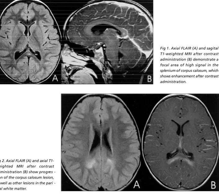
The initial brain MRI showed a focal area of high sig-nal on T2-weighted images and enhancem ent after con-trast administration at the splenium of the corpus calosum (Fig 1). The patient was scored one following the Loes’s cri-teria4.
One year later the patient retuned to perf o rmthe fol-low-up MRI. At this time he remained asymptomatic, with normal neurological examination. The MRI demonstrated a reas of high signal on T2-w eighted images with periph-eral enhancement after contrast administration on the sple-nium of the corpus calosum, as well as on the white mat-ter of the parietal and occipital lobes (Fig 2). The lesion at the corpus calosum was considerably large in this second MRI. The patient was then scored as three considering the L o e s ’s criteria4and he was re f e rred to the bone marro w transplantation (BMT) unit of the Hospital de Clínicas of the University of Paraná f or evaluati on. At this moment, the parents agreed with the publication of the case and signed the consent.
DISCUSSION
Childhood cerebral X-linked ALD is the most fre-quently and severe form of ALD, being present in 35% to 40% of boys with the biochemical diagnosis of X-ALD. Patients usually show normal development
Fig 1. Axial FLAIR (A) and sagital T1-weighted MRI after contrast administration (B) demonstrate a focal area of high signal in the splenium of corpus calosum, which shows enhancement after contrast administration.
Arq Neuropsiquiatr 2006;64(4) 1035
until 4-10 years of age. Clinical manifestations are characterized by adrenal insufficiency and neuro l o g-ical deterioration that leads to death within months to years1. BMT can an effective way of tre a t m e n t ,
which can achieve long term stabilization for patients who are at risk of rapid pro g ression, only if under-went at an early enough stage1 , 2. The monitoring of
asymptomatic patients with noninvasive neuro i m a g-ing and neuropsychological studies may conduct to the early detection of cerebral involvement1. In the
p resent case, the diagnosis of X-ADL was based on biochemical and genetic methods. The investigation was perf o rmed because the familiar history of X-ADL (two maternal uncles).
M R I is essential for both initial and follow-up eval-uation of childhood cerebral X-linked ALD. The CNS characteristics lesions include extensive demyeliza-tion in the periventricular deep white matter, cavi-tations, and perivascular lymphocyte infiltrates. The white matter demyelization most often begins in the parietal and occiptal regions and pro g ress to the an-terior and lateral regions. The follow-up MRI may show improvement, stabilization, or aggravation of the lesions5. In our case, the initial MRI showed a
small lesion in the splenium of the corpus calosum, and the follow-up MRI demonstrated enlarg e m e n t of this lesion, as well as the presence of other lesions in the parietal and occipital lobes.
A MRI severity scale score for patients with X-ADL was proposed by Loes et al.4, based on the location
and extension of the lesions, and presence of focal and/or global atro p h y. The scale consists in a point system (0 to 34), where each region of the brain is s c o red as 0 if normal; 0.5 if there is an unilateral lesion or if the involvement is questionable and the patient had no other abnormalities; and 1 if the le-sion or atrophy is bilateral. The present case was scor-ed as one at the initial evaluation, considering the corpus calosum lesion, and as three on the follow-up MRI because of the several brain abnormalities.
E m e rging MRI techniques such as proton MR spec-t roscopy idenspec-tify impending or beginning lesions spec-thaspec-t still appears normal on conventional MRI. It can be considered suitable for the prediction of lesion pro-g ression on MRI in X-ADL. Accordinpro-g to Eichler at al.6,
a N-acetylaspartate (NAA) to choline ratio in norm a l appearing white matter of less than 5.0 is stro n g l y p redictive of subsequent lesion pro g ression on MRI.
Loes et al.7re p o rted five patterns of X-ADL based
on the anatomic location, age of the patient and ini-tial MRI severity scale score. The suggested that those p a t t e rns could allow predicting the disease pro g re s-sion. Pattern 1, the most common, was defined as p r i m a ryinvolvement of posterior white matter, main-ly on boys younger than 10 and has re s e rved pro g-nosis especially if contrast enhancement is pre s e n t and if the abnormalities show up at an early age. The second most common (pattern 2) in the 10- to 16-y e a r-old patients has a frontal involvement, the pro-g ression vary with the apro-ge and severity score but also with a bad prognosis. The pattern 3, most common in adults, involving the frontopontine or cort i c o s p i n a l projection fibers, and pattern 4, which primarily in-volves the cerebellum in late teenage years, have both a much slower pro g ression. Pattern 5 defined as combined but separate parieto-occciptal and fro n-tal white matter lesions at an early age pro g re s s e d v e ry rapidly. Our case presented with the Loes’s pat-t e rn1. The inipat-tial MRI was scored as one and pat-the fol-low-up MRI, one year late, as three.
In conclusion, patients with childhood cerebral X-linked ALD may present areas of high signal on T2-weighted images and enhancement after contrast administration. These lesions usually predominate in the splenium of the corpus calosum and in the pari-eto-occipital regions. The application of the Loes’s criteria is important at the initial MRI evaluation of patients with X-ADL, as well as it is helpful in the fol-low-up of affected patients.
REFERENCES
1. Peters C, Charnas RL, Tan Y, et al. Cerebral X-linked adre n o l e u k o d y-s t rophy: the international hematopoietic cell trany-splantation experi-ence from 1982 to 1999. Blood 2004;104:1881-1888
2. Loes DJ, Stillman AE, Hite S, et al. Childhood cerebral form of adre n o-l e u k o d y s t rophy: short-term effect of bone marrow transpo-lantation on brain MR observations. AJNR 1994;15:1767-1771.
3. Melhem ER, Loes DJ, Georgiades CS, Raymond GV, Moser HW. X-linked adre n o l e u k o d y s t rophy: the role of contrast-enhanced MR imag-ing in predictimag-ing disease progression. AJNR 2000;21:839-844. 4. Loes DJ, Hite S, Moser H, et al. A d re n o l e u k o d y s t rophy: a scoring
meth-od for brain MR observations. AJNR 1994;15:1761-1766.
5. Kim JH, Kim HJ. Childhood X-linked A d re n o l e u k o d y s t rophy: clinical-pathologic overview and MR imaging manifestation at initial evalua-tion and follow-up. RadioGraphics 2005;25:619-631.
6. Eichler FS, Barker PB, Cox C, et al. Proton MR spectroscopic imaging p redicts lesion pro g ression on MRI in X-linked adre n o l e u k o d y s t ro p h y. Neurology 2002;58:901-907.